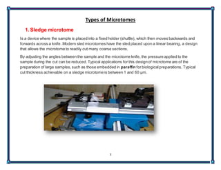
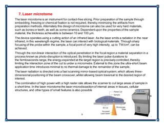
SlideShare a Scribd company logo
1
Ain shams university
Faculty of science
Zoology department
Prepared and presented by
GeorgeMagdy
2
Notes on Microtome
A microtome (from the Greek mikros, meaning "small", and temnein, meaning "to cut") is a
device used to cut extremely thin slices of material, known as sections. Important in science,
microtomes are used in microscopy, allowing for the preparation of samples for observation
under transmitted light or electron radiation.
Microtomes use steel, glass, or diamond blades depending upon the specimen being sliced and
the desired thickness of the sections being cut.
1. Steel blades are used to prepare sections of animal or plant tissues for light
microscopy histology.
2. Glass knives are used to slice sections for light microscopy and to slice very thin sections
for electron microscopy
3. Diamond knives are used to slice hard materials such as bone, teeth and plant matter for
both light microscopy and for electron microscopy.
Gem quality diamond knives are used for slicing thin sections for electron microscopy.
Microtomy is a method for the preparation of thin sections for materials such as bones,
minerals and teeth, and an alternative to electropolishing and ion milling. Microtome sections
can be made thin enough to section a human hair across its breadth, with section thickness
between 50 nm and 100 µm.
3
Types of Microtomes
1. Sledge microtome
Is a device where the sample is placed into a fixed holder (shuttle), which then moves backwards and
forwards across a knife. Modern sled microtomes have the sled placed upon a linear bearing, a design
that allows the microtome to readily cut many coarse sections.
By adjusting the angles between the sample and the microtome knife, the pressure applied to the
sample during the cut can be reduced. Typical applications for this designof microtome are of the
preparation of large samples,such as those embedded in paraffin forbiologicalpreparations. Typical
cut thickness achievable on a sledge microtome is between 1 and 60 µm.
4
2. Rotary microtome
This instrument is a commonmicrotome design.This device operates with a staged rotary action such
that the actual cutting is part of the rotary motion. In a rotary microtome,the knife is typically fixed in a
horizontal position, the principle of the cut is explained. Through the motion of the sample holder, the
sample is cut by the knife, at which point the freshsection remains on the knife.
At the highest point of the rotary motion, the sample holder is advanced by the same thickness as the
sectionthat is to be made, allowing the next sectionto be made.
The flywheel in many microtomescan be operated by hand. This has the advantage that a clean cut can
be made, as the relatively large mass of the flywheel prevents the sample from being stoppedduring the
sample cut. The flywheel in newer models is oftenintegrated inside the microtome casing.The typical
cut thickness for a rotary microtome is between1 and 60 µm.
For hard materials, such as a sample embedded in a synthetic resin, this designof microtome can allow
good "semi-thin" sections with a thickness of as low as 0.5 µm
5
3. Cryomicrotome
For the cutting of frozen samples,many rotary microtomes can be adapted to cut in a liquid-nitrogen
chamber, in a so-called cryomicrotomesetup.
The reduced temperature allows the hardness of the sample to be increased, such as by undergoing a
glass transition, which allows the preparation of semi-thin samples.
However the sample temperature and the knife temperature must be controlled in orderto optimise the
resultant sample thickness.
6
4. Ultramicrotome
It allows the preparation of extremely thin sections,with the device functioning in the same manner as a
rotational microtome,but with very tight tolerances on the mechanical construction.
As a result of the careful mechanical construction, the linear thermal expansion of the mounting is used
to provide very fine control of the thickness.
These extremely thin cuts are important for use with transmission electron microscope(TEM)and serial
block-face scanning electron microscopy (SBFSEM),and are sometimesalso important for light-optical
microscopy
The typical thickness of these cuts is between 40 and 100 nm for transmission electronmicroscopyand
often between 30 and 50 nm for SBFSEM.Thicker sections up to 500 nm thick are also taken for
specialized TEM applications or for light-microscopysurvey sections to selectan area for the final thin
sections.
Diamond knives (preferably)and glass knives are used with ultramicrotomes.To collectthe sections,
they are floated on top of a liquid as they are cut and are carefully picked up onto grids suitable for TEM
specimenviewing. The thickness of the sectioncan be estimated by the thin-film interference colors of
reflected light that are seenas a result of the extremely low sample thickness.
7
5. Vibrating microtome
The vibrating microtome operates by cutting using a vibrating blade, allowing the resultant cut to be
made with less pressure than would be required for a stationary blade.
The vibrating microtome is usually used for difficultbiological samples.
The cut thickness is usually around 30–500 µm forlive tissue and 10–500µm for fixed tissue.
8
6. Saw microtome
The saw microtome is especiallyfor hard materials such as teeth or bones.
The microtome of this type has a recessed rotating saw, which slices through the sample.
The minimal cut thickness is approximately 30 µm and can be made for comparatively large samples.
9
7. Laser microtome
The laser microtome is an instrument for contact-free slicing. Prior preparation of the sample through
embedding,freezing or chemical fixation is not required, thereby minimizing the artifacts from
preparation methods.Alternately this design of microtome can also be used for very hard materials,
such as bones or teeth, as well as some ceramics.Dependentupon the properties of the sample
material, the thickness achievable is between 10 and 100 µm.
The device operates using a cutting action of an infrared laser. As the laser emits a radiation in the near
infrared, in this wavelength regime, the laser can interact with biologicalmaterials. Through sharp
focusing of the probe within the sample,a focalpoint of very high intensity, up to TW/cm2
,can be
achieved.
Through the non-linear interaction of the optical penetration in the focal region a material separation in a
process known as photo-disruptionis introduced. By limiting the laser pulse durations to
the femtoseconds range,the energy expended at the target region is preciselycontrolled,thereby
limiting the interaction zone of the cut to under a micrometre.External to this zone the ultra-short beam
application time introduces minimal to no thermal damage to the remainder of the sample.
The laser radiation is directed onto a fast scanning mirror-based optical system,which allows three-
dimensional positioning of the beam crossover,whilst allowing beam traversal to the desired region of
interest.
The combination of high power with a high raster rate allows the scanner to cut large areas of sample in
a short time. In the laser microtome the laser-microdissectionof internal areas in tissues, cellular
structures, and other types of small features is also possible.

More Related Content

What's hot

1. Microtome
1. Microtome1. Microtome
1. Microtome
Kishor Bahadur Bist
 
Microtome knife profile
Microtome knife profileMicrotome knife profile
Microtome knife profile
Faris K
 
Microtomy
MicrotomyMicrotomy
Microtomy
Ekta Jajodia
 
Cryostat and frozen section
Cryostat and frozen sectionCryostat and frozen section
Cryostat and frozen section
SUNIL KUMAR PEDDANA
 
Embedding
EmbeddingEmbedding
Fixatives
FixativesFixatives
Fixatives
Snigdha Das
 
Microtomy
MicrotomyMicrotomy
Microtomy
Swati Wadhai
 
Histotechnique
HistotechniqueHistotechnique
Histotechnique
DrSyed Asif
 
Microtomes, Section Cutting and Sharpening of razor
Microtomes, Section Cutting and Sharpening of razorMicrotomes, Section Cutting and Sharpening of razor
Microtomes, Section Cutting and Sharpening of razor
Vikash Prasad
 
Fixation & fixatives in histopathology, dr naveen reddy
Fixation & fixatives in histopathology, dr naveen reddyFixation & fixatives in histopathology, dr naveen reddy
Fixation & fixatives in histopathology, dr naveen reddy
Naveen Parvathareddy
 
MICROTOME and it's TYPES
MICROTOME and it's TYPES MICROTOME and it's TYPES
MICROTOME and it's TYPES
ShahzebHUSSAIN5
 
Fixation
Fixation Fixation
Types Of Microtomes In Histopathology
Types Of Microtomes In Histopathology Types Of Microtomes In Histopathology
Types Of Microtomes In Histopathology
Omnia Elamin
 
Fixatives in Histopathology
Fixatives in HistopathologyFixatives in Histopathology
Fixatives in Histopathology
Ishwar9
 
2. fixatives and fixation seminar
2. fixatives and fixation seminar2. fixatives and fixation seminar
2. fixatives and fixation seminar
Dr SANTHIPRIYA GOPASANA
 
Lecture 2. tissue processing
Lecture 2. tissue processingLecture 2. tissue processing
Lecture 2. tissue processing
Catholic university of Rwanda
 
Gross Examination, Selection, Collection and Fixation of Specimen
Gross Examination, Selection, Collection and Fixation of Specimen Gross Examination, Selection, Collection and Fixation of Specimen
Gross Examination, Selection, Collection and Fixation of Specimen
ghulam abbas
 
Microtome
MicrotomeMicrotome
Microtome
Gaurav Kaushik
 
Automation histopathology
Automation histopathologyAutomation histopathology
Automation histopathology
Ankita Sain
 
Tissue processing
Tissue processingTissue processing
Tissue processing
Akash Dhiman
 

What's hot (20)

1. Microtome
1. Microtome1. Microtome
1. Microtome
 
Microtome knife profile
Microtome knife profileMicrotome knife profile
Microtome knife profile
 
Microtomy
MicrotomyMicrotomy
Microtomy
 
Cryostat and frozen section
Cryostat and frozen sectionCryostat and frozen section
Cryostat and frozen section
 
Embedding
EmbeddingEmbedding
Embedding
 
Fixatives
FixativesFixatives
Fixatives
 
Microtomy
MicrotomyMicrotomy
Microtomy
 
Histotechnique
HistotechniqueHistotechnique
Histotechnique
 
Microtomes, Section Cutting and Sharpening of razor
Microtomes, Section Cutting and Sharpening of razorMicrotomes, Section Cutting and Sharpening of razor
Microtomes, Section Cutting and Sharpening of razor
 
Fixation & fixatives in histopathology, dr naveen reddy
Fixation & fixatives in histopathology, dr naveen reddyFixation & fixatives in histopathology, dr naveen reddy
Fixation & fixatives in histopathology, dr naveen reddy
 
MICROTOME and it's TYPES
MICROTOME and it's TYPES MICROTOME and it's TYPES
MICROTOME and it's TYPES
 
Fixation
Fixation Fixation
Fixation
 
Types Of Microtomes In Histopathology
Types Of Microtomes In Histopathology Types Of Microtomes In Histopathology
Types Of Microtomes In Histopathology
 
Fixatives in Histopathology
Fixatives in HistopathologyFixatives in Histopathology
Fixatives in Histopathology
 
2. fixatives and fixation seminar
2. fixatives and fixation seminar2. fixatives and fixation seminar
2. fixatives and fixation seminar
 
Lecture 2. tissue processing
Lecture 2. tissue processingLecture 2. tissue processing
Lecture 2. tissue processing
 
Gross Examination, Selection, Collection and Fixation of Specimen
Gross Examination, Selection, Collection and Fixation of Specimen Gross Examination, Selection, Collection and Fixation of Specimen
Gross Examination, Selection, Collection and Fixation of Specimen
 
Microtome
MicrotomeMicrotome
Microtome
 
Automation histopathology
Automation histopathologyAutomation histopathology
Automation histopathology
 
Tissue processing
Tissue processingTissue processing
Tissue processing
 

Similar to Microtome

Botany:Microtome And Their Types
Botany:Microtome And Their TypesBotany:Microtome And Their Types
Botany:Microtome And Their Types
St Mary's College,Thrissur,Kerala
 
Microtomy.pptx
Microtomy.pptxMicrotomy.pptx
Microtomy.pptx
AliMuhammad210109
 
microtomy-161016082109.pdf
microtomy-161016082109.pdfmicrotomy-161016082109.pdf
microtomy-161016082109.pdf
VandanaBadaliya
 
Microtome
Microtome Microtome
Microtomy
MicrotomyMicrotomy
microtomy.pptx
microtomy.pptxmicrotomy.pptx
microtomy.pptx
FraishuFrancis1
 
Beam modifying devices
Beam modifying devicesBeam modifying devices
Beam modifying devices
Mariya Kikali
 
Laser cutting machine | Metal sheet & tube laser Cutter‎, Amtec inc.
Laser cutting machine | Metal sheet & tube laser Cutter‎, Amtec inc.Laser cutting machine | Metal sheet & tube laser Cutter‎, Amtec inc.
Laser cutting machine | Metal sheet & tube laser Cutter‎, Amtec inc.
Amtecinc Product
 
Seminar 2 .beam modification devices
Seminar 2 .beam modification devicesSeminar 2 .beam modification devices
Seminar 2 .beam modification devices
Dr ABDUL WAHEED DAR
 
beam modifying devises
beam modifying devisesbeam modifying devises
beam modifying devises
saikishore15
 
Microtome
MicrotomeMicrotome
Microtome
ariva zhagan
 
size reduction equipments
size reduction equipmentssize reduction equipments
size reduction equipments
university of gujarat
 
Product manufacturing
Product manufacturingProduct manufacturing
Ajm(Abrasive jet machining)
Ajm(Abrasive jet machining)Ajm(Abrasive jet machining)
Ajm(Abrasive jet machining)
Samandeep Singh
 
Beam modification in radiotherapy
Beam modification in radiotherapyBeam modification in radiotherapy
Beam modification in radiotherapy
Tata Memorial Centre
 
Beam modifying devices
Beam modifying devices Beam modifying devices
Beam modifying devices
Neena John
 
2. Microtomy Bio Techniques.pptx
2. Microtomy Bio Techniques.pptx2. Microtomy Bio Techniques.pptx
2. Microtomy Bio Techniques.pptx
MehruMaher
 
Beam modifications
Beam modificationsBeam modifications
BEAM MODIFYING DEVICES IN RADIOTHERAPY
BEAM MODIFYING DEVICES IN RADIOTHERAPYBEAM MODIFYING DEVICES IN RADIOTHERAPY
BEAM MODIFYING DEVICES IN RADIOTHERAPY
vaisal gopan
 
ultrasonic maching
ultrasonic machingultrasonic maching
ultrasonic maching
Mann Harr
 

Similar to Microtome (20)

Botany:Microtome And Their Types
Botany:Microtome And Their TypesBotany:Microtome And Their Types
Botany:Microtome And Their Types
 
Microtomy.pptx
Microtomy.pptxMicrotomy.pptx
Microtomy.pptx
 
microtomy-161016082109.pdf
microtomy-161016082109.pdfmicrotomy-161016082109.pdf
microtomy-161016082109.pdf
 
Microtome
Microtome Microtome
Microtome
 
Microtomy
MicrotomyMicrotomy
Microtomy
 
microtomy.pptx
microtomy.pptxmicrotomy.pptx
microtomy.pptx
 
Beam modifying devices
Beam modifying devicesBeam modifying devices
Beam modifying devices
 
Laser cutting machine | Metal sheet & tube laser Cutter‎, Amtec inc.
Laser cutting machine | Metal sheet & tube laser Cutter‎, Amtec inc.Laser cutting machine | Metal sheet & tube laser Cutter‎, Amtec inc.
Laser cutting machine | Metal sheet & tube laser Cutter‎, Amtec inc.
 
Seminar 2 .beam modification devices
Seminar 2 .beam modification devicesSeminar 2 .beam modification devices
Seminar 2 .beam modification devices
 
beam modifying devises
beam modifying devisesbeam modifying devises
beam modifying devises
 
Microtome
MicrotomeMicrotome
Microtome
 
size reduction equipments
size reduction equipmentssize reduction equipments
size reduction equipments
 
Product manufacturing
Product manufacturingProduct manufacturing
Product manufacturing
 
Ajm(Abrasive jet machining)
Ajm(Abrasive jet machining)Ajm(Abrasive jet machining)
Ajm(Abrasive jet machining)
 
Beam modification in radiotherapy
Beam modification in radiotherapyBeam modification in radiotherapy
Beam modification in radiotherapy
 
Beam modifying devices
Beam modifying devices Beam modifying devices
Beam modifying devices
 
2. Microtomy Bio Techniques.pptx
2. Microtomy Bio Techniques.pptx2. Microtomy Bio Techniques.pptx
2. Microtomy Bio Techniques.pptx
 
Beam modifications
Beam modificationsBeam modifications
Beam modifications
 
BEAM MODIFYING DEVICES IN RADIOTHERAPY
BEAM MODIFYING DEVICES IN RADIOTHERAPYBEAM MODIFYING DEVICES IN RADIOTHERAPY
BEAM MODIFYING DEVICES IN RADIOTHERAPY
 
ultrasonic maching
ultrasonic machingultrasonic maching
ultrasonic maching
 

Microtome

  • 1. 1 Ain shams university Faculty of science Zoology department Prepared and presented by GeorgeMagdy
  • 2. 2 Notes on Microtome A microtome (from the Greek mikros, meaning "small", and temnein, meaning "to cut") is a device used to cut extremely thin slices of material, known as sections. Important in science, microtomes are used in microscopy, allowing for the preparation of samples for observation under transmitted light or electron radiation. Microtomes use steel, glass, or diamond blades depending upon the specimen being sliced and the desired thickness of the sections being cut. 1. Steel blades are used to prepare sections of animal or plant tissues for light microscopy histology. 2. Glass knives are used to slice sections for light microscopy and to slice very thin sections for electron microscopy 3. Diamond knives are used to slice hard materials such as bone, teeth and plant matter for both light microscopy and for electron microscopy. Gem quality diamond knives are used for slicing thin sections for electron microscopy. Microtomy is a method for the preparation of thin sections for materials such as bones, minerals and teeth, and an alternative to electropolishing and ion milling. Microtome sections can be made thin enough to section a human hair across its breadth, with section thickness between 50 nm and 100 µm.
  • 3. 3 Types of Microtomes 1. Sledge microtome Is a device where the sample is placed into a fixed holder (shuttle), which then moves backwards and forwards across a knife. Modern sled microtomes have the sled placed upon a linear bearing, a design that allows the microtome to readily cut many coarse sections. By adjusting the angles between the sample and the microtome knife, the pressure applied to the sample during the cut can be reduced. Typical applications for this designof microtome are of the preparation of large samples,such as those embedded in paraffin forbiologicalpreparations. Typical cut thickness achievable on a sledge microtome is between 1 and 60 µm.
  • 4. 4 2. Rotary microtome This instrument is a commonmicrotome design.This device operates with a staged rotary action such that the actual cutting is part of the rotary motion. In a rotary microtome,the knife is typically fixed in a horizontal position, the principle of the cut is explained. Through the motion of the sample holder, the sample is cut by the knife, at which point the freshsection remains on the knife. At the highest point of the rotary motion, the sample holder is advanced by the same thickness as the sectionthat is to be made, allowing the next sectionto be made. The flywheel in many microtomescan be operated by hand. This has the advantage that a clean cut can be made, as the relatively large mass of the flywheel prevents the sample from being stoppedduring the sample cut. The flywheel in newer models is oftenintegrated inside the microtome casing.The typical cut thickness for a rotary microtome is between1 and 60 µm. For hard materials, such as a sample embedded in a synthetic resin, this designof microtome can allow good "semi-thin" sections with a thickness of as low as 0.5 µm
  • 5. 5 3. Cryomicrotome For the cutting of frozen samples,many rotary microtomes can be adapted to cut in a liquid-nitrogen chamber, in a so-called cryomicrotomesetup. The reduced temperature allows the hardness of the sample to be increased, such as by undergoing a glass transition, which allows the preparation of semi-thin samples. However the sample temperature and the knife temperature must be controlled in orderto optimise the resultant sample thickness.
  • 6. 6 4. Ultramicrotome It allows the preparation of extremely thin sections,with the device functioning in the same manner as a rotational microtome,but with very tight tolerances on the mechanical construction. As a result of the careful mechanical construction, the linear thermal expansion of the mounting is used to provide very fine control of the thickness. These extremely thin cuts are important for use with transmission electron microscope(TEM)and serial block-face scanning electron microscopy (SBFSEM),and are sometimesalso important for light-optical microscopy The typical thickness of these cuts is between 40 and 100 nm for transmission electronmicroscopyand often between 30 and 50 nm for SBFSEM.Thicker sections up to 500 nm thick are also taken for specialized TEM applications or for light-microscopysurvey sections to selectan area for the final thin sections. Diamond knives (preferably)and glass knives are used with ultramicrotomes.To collectthe sections, they are floated on top of a liquid as they are cut and are carefully picked up onto grids suitable for TEM specimenviewing. The thickness of the sectioncan be estimated by the thin-film interference colors of reflected light that are seenas a result of the extremely low sample thickness.
  • 7. 7 5. Vibrating microtome The vibrating microtome operates by cutting using a vibrating blade, allowing the resultant cut to be made with less pressure than would be required for a stationary blade. The vibrating microtome is usually used for difficultbiological samples. The cut thickness is usually around 30–500 µm forlive tissue and 10–500µm for fixed tissue.
  • 8. 8 6. Saw microtome The saw microtome is especiallyfor hard materials such as teeth or bones. The microtome of this type has a recessed rotating saw, which slices through the sample. The minimal cut thickness is approximately 30 µm and can be made for comparatively large samples.
  • 9. 9 7. Laser microtome The laser microtome is an instrument for contact-free slicing. Prior preparation of the sample through embedding,freezing or chemical fixation is not required, thereby minimizing the artifacts from preparation methods.Alternately this design of microtome can also be used for very hard materials, such as bones or teeth, as well as some ceramics.Dependentupon the properties of the sample material, the thickness achievable is between 10 and 100 µm. The device operates using a cutting action of an infrared laser. As the laser emits a radiation in the near infrared, in this wavelength regime, the laser can interact with biologicalmaterials. Through sharp focusing of the probe within the sample,a focalpoint of very high intensity, up to TW/cm2 ,can be achieved. Through the non-linear interaction of the optical penetration in the focal region a material separation in a process known as photo-disruptionis introduced. By limiting the laser pulse durations to the femtoseconds range,the energy expended at the target region is preciselycontrolled,thereby limiting the interaction zone of the cut to under a micrometre.External to this zone the ultra-short beam application time introduces minimal to no thermal damage to the remainder of the sample. The laser radiation is directed onto a fast scanning mirror-based optical system,which allows three- dimensional positioning of the beam crossover,whilst allowing beam traversal to the desired region of interest. The combination of high power with a high raster rate allows the scanner to cut large areas of sample in a short time. In the laser microtome the laser-microdissectionof internal areas in tissues, cellular structures, and other types of small features is also possible.