The subclavian artery arises from the brachiocephalic artery and travels upward and laterally in the thorax before becoming the axillary artery at the outer border of the first rib. It is divided into three parts by the scalenus anterior muscle. The first part gives rise to the vertebral, thyrocervical, and internal thoracic arteries. The second part lies behind the scalenus anterior and gives off the costocervical trunk. The third part extends to the lateral border of the first rib before becoming the axillary artery.
Join live classes, download study aids, sell your documents, join or host your own classes online, get tutoring, tutor students, take practices tests and more at Examville.com
Join live classes, download study aids, sell your documents, join or host your own classes online, get tutoring, tutor students, take practices tests and more at Examville.com
Aorta is the main artery of systemic circulation.
Aorta is divided into 4 parts - 1) Ascending aorta
2) Arch of aorta 3) Descending thoracic aorta 4) Abdominal aorta
1) Ascending aorta - branches - Right and Left coronary arteries . At the level of sternal angle it is continuous with arch of aorta.
2) Arch of aorta - branches - Brachiocephalic artery, Left common carotid artery, Left subclavian artery. Ligamentum arteriosus is attached to arch of aorta and pulmonary trunk. for details about ligamentum arteriosus please go through the video of fetal circulation
https://youtu.be/kBR6p7-GmaE
3) Descending thoracic aorta - is continuation of arch of aorta from the level of T4 vertebra. it descends downwards by giving branches - 9 pairs of Posterior intercostal arteries, esophageal artery, left bronchial artery, pericardial branches, superior thoracic artery.
4) Abdominal aorta - at the level of T12 vertebra thoracic aorta enters into abdomen through aortic opening of diaphragm. Abdominal aorta divides into its terminal branches Left and Right common iliac arteries at the level of L4 vertebra.
Anatomy of axilla with Dr- Ameera Al-Humidi .pptxAmeera Al-Humidi
The axilla is the anatomical region under the shoulder joint where the arm connects to the shoulder.
The axilla has five anatomic borders: superior, anterior, posterior, lateral, and medial walls.
The borders of the axilla are composed of muscles, including the serratus anterior, coracobrachialis, and short head of the biceps
The axillary walls are used as landmarks by surgeons to prevent damage to the neurovascular structures within the axilla during surgery
The contents of the axilla include muscles, nerves, vessels, and lymphatics
The axillary artery and vein, brachial plexus, and axillary lymph nodes are some of the neurovascular structures found in the axilla
Explore natural remedies for syphilis treatment in Singapore. Discover alternative therapies, herbal remedies, and lifestyle changes that may complement conventional treatments. Learn about holistic approaches to managing syphilis symptoms and supporting overall health.
- Video recording of this lecture in English language: https://youtu.be/lK81BzxMqdo
- Video recording of this lecture in Arabic language: https://youtu.be/Ve4P0COk9OI
- Link to download the book free: https://nephrotube.blogspot.com/p/nephrotube-nephrology-books.html
- Link to NephroTube website: www.NephroTube.com
- Link to NephroTube social media accounts: https://nephrotube.blogspot.com/p/join-nephrotube-on-social-media.html
These lecture slides, by Dr Sidra Arshad, offer a quick overview of physiological basis of a normal electrocardiogram.
Learning objectives:
1. Define an electrocardiogram (ECG) and electrocardiography
2. Describe how dipoles generated by the heart produce the waveforms of the ECG
3. Describe the components of a normal electrocardiogram of a typical bipolar leads (limb II)
4. Differentiate between intervals and segments
5. Enlist some common indications for obtaining an ECG
Study Resources:
1. Chapter 11, Guyton and Hall Textbook of Medical Physiology, 14th edition
2. Chapter 9, Human Physiology - From Cells to Systems, Lauralee Sherwood, 9th edition
3. Chapter 29, Ganong’s Review of Medical Physiology, 26th edition
4. Electrocardiogram, StatPearls - https://www.ncbi.nlm.nih.gov/books/NBK549803/
5. ECG in Medical Practice by ABM Abdullah, 4th edition
6. ECG Basics, http://www.nataliescasebook.com/tag/e-c-g-basics
These simplified slides by Dr. Sidra Arshad present an overview of the non-respiratory functions of the respiratory tract.
Learning objectives:
1. Enlist the non-respiratory functions of the respiratory tract
2. Briefly explain how these functions are carried out
3. Discuss the significance of dead space
4. Differentiate between minute ventilation and alveolar ventilation
5. Describe the cough and sneeze reflexes
Study Resources:
1. Chapter 39, Guyton and Hall Textbook of Medical Physiology, 14th edition
2. Chapter 34, Ganong’s Review of Medical Physiology, 26th edition
3. Chapter 17, Human Physiology by Lauralee Sherwood, 9th edition
4. Non-respiratory functions of the lungs https://academic.oup.com/bjaed/article/13/3/98/278874
micro teaching on communication m.sc nursing.pdfAnurag Sharma
Microteaching is a unique model of practice teaching. It is a viable instrument for the. desired change in the teaching behavior or the behavior potential which, in specified types of real. classroom situations, tends to facilitate the achievement of specified types of objectives.
Flu Vaccine Alert in Bangalore Karnatakaaddon Scans
As flu season approaches, health officials in Bangalore, Karnataka, are urging residents to get their flu vaccinations. The seasonal flu, while common, can lead to severe health complications, particularly for vulnerable populations such as young children, the elderly, and those with underlying health conditions.
Dr. Vidisha Kumari, a leading epidemiologist in Bangalore, emphasizes the importance of getting vaccinated. "The flu vaccine is our best defense against the influenza virus. It not only protects individuals but also helps prevent the spread of the virus in our communities," he says.
This year, the flu season is expected to coincide with a potential increase in other respiratory illnesses. The Karnataka Health Department has launched an awareness campaign highlighting the significance of flu vaccinations. They have set up multiple vaccination centers across Bangalore, making it convenient for residents to receive their shots.
To encourage widespread vaccination, the government is also collaborating with local schools, workplaces, and community centers to facilitate vaccination drives. Special attention is being given to ensuring that the vaccine is accessible to all, including marginalized communities who may have limited access to healthcare.
Residents are reminded that the flu vaccine is safe and effective. Common side effects are mild and may include soreness at the injection site, mild fever, or muscle aches. These side effects are generally short-lived and far less severe than the flu itself.
Healthcare providers are also stressing the importance of continuing COVID-19 precautions. Wearing masks, practicing good hand hygiene, and maintaining social distancing are still crucial, especially in crowded places.
Protect yourself and your loved ones by getting vaccinated. Together, we can help keep Bangalore healthy and safe this flu season. For more information on vaccination centers and schedules, residents can visit the Karnataka Health Department’s official website or follow their social media pages.
Stay informed, stay safe, and get your flu shot today!
Ethanol (CH3CH2OH), or beverage alcohol, is a two-carbon alcohol
that is rapidly distributed in the body and brain. Ethanol alters many
neurochemical systems and has rewarding and addictive properties. It
is the oldest recreational drug and likely contributes to more morbidity,
mortality, and public health costs than all illicit drugs combined. The
5th edition of the Diagnostic and Statistical Manual of Mental Disorders
(DSM-5) integrates alcohol abuse and alcohol dependence into a single
disorder called alcohol use disorder (AUD), with mild, moderate,
and severe subclassifications (American Psychiatric Association, 2013).
In the DSM-5, all types of substance abuse and dependence have been
combined into a single substance use disorder (SUD) on a continuum
from mild to severe. A diagnosis of AUD requires that at least two of
the 11 DSM-5 behaviors be present within a 12-month period (mild
AUD: 2–3 criteria; moderate AUD: 4–5 criteria; severe AUD: 6–11 criteria).
The four main behavioral effects of AUD are impaired control over
drinking, negative social consequences, risky use, and altered physiological
effects (tolerance, withdrawal). This chapter presents an overview
of the prevalence and harmful consequences of AUD in the U.S.,
the systemic nature of the disease, neurocircuitry and stages of AUD,
comorbidities, fetal alcohol spectrum disorders, genetic risk factors, and
pharmacotherapies for AUD.
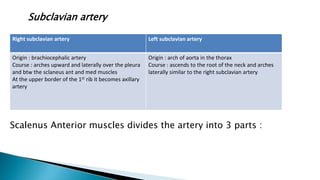
1. Subclavian artery
Scalenus Anterior muscles divides the artery into 3 parts :
Right subclavian artery Left subclavian artery
Origin : brachiocephalic artery
Course : arches upward and laterally over the pleura
and btw the sclaneus ant and med muscles
At the upper border of the 1st rib it becomes axillary
artery
Origin : arch of aorta in the thorax
Course : ascends to the root of the neck and arches
laterally similar to the right subclavian artery
2. 1st part
Extends fro the origin of the subclavian artery to MEDIAL
border of scal. Anterior muscle
Giving 3 branches
Vertebral artery
Thyrocervical trunk
internal thoracic artery
2nd part
Lies behind the scal. Anterior muscle
Gives following branches
Cost cervical trunk (backward over the dome of the plura
dividing into
Superior intercostal artery
Deep cervical artery
3rd part
Extends from the Lateral Border of Scal. Anterior Muscle
Cross the post triangle of the neck to the lateral border of the
1st rib becoming Axillary artery , closely related to the nerves
of brachial plexus
Occasionally giving branches
• Superficial cervical arteries
• Suprascapular arteries
3.
4. Continuation of subclavian
artery
Exteds from outer border of
1st rib upto lower border of
Teres major muscle
Continues as brachial artery
Closely related to brachial
plexus cords
Pectoralis minor muscle
divides the artery into 3 parts.
5. Axial artery pulsastion can be felt against
the lower part of the lateral wall of axilla
In order to check bleeding from the distal part of limb
- The artery can be compressed against the humerus in the
lower part of the lateral wall of axilla