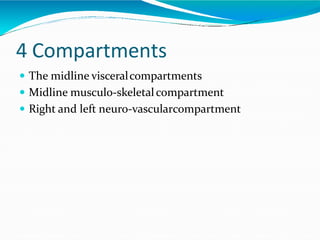
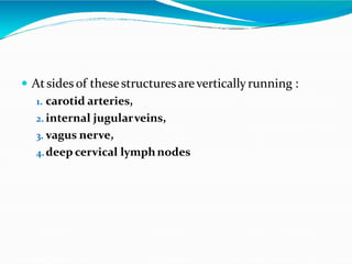
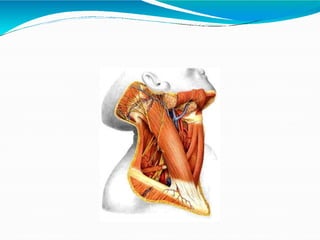
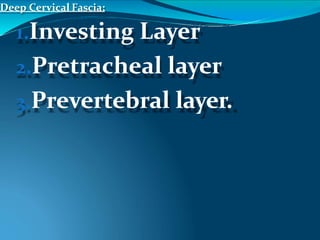
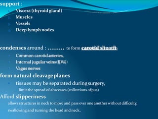
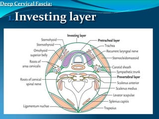
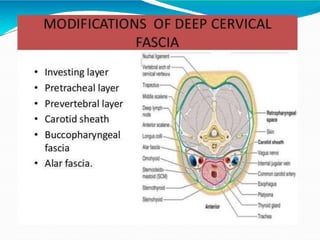
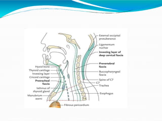
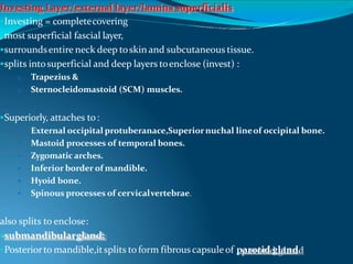
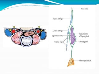
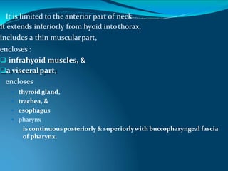
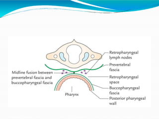
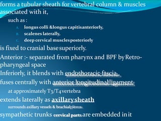
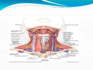
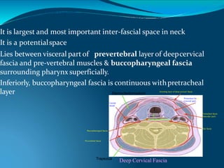
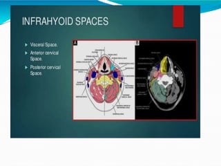
Deep fascia of theneck
Contents
 Introduction
 Boundaries
 Superficial fascia
 Layers of Deep fascia
 Spaces around theneck
 Applied anatomy
Introduction
 Neck is the transition andconducting zone between
head and rest of thebody
 It holds importantvisceraof theendocrine, respiratory
and digestivesystems
 The structure occupying the neck are crowded : as
there is no bonycovering orcage, thesestructuresalso
are prone to injury anddamage.
Contd.
 The subcutaneous tissue nerves, veinsof the neck,
 The main anatomical potential space in the neck
which lead to Spread of infections to themediastinum.
 Four major fascial compartmentsof the neck
 Where thevisceraof the neck are located.
The Neck
• lies between lower margin of mandible above & base of cranium
• suprasternal notch & upper border of clavicle below/ superior thoracicaperture
It is strengthened bycervical partof vertebral column,
• is convex forward
• supports skull.
Behind thevertebrae is …………..a massof extensormuscles
In front is ………………………………..asmallergroupof flexor muscles .
In central regionare :…………………partsof respiratorysystem,
larynx & trachea,
behind are partsof alimentary system, pharynx & esophagus.
Cutaneous nerves and superficial veins
External jugular vein
Anterior jugular vein
Lesser occipital n.
Greater auricular n.
Transverse nerve of neck
Supraclavicular n.
4 Compartments
 The midline visceralcompartments
 Midline musculo-skeletalcompartment
 Right and left neuro-vascularcompartment
 Atsides of thesestructuresareverticallyrunning :
1. carotid arteries,
2. internal jugularveins,
3. vagus nerve,
4.deep cervical lymphnodes
Landmarks of the neck
 Sternocleidomastoid
 Suprasternal fossa
 Greater supraclaviclarfossa
Landmarks of the neck
 Hyoid bone
 Thyroid cartilage
 Cricoid cartilage
The natural line of cleavage of the skin are
constant and run almost horizontally
around the neck
Fascia of Neck
1-Superficial Fascia
2-deep Fascia
Superficial Fascia
Superficial Fascia
o forms a thin layerand has no specific features
oAntero-lateral aspects on both sidesencloses Platysma muscle.
embedded in it are:
 cutaneous nerves,
 superficial veins,
 superficial lymph nodes.
 Especially in females fat tissuepresent.
Ext. jugular
Int. jugular
Ant. jugular
Sup. thyroid
Middle
thyroid
Inf. thyroid
Structures in neck:
 are surrounded bya layerof subcutaneous tissue (superficial fascia)
 are compartmentalized by layers of deepcervical fascia.
 fascial planes determine direction in which an infection in neck mayspread.
Cervical Subcutaneous Tissue&Platysma
 superficial cervical fascia
 is a layerof fattyconnectivetissue
 lies between dermisof skin & investing layerof deepcervical fascia
 It is usually thinnerthan in otherregions, anteriorly.
 Platysma……..Anterolaterally
external jugular vein(EJV)
• Descending from angleof mandible to middleof clavicle
• are superficial to main cutaneous nerves ofneck.
• covers anterolateral aspect of neck.
• Help to measureCVP
Platysma/Musculus platysma myoids/subcutaneous
collis/Tetragonus.
• Flat plate isa broad, thin sheetof muscle in subcutaneous tissue
of neck and it is a remnant of Panniculosuscarnosus.
• is supplied by cervical branch of CNVII.
• Its fibers arise in deep fasciacovering superiorparts of deltoid &
pectoralis major muscles
• sweep superomediallyoverclavicle to inferior borderof mandible
and merge with fascia of theface.
anterior borders of the two musclesdecussate overchin
blend with facial muscles.
Inferiorly, fibers diverge, leaving agapanteriorto larynx & trachea
• tenses skin,
 producing vertical skinridges
 Expression of horror, tension and stress
 releasing pressure on superficialveins.
• use inshaving
• It is a muscle ofgrimace.
• depress the mandibleand drawcornersof mouth inferiorly
• Acting its inferiorattachment
Its superficial to all structures. 2 bones and doesn’t cover upper
part of the anterior triangle but lower part covers and the posterior
triangle.
Contents
 Platysma
 Superficial veins
 Anterior jugularv.
 External jugularv.
 Cutaneous nerves
 Lesser occipital n.
 Greator auricularn.
 Transverse nerve of neck
 Supraclavicular n.
 Cervical branch of facial n.
Regions of neck
 Neck
 Anterior region of neck
 Sternocleidomastoid region
 Lateral region of neck
Triangles of posterior (lateral) region of
neck
Occipital triangle
supraclavicular triangle
(greater supraclavicular fossa)
Deep Cervical
Fascia / colli
Deep Cervical Fascia:
1.Investing Layer
2.Pretracheal layer
3.Prevertebral layer.
support :
1. Viscera (thyroid gland)
2. Muscles
3. Vessels
4. Deep lymph nodes
condenses around : ……… to form carotidsheath
 Common carotidarteries,
 Internal jugular veins(IJVs)
 Vagus nerves
form natural cleavageplanes
 tissues may be separated duringsurgery,
• limit the spread of abscesses (collections ofpus)
Afford slipperiness
allowsstructures in neck to move and pass overone anotherwithoutdifficulty,
swallowing and turning the head and neck.
Deep Cervical Fascia:
1.Investing layer
Investing Layer/external layer/lamina superficialis
Investing = completecovering
most superficial fascial layer,
surroundsentire neck deep toskin and subcutaneous tissue.
splits intosuperficial and deep layers toenclose (invest) :
1. Trapezius &
2. Sternocleidomastoid (SCM) muscles.
Superiorly, attaches to:
 External occipital protuberanace,Superiornuchal lineof occipital bone.
 Mastoid processes of temporal bones.
 Zygomatic arches.
 Inferior border of mandible.
 Hyoid bone.
 Spinous processes of cervicalvertebrae.
also splits to enclose:
submandibulargland;
Posteriorto mandible,itsplits to form fibrouscapsuleof parotid gland.
Investing Layer
stylomandibular ligament is a thickenedmodification
Inferiorly, attaches to:
 manubrium,
 clavicles, &
 acromions
 spines of scapulae.
continuous posteriorly
with :
 Periosteum covering C7 spinousprocess
 nuchal ligament
 a triangularmembrane
 formsa median fibrous septum between muscles of two sides of
neck
Horizontal disposition
 This traced from posterior attachment, it splits to enclose the trapezius andit
form a single layer that forms the roof of the posteriortriangle
 In its lower attachment it splits twice to enclose 2 spaces
 Above the manubrium sterni it splits into 2
 Anterior layer attaches to anterior border of manubrium sterni and posterior
layer to posterior border
 Space called supra-sternal space of Burns.
contents :- sternal head of SCM and anterior jugular vein,jugular
venous arch , fat and lymph node .
• above the clavicle near the lower part of roof of the posteriortriangle
• Splits into 2 antero-lateral aspect of the clavicle
posterior aspect of the clavicle
Space called Supra- clavicular space
contents :- EJV. Subclavian vein, supra- clavicular nerves.
Vertical disposition
 Traced upwards it covers sub-mandibular region and enclose
the gland
 Attaches to lower border of mandible and deep layer of
mylohyoid line of mandible
 Posterior to the gland again splits into enclose the parotid gland
and attach to zygomatic arch to form parotido masseteric fascia
deeper layer goes to Tympanic plate
 Between angle of mandible and styloid process form stylo-
mandibular ligament
 Spinal XI nerve closely related to this layer.
Deep Cervical Fascia:
Pretracheal/middle cervical fascia/porter’s fascia/
lamina pretrachealis
Anterior- thin and encloses thyroid gland and stretches in front of
trachea
Superior – hyoid bone, arch of cricoid cartilage and oblique line of
thyroid cartilage
Inferior - passes along the trachea , superior mediastinum and mergs
with fibrous pericardium of heart, movements of thyroid
gland with deglutition, sibson’s fascia .
Laterally – antero-laterally of carotid sheath between 2 lateral limits,
the pretracheal layer encloses the infra- hyoid muscles .
and they have 2 parts
Muscular part and visceral part
 Posterosuperiorly :- pre-tracheal layer is continues with of the
pharynx
 Ligament of Berry is derived from this fascia and connects
the lobes of thyroid gland with cricoid cartilage
 Dysphagia :- postero-lateral surface of Thyroid lobe is ill-
defind.
It is limited to the anterior part of neck
It extends inferiorly from hyoid intothorax,
includes a thin muscularpart,
encloses :
 infrahyoid muscles, &
a visceralpart,
encloses
 thyroid gland,
 trachea, &
 esophagus
 pharynx
iscontinuousposteriorly & superiorlywith buccopharyngeal fascia
of pharynx.
In hyoid,
a thickening of pre tracheal fascia formsa
pulley or trochleathrough
intermediate tendon of digastric musclepasses,
 suspending hyoid.
tethers two-bellied omohyoid muscle,
redirecting course of muscle betweenbellies.
Prevertebral Layer/ PV
Fascia/lamina prevertebralis
forms a tubular sheath for vertebral column & muscles
associated with it,
such as :
A. longus colli &longus capitisanteriorly,
B. scalenes laterally,
C. deep cervical musclesposteriorly
is fixed to cranial basesuperiorly.
Anterior :- separated from pharynx and BPF byRetro-
pharyngeal space
Inferiorly, it blends with endothoracic fascia .
fuses centrally with anterior longitudinalligament
at approximately T3/T4vertebra
extends laterally as axillarysheath
surrounds axillary vessels & brachialplexus.
sympathetic trunks cervical parts are embedded init
Carotid Sheath
Submendibular gland
OmohyoidSternohyoid
Sternothyroid
Cervical plexus
Phrenic n.
Superior thyroid a.
Ansa cervicalis
Vagus n.
Hypoglossal n.
Accessory n.Digastric
Condensation of the connective tissue around great vessel and
tubular fascial investment
extends from cranial base to root ofneck.
blends :
 anteriorly with investing and pretracheallayers
Posteriorly with prevertebral layer
contains :
(1) common and internal carotidarteries,
(2) internal jugularvein,
(3) vagus nerve (CN X),
(4) deep cervical lymphnodes,
(5) carotid sinus ,
(6) sympathetic nerve fibers (carotid periarterialplexuses).
carotid sheath and pretracheal fascia communicate freelywith:
 mediastinum of thorax inferiorly&
 cranial cavitysuperiorly.
representpotential pathways for spread of infection and extravasated blood.
 Superior :- base ofskull
 Inferior :- mergewith covering thearch of
aorta
 Posterior :- symphathictrunk
 Anterior :- ansacervicalis tocarotid sheath
 Str piercing :- ECA,IJV, IX, XI,XII,XCN
AA:- Block dissection of neck, during surgical removal
of deep cervical LN
Spaces around the Neck
 Disposition of various layers of fascia result in the formation of
the potential tissue spaces in the neck
 They are not real spaces but are potential
 In healthy persons- filled with CT
in pathologically secretions are noted and there is no tissue
barrier, spread of infections
 They are 2 groups
1. Supra- hyoid spaces
2. Infra- hyoid spaces
Supra- hyoid spaces
 Continuity with regions of head
 Infra temporal fossa, sub mandibularfossaand
tonsillarfossa
 Above the hyoid are in SM regions of head and pre-
vertebral region
1. Spaces around the lowerjaw
2. Pharyngeal spaces
3. Pre-vertebral spaces
Spaces around lower jaw
 Sub-mental spaces
 Sub-mandibularspaces
 Sub-lingual spaces
 Retro-pharyngeal space
 Peri-pharyngeal spaces
 Peri-tonsilarspaces
Sub-mental space :- deep to upper part of inv.layer of
DCF and S/F to mylohyoid muscle bounded by
anterior belly of digastric muscle
Sub- mandibular spaces :- these are paired, deep to
concerned half of mandible and both bellys of
digastric muscle.
Sub-lingual spaces :- lies deep to mylohyoid in the floor
of mouth and paired
its free communication with sub mental space which is
anatomically a cervical tissue space.
Retropharyngeal Space
It is largest and most important inter-fascial space in neck
It is a potentialspace
Lies between visceral part of prevertebral layer of deepcervical
fascia and pre-vertebral muscles & buccopharyngeal fascia
surrounding pharynx superficially.
Inferiorly, buccopharyngeal fascia is continuous withpretracheal
layer Sternocleidomastoid
Trapezius
Deep Cervical Fascia
Investing layer of deep cervical fascia
Prevertebral fascia
Pretracheal fascia
(visceral part)
Carotid
sheath
Buccopharyngeal fascia
Alar fascia
Pretracheal fascia
(muscular part)
T
E
Alar fascia:
• formsa furthersubdivisionof retropharyngeal space.
• is attached along midlineof buccopharyngeal fascia from cranium to
level of the C7vertebra.
• itextends laterallyand terminates in carotid sheath.
 permits movement of pharynx, esophagus, larynx, and
trachearelative tovertebral column during swallowing.
 is closed :
superiorly by cranial baseand
on each side by carotidsheath.
 It opens inferiorly into superiormediastinum
 Contents :- LCT, retro-pharyngeal LN, pharyngeal
Plexuses and vessels.
 A.A :- Cold abscess and para- medianswelling.
Peri-pharyngeal/ para pharyngeal space
 These are paired.
 Location:- on each side of posterolateral aspect of pharynx
 Medially- sup constrictor muscle of pharynx and palatine tonsil
 Antero-laterally- medial pterygoid with ramus of mandible
 Postero-laterally- parotid gland with parotid fascia and
communicates with retro-pharyngeal space
 Posterior – carotid sheath
Contents :-
branch of maxillary nerve and maxillary
artery, fibro-fatty tissue
Peri- tonsillar space :- actually a part of intra-pharyngealspace
location :- around palatine tonsils and medial surface of
superior constrictor.
Infra-hyoid spaces
Theyare
 Pretracheal space,
 Retro-visceral space,
 Pre-vertebral space
 Carotid space
Pre- tracheal space/ para tracheal space
 Location :- behind pre-tracheal fasciaand
infra-hyoid muscle anterior wall of oesophagus
 Superior :- infrahyoid muscles to thyroidcartilage
 Inferior :- space open into superiormediastinum
Retro- visceral spaces
 locaction :- between posterior wall oesophagusand
prevertebral fascia
 Superior
 Inferior
:- retro-pharyngeal space
:- superiormediastinum
Pre- vertebral space
 Location :- between prevertebral fascia and vertebral column
laterally prevertbral muscle are closely packed can’t extends
laterally
 Superior :- base of skull
 Inferior :- continues posterior mediastinum
 Anterior :- prevertebral fascia weakest especially time of
infection.
Carotid space
 Space around the contents of carotid sheath
 Above and below closed because of adhesion of the fascia to
the adventitia of vessels.
Applied anatomy
 Arrangements of layers in neck determine the direction of
spread of infections in and around the neck
 Investing layer :- prevents spread of purulent infection from s/f
aspect of neck to deep areas of thorax,
 Pretracheal muscular part doesn’t spread to thorax but visceral
part spreads down easily.
 In Tuberculosis of cervical vertebrae – pus between
vertebral column and prevertebral fascia and produce median
swelling of posteior wall of pharynx are called chronic
retropharyngeal abscess. Infection spread to prevertebral
muscles and skin of posterior triangle , axilla and axillary
sheath.
Contd..,
Collar stud abcess :- in tuberculosis deep cervical LN affected.
pus spread from superficial to deep.
Retro pharyngeal abscess :- retropharyngeal LN gets infected
and BPF also infected.
Ludwig’s angina :- an infection in sub-mandibular region is
limited to a triangular area 2 halves of mandible and hyoid
bone
Parotid fascia ;- is very dense, infection of parotid gland is
very painful.
Thank you

Deepfasciaofneck by dr.meher

  • 1.
  • 2.
    Contents  Introduction  Boundaries Superficial fascia  Layers of Deep fascia  Spaces around theneck  Applied anatomy
  • 3.
    Introduction  Neck isthe transition andconducting zone between head and rest of thebody  It holds importantvisceraof theendocrine, respiratory and digestivesystems  The structure occupying the neck are crowded : as there is no bonycovering orcage, thesestructuresalso are prone to injury anddamage.
  • 4.
    Contd.  The subcutaneoustissue nerves, veinsof the neck,  The main anatomical potential space in the neck which lead to Spread of infections to themediastinum.  Four major fascial compartmentsof the neck  Where thevisceraof the neck are located.
  • 6.
    The Neck • liesbetween lower margin of mandible above & base of cranium • suprasternal notch & upper border of clavicle below/ superior thoracicaperture It is strengthened bycervical partof vertebral column, • is convex forward • supports skull. Behind thevertebrae is …………..a massof extensormuscles In front is ………………………………..asmallergroupof flexor muscles . In central regionare :…………………partsof respiratorysystem, larynx & trachea, behind are partsof alimentary system, pharynx & esophagus.
  • 7.
    Cutaneous nerves andsuperficial veins External jugular vein Anterior jugular vein Lesser occipital n. Greater auricular n. Transverse nerve of neck Supraclavicular n.
  • 8.
    4 Compartments  Themidline visceralcompartments  Midline musculo-skeletalcompartment  Right and left neuro-vascularcompartment
  • 9.
     Atsides ofthesestructuresareverticallyrunning : 1. carotid arteries, 2. internal jugularveins, 3. vagus nerve, 4.deep cervical lymphnodes
  • 10.
    Landmarks of theneck  Sternocleidomastoid  Suprasternal fossa  Greater supraclaviclarfossa
  • 11.
    Landmarks of theneck  Hyoid bone  Thyroid cartilage  Cricoid cartilage
  • 12.
    The natural lineof cleavage of the skin are constant and run almost horizontally around the neck
  • 13.
  • 14.
  • 15.
  • 16.
    Superficial Fascia o formsa thin layerand has no specific features oAntero-lateral aspects on both sidesencloses Platysma muscle. embedded in it are:  cutaneous nerves,  superficial veins,  superficial lymph nodes.  Especially in females fat tissuepresent.
  • 19.
    Ext. jugular Int. jugular Ant.jugular Sup. thyroid Middle thyroid Inf. thyroid
  • 20.
    Structures in neck: are surrounded bya layerof subcutaneous tissue (superficial fascia)  are compartmentalized by layers of deepcervical fascia.  fascial planes determine direction in which an infection in neck mayspread. Cervical Subcutaneous Tissue&Platysma  superficial cervical fascia  is a layerof fattyconnectivetissue  lies between dermisof skin & investing layerof deepcervical fascia  It is usually thinnerthan in otherregions, anteriorly.  Platysma……..Anterolaterally
  • 21.
    external jugular vein(EJV) •Descending from angleof mandible to middleof clavicle • are superficial to main cutaneous nerves ofneck. • covers anterolateral aspect of neck. • Help to measureCVP
  • 22.
    Platysma/Musculus platysma myoids/subcutaneous collis/Tetragonus. •Flat plate isa broad, thin sheetof muscle in subcutaneous tissue of neck and it is a remnant of Panniculosuscarnosus. • is supplied by cervical branch of CNVII. • Its fibers arise in deep fasciacovering superiorparts of deltoid & pectoralis major muscles • sweep superomediallyoverclavicle to inferior borderof mandible and merge with fascia of theface. anterior borders of the two musclesdecussate overchin blend with facial muscles. Inferiorly, fibers diverge, leaving agapanteriorto larynx & trachea
  • 23.
    • tenses skin, producing vertical skinridges  Expression of horror, tension and stress  releasing pressure on superficialveins. • use inshaving • It is a muscle ofgrimace. • depress the mandibleand drawcornersof mouth inferiorly • Acting its inferiorattachment Its superficial to all structures. 2 bones and doesn’t cover upper part of the anterior triangle but lower part covers and the posterior triangle.
  • 25.
    Contents  Platysma  Superficialveins  Anterior jugularv.  External jugularv.  Cutaneous nerves  Lesser occipital n.  Greator auricularn.  Transverse nerve of neck  Supraclavicular n.  Cervical branch of facial n.
  • 26.
    Regions of neck Neck  Anterior region of neck  Sternocleidomastoid region  Lateral region of neck
  • 27.
    Triangles of posterior(lateral) region of neck Occipital triangle supraclavicular triangle (greater supraclavicular fossa)
  • 29.
  • 30.
    Deep Cervical Fascia: 1.InvestingLayer 2.Pretracheal layer 3.Prevertebral layer.
  • 31.
    support : 1. Viscera(thyroid gland) 2. Muscles 3. Vessels 4. Deep lymph nodes condenses around : ……… to form carotidsheath  Common carotidarteries,  Internal jugular veins(IJVs)  Vagus nerves form natural cleavageplanes  tissues may be separated duringsurgery, • limit the spread of abscesses (collections ofpus) Afford slipperiness allowsstructures in neck to move and pass overone anotherwithoutdifficulty, swallowing and turning the head and neck.
  • 32.
  • 35.
    Investing Layer/external layer/laminasuperficialis Investing = completecovering most superficial fascial layer, surroundsentire neck deep toskin and subcutaneous tissue. splits intosuperficial and deep layers toenclose (invest) : 1. Trapezius & 2. Sternocleidomastoid (SCM) muscles. Superiorly, attaches to:  External occipital protuberanace,Superiornuchal lineof occipital bone.  Mastoid processes of temporal bones.  Zygomatic arches.  Inferior border of mandible.  Hyoid bone.  Spinous processes of cervicalvertebrae. also splits to enclose: submandibulargland; Posteriorto mandible,itsplits to form fibrouscapsuleof parotid gland.
  • 36.
    Investing Layer stylomandibular ligamentis a thickenedmodification Inferiorly, attaches to:  manubrium,  clavicles, &  acromions  spines of scapulae. continuous posteriorly with :  Periosteum covering C7 spinousprocess  nuchal ligament  a triangularmembrane  formsa median fibrous septum between muscles of two sides of neck
  • 37.
    Horizontal disposition  Thistraced from posterior attachment, it splits to enclose the trapezius andit form a single layer that forms the roof of the posteriortriangle  In its lower attachment it splits twice to enclose 2 spaces  Above the manubrium sterni it splits into 2  Anterior layer attaches to anterior border of manubrium sterni and posterior layer to posterior border  Space called supra-sternal space of Burns. contents :- sternal head of SCM and anterior jugular vein,jugular venous arch , fat and lymph node . • above the clavicle near the lower part of roof of the posteriortriangle • Splits into 2 antero-lateral aspect of the clavicle posterior aspect of the clavicle Space called Supra- clavicular space contents :- EJV. Subclavian vein, supra- clavicular nerves.
  • 38.
    Vertical disposition  Tracedupwards it covers sub-mandibular region and enclose the gland  Attaches to lower border of mandible and deep layer of mylohyoid line of mandible  Posterior to the gland again splits into enclose the parotid gland and attach to zygomatic arch to form parotido masseteric fascia deeper layer goes to Tympanic plate  Between angle of mandible and styloid process form stylo- mandibular ligament  Spinal XI nerve closely related to this layer.
  • 39.
    Deep Cervical Fascia: Pretracheal/middlecervical fascia/porter’s fascia/ lamina pretrachealis Anterior- thin and encloses thyroid gland and stretches in front of trachea Superior – hyoid bone, arch of cricoid cartilage and oblique line of thyroid cartilage Inferior - passes along the trachea , superior mediastinum and mergs with fibrous pericardium of heart, movements of thyroid gland with deglutition, sibson’s fascia . Laterally – antero-laterally of carotid sheath between 2 lateral limits, the pretracheal layer encloses the infra- hyoid muscles . and they have 2 parts Muscular part and visceral part
  • 41.
     Posterosuperiorly :-pre-tracheal layer is continues with of the pharynx  Ligament of Berry is derived from this fascia and connects the lobes of thyroid gland with cricoid cartilage  Dysphagia :- postero-lateral surface of Thyroid lobe is ill- defind.
  • 42.
    It is limitedto the anterior part of neck It extends inferiorly from hyoid intothorax, includes a thin muscularpart, encloses :  infrahyoid muscles, & a visceralpart, encloses  thyroid gland,  trachea, &  esophagus  pharynx iscontinuousposteriorly & superiorlywith buccopharyngeal fascia of pharynx.
  • 43.
    In hyoid, a thickeningof pre tracheal fascia formsa pulley or trochleathrough intermediate tendon of digastric musclepasses,  suspending hyoid. tethers two-bellied omohyoid muscle, redirecting course of muscle betweenbellies.
  • 44.
  • 46.
    forms a tubularsheath for vertebral column & muscles associated with it, such as : A. longus colli &longus capitisanteriorly, B. scalenes laterally, C. deep cervical musclesposteriorly is fixed to cranial basesuperiorly. Anterior :- separated from pharynx and BPF byRetro- pharyngeal space Inferiorly, it blends with endothoracic fascia . fuses centrally with anterior longitudinalligament at approximately T3/T4vertebra extends laterally as axillarysheath surrounds axillary vessels & brachialplexus. sympathetic trunks cervical parts are embedded init
  • 47.
  • 48.
    Submendibular gland OmohyoidSternohyoid Sternothyroid Cervical plexus Phrenicn. Superior thyroid a. Ansa cervicalis Vagus n. Hypoglossal n. Accessory n.Digastric
  • 49.
    Condensation of theconnective tissue around great vessel and tubular fascial investment extends from cranial base to root ofneck. blends :  anteriorly with investing and pretracheallayers Posteriorly with prevertebral layer contains : (1) common and internal carotidarteries, (2) internal jugularvein, (3) vagus nerve (CN X), (4) deep cervical lymphnodes, (5) carotid sinus , (6) sympathetic nerve fibers (carotid periarterialplexuses). carotid sheath and pretracheal fascia communicate freelywith:  mediastinum of thorax inferiorly&  cranial cavitysuperiorly. representpotential pathways for spread of infection and extravasated blood.
  • 50.
     Superior :-base ofskull  Inferior :- mergewith covering thearch of aorta  Posterior :- symphathictrunk  Anterior :- ansacervicalis tocarotid sheath  Str piercing :- ECA,IJV, IX, XI,XII,XCN AA:- Block dissection of neck, during surgical removal of deep cervical LN
  • 51.
    Spaces around theNeck  Disposition of various layers of fascia result in the formation of the potential tissue spaces in the neck  They are not real spaces but are potential  In healthy persons- filled with CT in pathologically secretions are noted and there is no tissue barrier, spread of infections  They are 2 groups 1. Supra- hyoid spaces 2. Infra- hyoid spaces
  • 52.
    Supra- hyoid spaces Continuity with regions of head  Infra temporal fossa, sub mandibularfossaand tonsillarfossa  Above the hyoid are in SM regions of head and pre- vertebral region 1. Spaces around the lowerjaw 2. Pharyngeal spaces 3. Pre-vertebral spaces
  • 54.
    Spaces around lowerjaw  Sub-mental spaces  Sub-mandibularspaces  Sub-lingual spaces  Retro-pharyngeal space  Peri-pharyngeal spaces  Peri-tonsilarspaces
  • 57.
    Sub-mental space :-deep to upper part of inv.layer of DCF and S/F to mylohyoid muscle bounded by anterior belly of digastric muscle Sub- mandibular spaces :- these are paired, deep to concerned half of mandible and both bellys of digastric muscle. Sub-lingual spaces :- lies deep to mylohyoid in the floor of mouth and paired its free communication with sub mental space which is anatomically a cervical tissue space.
  • 58.
  • 59.
    It is largestand most important inter-fascial space in neck It is a potentialspace Lies between visceral part of prevertebral layer of deepcervical fascia and pre-vertebral muscles & buccopharyngeal fascia surrounding pharynx superficially. Inferiorly, buccopharyngeal fascia is continuous withpretracheal layer Sternocleidomastoid Trapezius Deep Cervical Fascia Investing layer of deep cervical fascia Prevertebral fascia Pretracheal fascia (visceral part) Carotid sheath Buccopharyngeal fascia Alar fascia Pretracheal fascia (muscular part) T E
  • 60.
    Alar fascia: • formsafurthersubdivisionof retropharyngeal space. • is attached along midlineof buccopharyngeal fascia from cranium to level of the C7vertebra. • itextends laterallyand terminates in carotid sheath.  permits movement of pharynx, esophagus, larynx, and trachearelative tovertebral column during swallowing.  is closed : superiorly by cranial baseand on each side by carotidsheath.  It opens inferiorly into superiormediastinum  Contents :- LCT, retro-pharyngeal LN, pharyngeal Plexuses and vessels.  A.A :- Cold abscess and para- medianswelling.
  • 61.
    Peri-pharyngeal/ para pharyngealspace  These are paired.  Location:- on each side of posterolateral aspect of pharynx  Medially- sup constrictor muscle of pharynx and palatine tonsil  Antero-laterally- medial pterygoid with ramus of mandible  Postero-laterally- parotid gland with parotid fascia and communicates with retro-pharyngeal space  Posterior – carotid sheath Contents :- branch of maxillary nerve and maxillary artery, fibro-fatty tissue Peri- tonsillar space :- actually a part of intra-pharyngealspace location :- around palatine tonsils and medial surface of superior constrictor.
  • 62.
    Infra-hyoid spaces Theyare  Pretrachealspace,  Retro-visceral space,  Pre-vertebral space  Carotid space
  • 64.
    Pre- tracheal space/para tracheal space  Location :- behind pre-tracheal fasciaand infra-hyoid muscle anterior wall of oesophagus  Superior :- infrahyoid muscles to thyroidcartilage  Inferior :- space open into superiormediastinum Retro- visceral spaces  locaction :- between posterior wall oesophagusand prevertebral fascia  Superior  Inferior :- retro-pharyngeal space :- superiormediastinum
  • 65.
    Pre- vertebral space Location :- between prevertebral fascia and vertebral column laterally prevertbral muscle are closely packed can’t extends laterally  Superior :- base of skull  Inferior :- continues posterior mediastinum  Anterior :- prevertebral fascia weakest especially time of infection. Carotid space  Space around the contents of carotid sheath  Above and below closed because of adhesion of the fascia to the adventitia of vessels.
  • 66.
    Applied anatomy  Arrangementsof layers in neck determine the direction of spread of infections in and around the neck  Investing layer :- prevents spread of purulent infection from s/f aspect of neck to deep areas of thorax,  Pretracheal muscular part doesn’t spread to thorax but visceral part spreads down easily.  In Tuberculosis of cervical vertebrae – pus between vertebral column and prevertebral fascia and produce median swelling of posteior wall of pharynx are called chronic retropharyngeal abscess. Infection spread to prevertebral muscles and skin of posterior triangle , axilla and axillary sheath.
  • 67.
    Contd.., Collar stud abcess:- in tuberculosis deep cervical LN affected. pus spread from superficial to deep. Retro pharyngeal abscess :- retropharyngeal LN gets infected and BPF also infected. Ludwig’s angina :- an infection in sub-mandibular region is limited to a triangular area 2 halves of mandible and hyoid bone Parotid fascia ;- is very dense, infection of parotid gland is very painful.
  • 72.