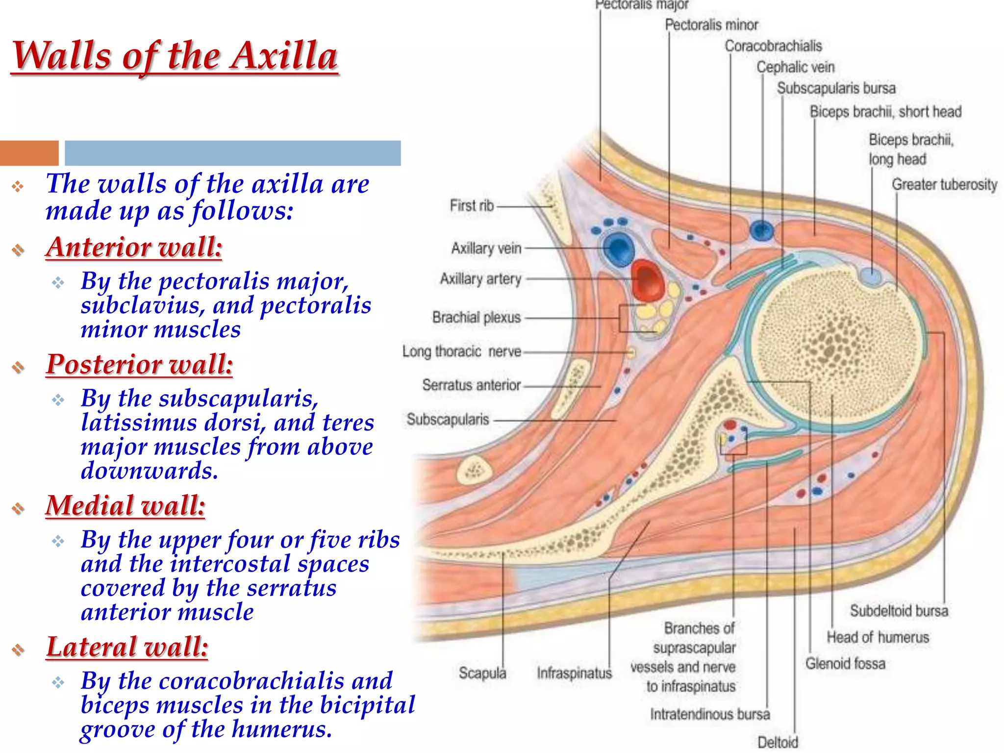
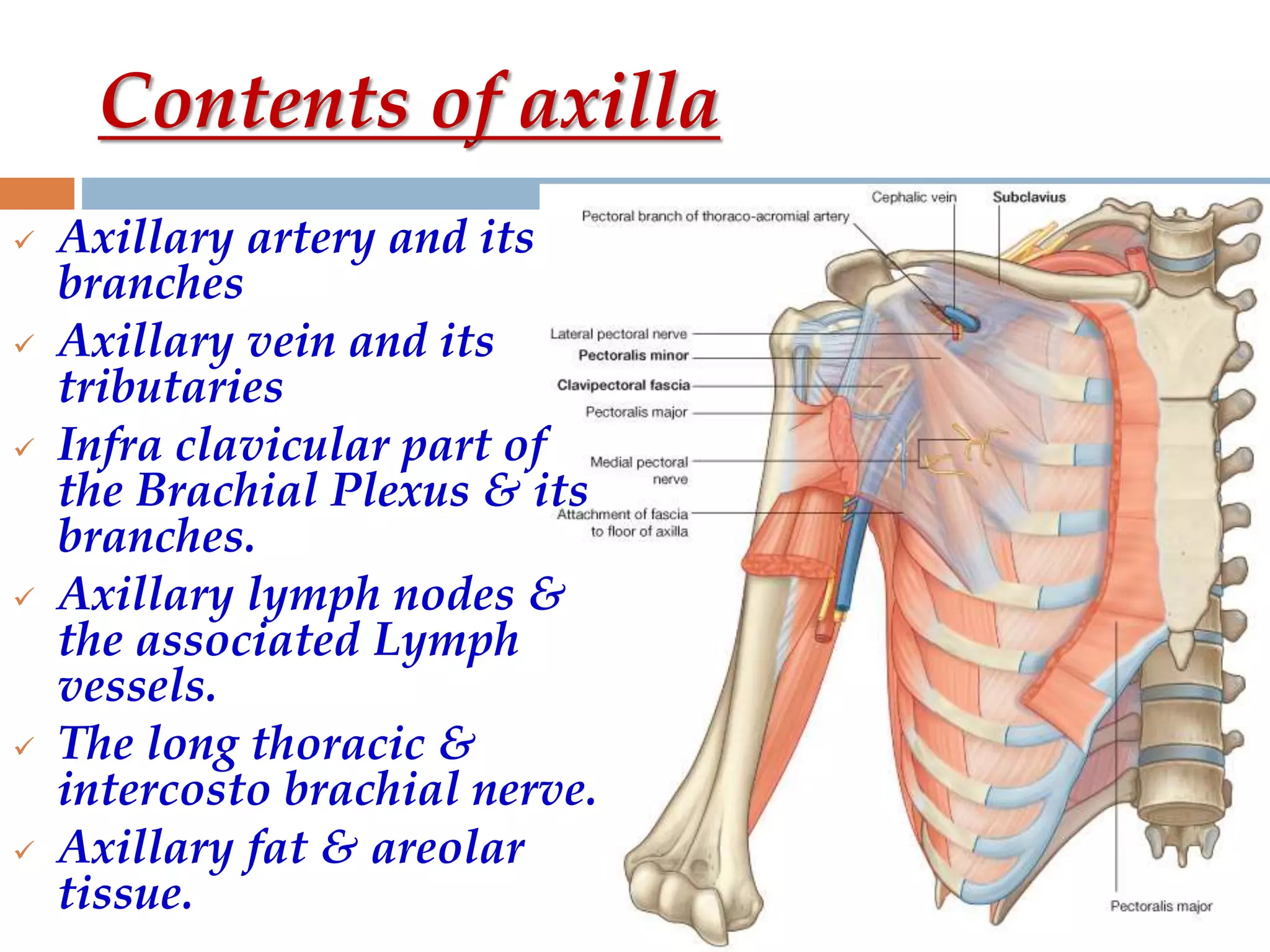
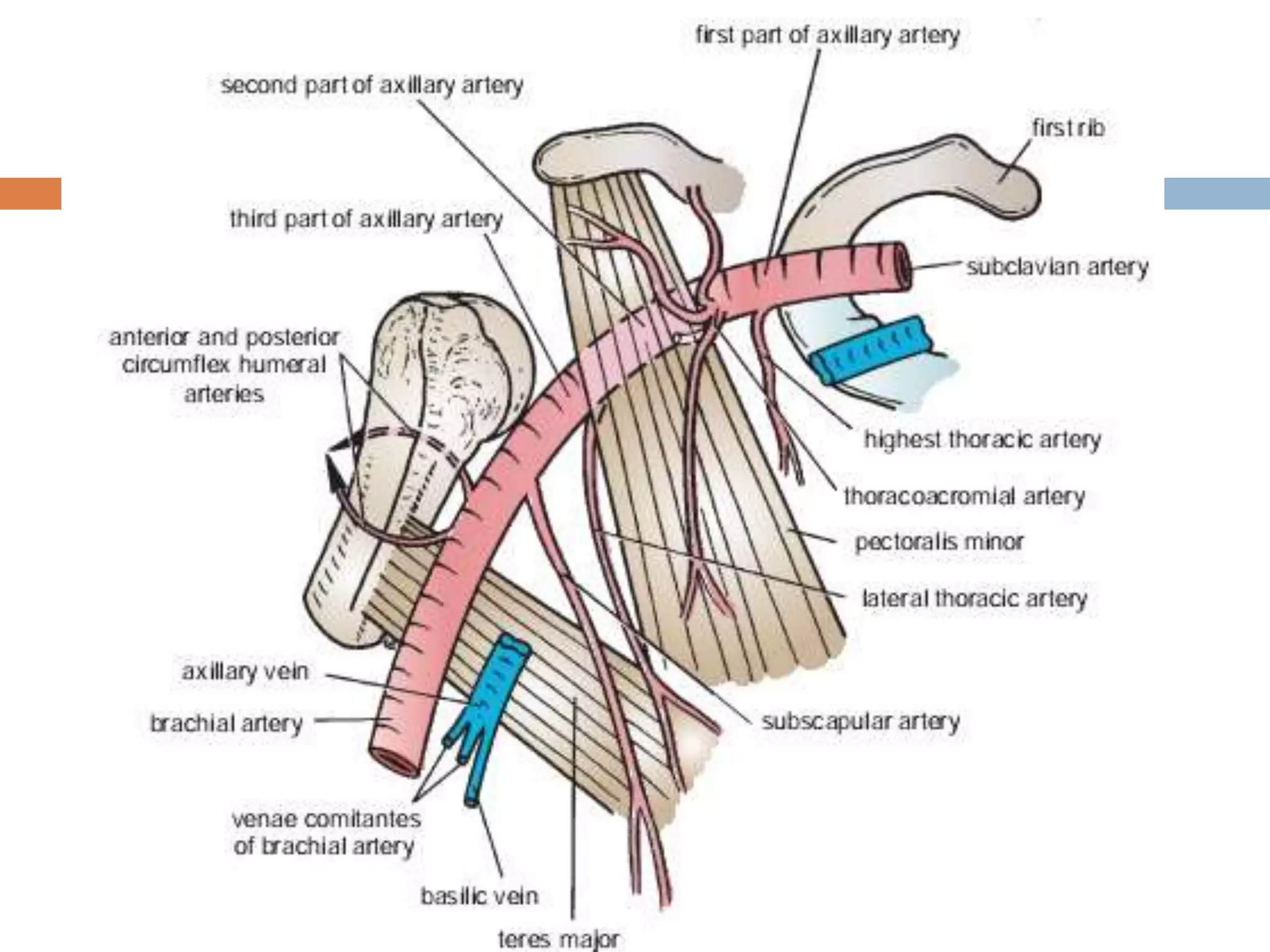
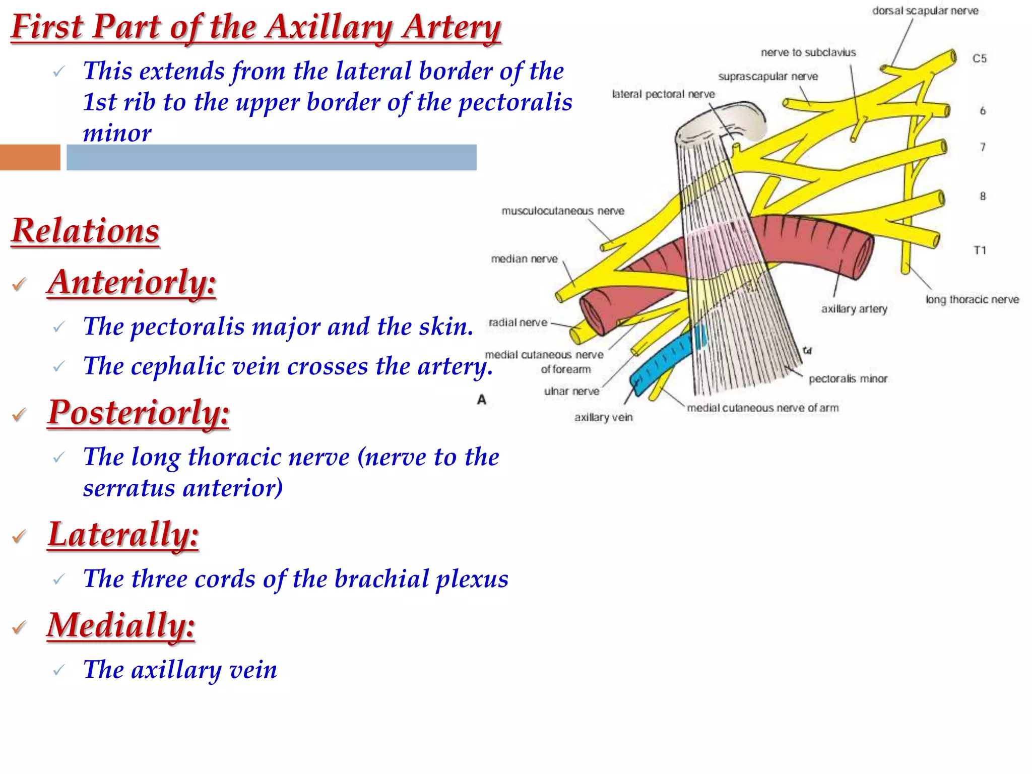
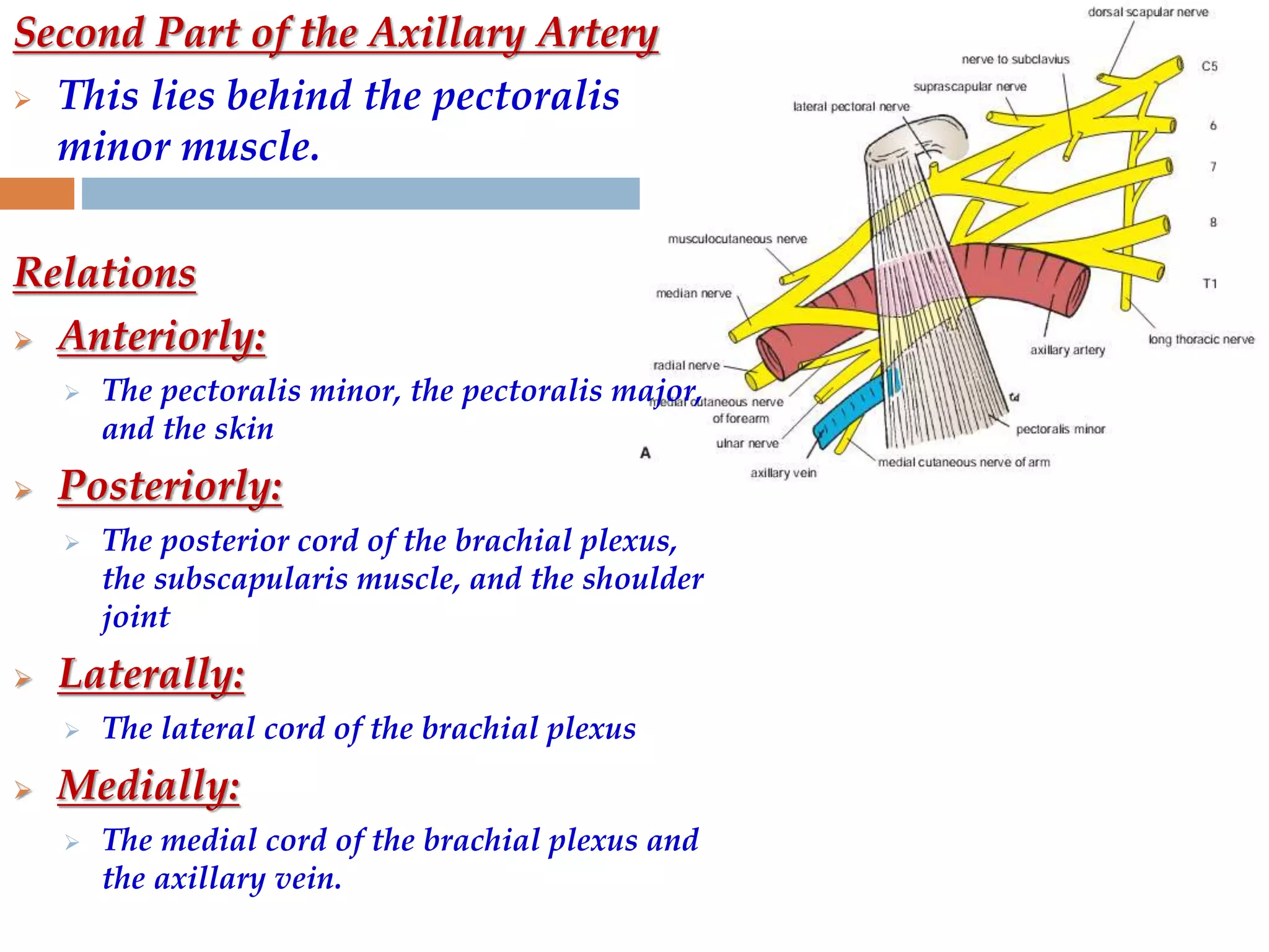
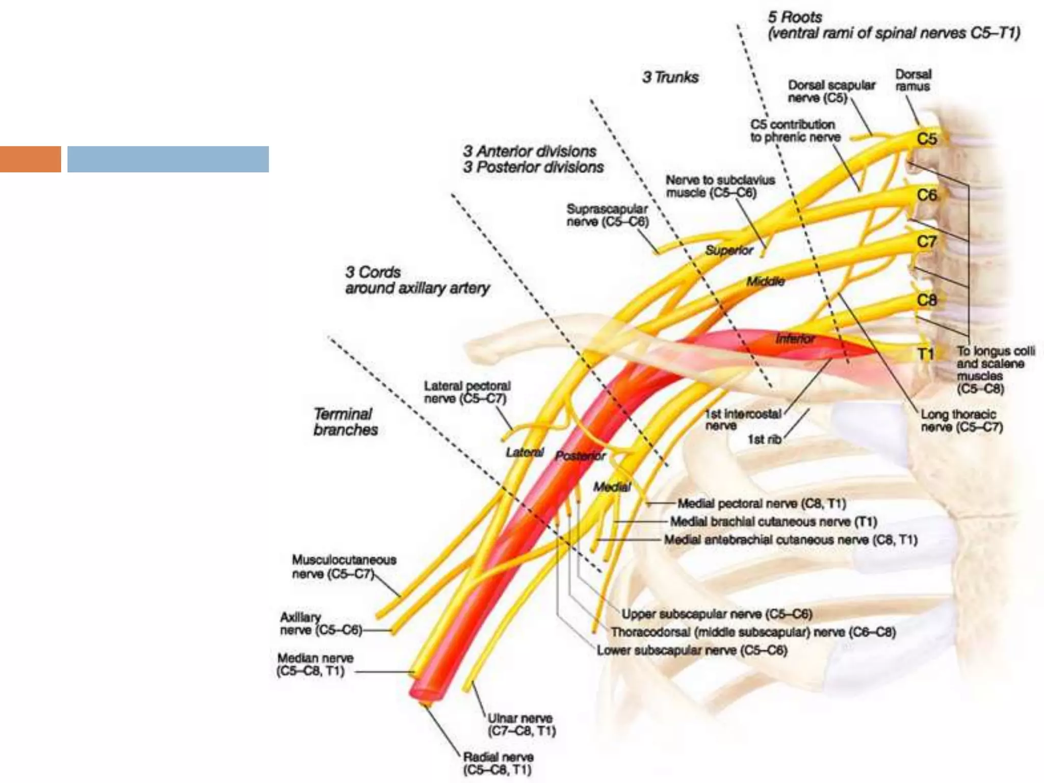
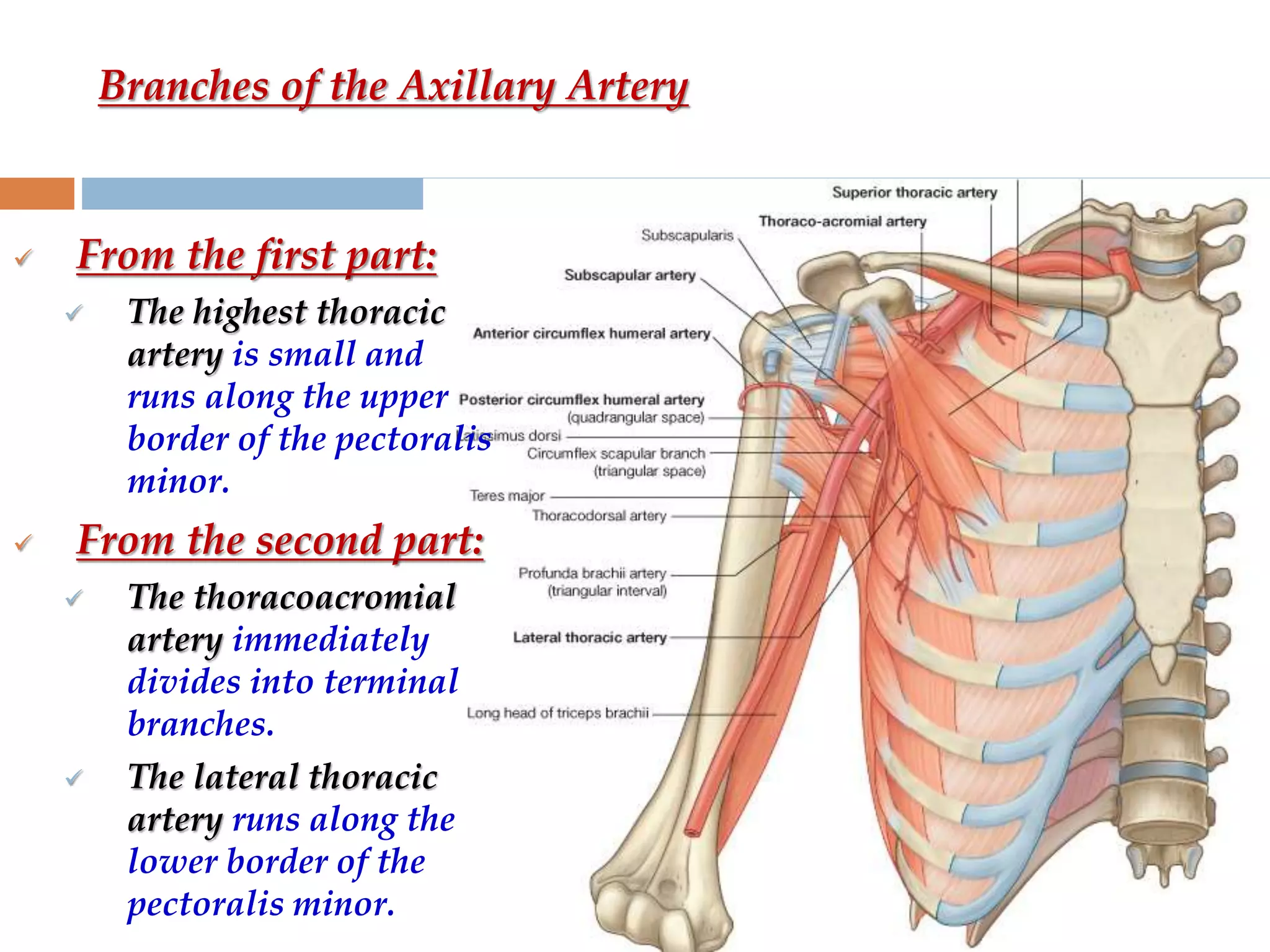
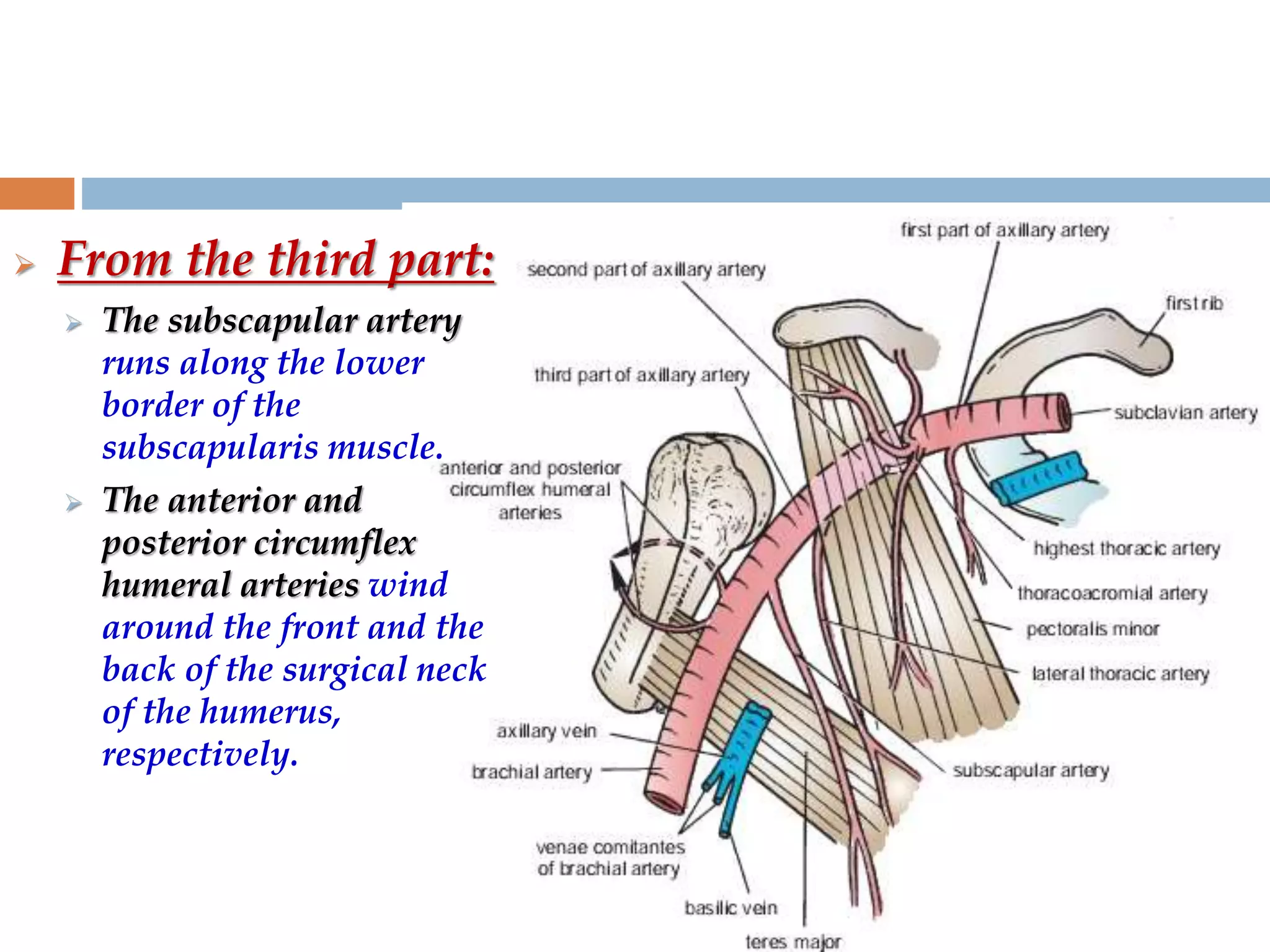
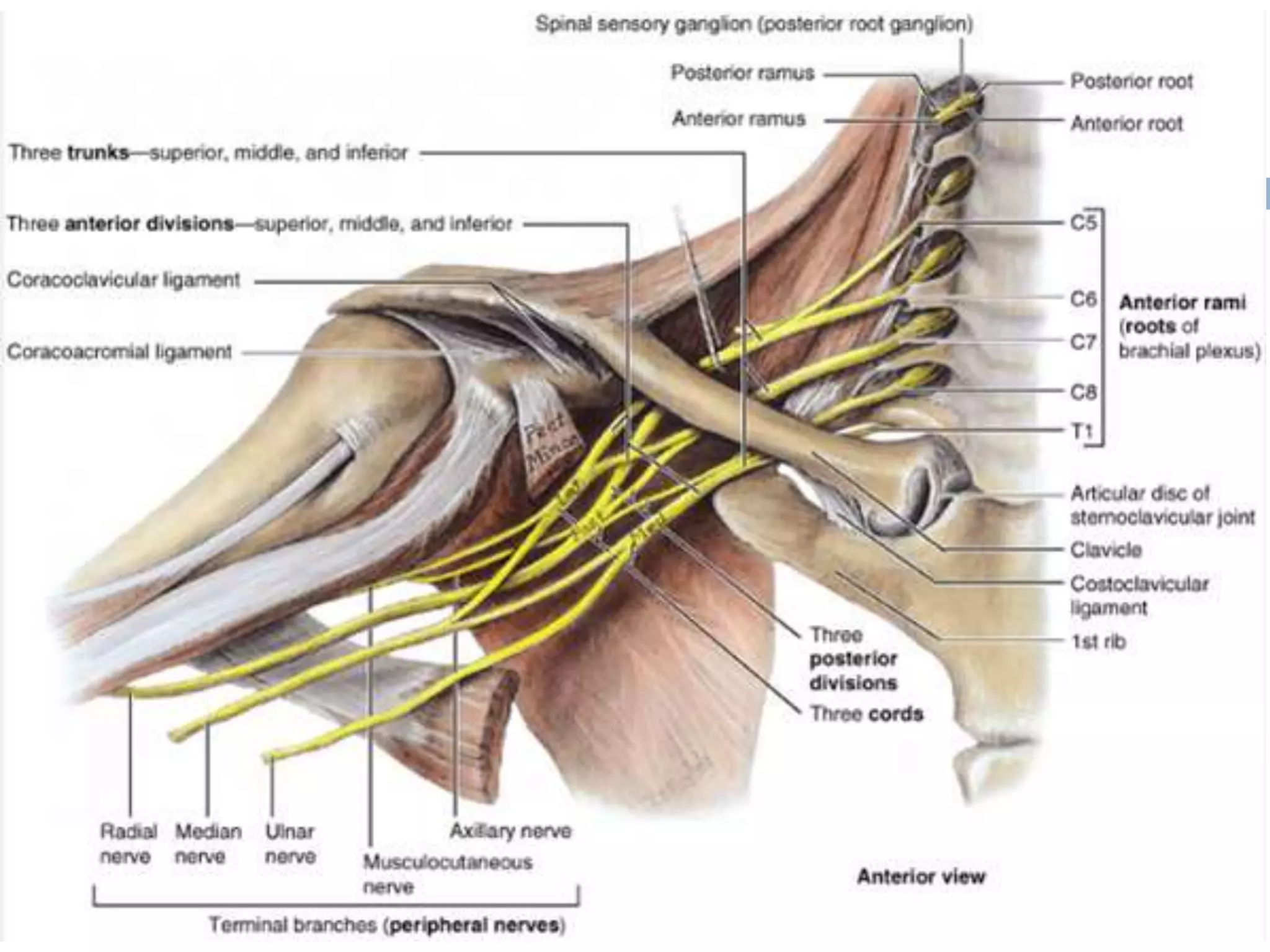
The axilla is a pyramid-shaped space located between the upper arm and chest. It contains nerves, blood vessels, lymph nodes, and fat, and serves as an important passageway. The brachial plexus, formed from cervical and thoracic spinal nerve roots, passes through the axilla, branching into nerves that supply the upper limb. The axillary artery and its branches, including the thoracoacromial, lateral thoracic, and subscapular arteries, also course through the axilla, along with the axillary vein and lymph nodes.
Seven cervical vertebrae
Identified by the presence of foramen in their transverse processes called foramen transversarium
3rd to 6th are typically have common features
1st, 2nd,and 7th are atypical
Ring-shaped and has no body and no spine
Consists of:
Right and left lateral masses
Short anterior arch and a long curved posterior arch
(c) Right and left transverse processes
Carpal Bone Anatomy Details PPT
Part-4 (UL Bone)
Carpal Bone names, attachments, clinical anatomy, General and specific points.
Carpal bones: 8
Like, share and comment.
Thank you
The hip joint is the hip bone (fusion of ilium, ischium, and pubis) fused with head of femur
The hip bone forms the bony connection between the sacrum and femur
Hip bone has 3 primary ossification centers which appear at 8wks, 4 mths, 5 mths for the ilium, ischium and pubis respectively.
At birth, these three are separated by a Y shaped cartilage and fusion occurs at age of 18
Anatomy of axilla with Dr- Ameera Al-Humidi .pptxAmeera Al-Humidi
The axilla is the anatomical region under the shoulder joint where the arm connects to the shoulder.
The axilla has five anatomic borders: superior, anterior, posterior, lateral, and medial walls.
The borders of the axilla are composed of muscles, including the serratus anterior, coracobrachialis, and short head of the biceps
The axillary walls are used as landmarks by surgeons to prevent damage to the neurovascular structures within the axilla during surgery
The contents of the axilla include muscles, nerves, vessels, and lymphatics
The axillary artery and vein, brachial plexus, and axillary lymph nodes are some of the neurovascular structures found in the axilla
Anti ulcer drugs and their Advance pharmacology ||
Anti-ulcer drugs are medications used to prevent and treat ulcers in the stomach and upper part of the small intestine (duodenal ulcers). These ulcers are often caused by an imbalance between stomach acid and the mucosal lining, which protects the stomach lining.
||Scope: Overview of various classes of anti-ulcer drugs, their mechanisms of action, indications, side effects, and clinical considerations.
MANAGEMENT OF ATRIOVENTRICULAR CONDUCTION BLOCK.pdfJim Jacob Roy
Cardiac conduction defects can occur due to various causes.
Atrioventricular conduction blocks ( AV blocks ) are classified into 3 types.
This document describes the acute management of AV block.
Explore natural remedies for syphilis treatment in Singapore. Discover alternative therapies, herbal remedies, and lifestyle changes that may complement conventional treatments. Learn about holistic approaches to managing syphilis symptoms and supporting overall health.
TEST BANK for Operations Management, 14th Edition by William J. Stevenson, Ve...kevinkariuki227
TEST BANK for Operations Management, 14th Edition by William J. Stevenson, Verified Chapters 1 - 19, Complete Newest Version.pdf
Tom Selleck Health: A Comprehensive Look at the Iconic Actor’s Wellness Journeygreendigital
Tom Selleck, an enduring figure in Hollywood. has captivated audiences for decades with his rugged charm, iconic moustache. and memorable roles in television and film. From his breakout role as Thomas Magnum in Magnum P.I. to his current portrayal of Frank Reagan in Blue Bloods. Selleck's career has spanned over 50 years. But beyond his professional achievements. fans have often been curious about Tom Selleck Health. especially as he has aged in the public eye.
Follow us on: Pinterest
Introduction
Many have been interested in Tom Selleck health. not only because of his enduring presence on screen but also because of the challenges. and lifestyle choices he has faced and made over the years. This article delves into the various aspects of Tom Selleck health. exploring his fitness regimen, diet, mental health. and the challenges he has encountered as he ages. We'll look at how he maintains his well-being. the health issues he has faced, and his approach to ageing .
Early Life and Career
Childhood and Athletic Beginnings
Tom Selleck was born on January 29, 1945, in Detroit, Michigan, and grew up in Sherman Oaks, California. From an early age, he was involved in sports, particularly basketball. which played a significant role in his physical development. His athletic pursuits continued into college. where he attended the University of Southern California (USC) on a basketball scholarship. This early involvement in sports laid a strong foundation for his physical health and disciplined lifestyle.
Transition to Acting
Selleck's transition from an athlete to an actor came with its physical demands. His first significant role in "Magnum P.I." required him to perform various stunts and maintain a fit appearance. This role, which he played from 1980 to 1988. necessitated a rigorous fitness routine to meet the show's demands. setting the stage for his long-term commitment to health and wellness.
Fitness Regimen
Workout Routine
Tom Selleck health and fitness regimen has evolved. adapting to his changing roles and age. During his "Magnum, P.I." days. Selleck's workouts were intense and focused on building and maintaining muscle mass. His routine included weightlifting, cardiovascular exercises. and specific training for the stunts he performed on the show.
Selleck adjusted his fitness routine as he aged to suit his body's needs. Today, his workouts focus on maintaining flexibility, strength, and cardiovascular health. He incorporates low-impact exercises such as swimming, walking, and light weightlifting. This balanced approach helps him stay fit without putting undue strain on his joints and muscles.
Importance of Flexibility and Mobility
In recent years, Selleck has emphasized the importance of flexibility and mobility in his fitness regimen. Understanding the natural decline in muscle mass and joint flexibility with age. he includes stretching and yoga in his routine. These practices help prevent injuries, improve posture, and maintain mobilit
Ozempic: Preoperative Management of Patients on GLP-1 Receptor Agonists Saeid Safari
Preoperative Management of Patients on GLP-1 Receptor Agonists like Ozempic and Semiglutide
ASA GUIDELINE
NYSORA Guideline
2 Case Reports of Gastric Ultrasound
ARTIFICIAL INTELLIGENCE IN HEALTHCARE.pdfAnujkumaranit
Artificial intelligence (AI) refers to the simulation of human intelligence processes by machines, especially computer systems. It encompasses tasks such as learning, reasoning, problem-solving, perception, and language understanding. AI technologies are revolutionizing various fields, from healthcare to finance, by enabling machines to perform tasks that typically require human intelligence.
Title: Sense of Taste
Presenter: Dr. Faiza, Assistant Professor of Physiology
Qualifications:
MBBS (Best Graduate, AIMC Lahore)
FCPS Physiology
ICMT, CHPE, DHPE (STMU)
MPH (GC University, Faisalabad)
MBA (Virtual University of Pakistan)
Learning Objectives:
Describe the structure and function of taste buds.
Describe the relationship between the taste threshold and taste index of common substances.
Explain the chemical basis and signal transduction of taste perception for each type of primary taste sensation.
Recognize different abnormalities of taste perception and their causes.
Key Topics:
Significance of Taste Sensation:
Differentiation between pleasant and harmful food
Influence on behavior
Selection of food based on metabolic needs
Receptors of Taste:
Taste buds on the tongue
Influence of sense of smell, texture of food, and pain stimulation (e.g., by pepper)
Primary and Secondary Taste Sensations:
Primary taste sensations: Sweet, Sour, Salty, Bitter, Umami
Chemical basis and signal transduction mechanisms for each taste
Taste Threshold and Index:
Taste threshold values for Sweet (sucrose), Salty (NaCl), Sour (HCl), and Bitter (Quinine)
Taste index relationship: Inversely proportional to taste threshold
Taste Blindness:
Inability to taste certain substances, particularly thiourea compounds
Example: Phenylthiocarbamide
Structure and Function of Taste Buds:
Composition: Epithelial cells, Sustentacular/Supporting cells, Taste cells, Basal cells
Features: Taste pores, Taste hairs/microvilli, and Taste nerve fibers
Location of Taste Buds:
Found in papillae of the tongue (Fungiform, Circumvallate, Foliate)
Also present on the palate, tonsillar pillars, epiglottis, and proximal esophagus
Mechanism of Taste Stimulation:
Interaction of taste substances with receptors on microvilli
Signal transduction pathways for Umami, Sweet, Bitter, Sour, and Salty tastes
Taste Sensitivity and Adaptation:
Decrease in sensitivity with age
Rapid adaptation of taste sensation
Role of Saliva in Taste:
Dissolution of tastants to reach receptors
Washing away the stimulus
Taste Preferences and Aversions:
Mechanisms behind taste preference and aversion
Influence of receptors and neural pathways
Impact of Sensory Nerve Damage:
Degeneration of taste buds if the sensory nerve fiber is cut
Abnormalities of Taste Detection:
Conditions: Ageusia, Hypogeusia, Dysgeusia (parageusia)
Causes: Nerve damage, neurological disorders, infections, poor oral hygiene, adverse drug effects, deficiencies, aging, tobacco use, altered neurotransmitter levels
Neurotransmitters and Taste Threshold:
Effects of serotonin (5-HT) and norepinephrine (NE) on taste sensitivity
Supertasters:
25% of the population with heightened sensitivity to taste, especially bitterness
Increased number of fungiform papillae
micro teaching on communication m.sc nursing.pdfAnurag Sharma
Microteaching is a unique model of practice teaching. It is a viable instrument for the. desired change in the teaching behavior or the behavior potential which, in specified types of real. classroom situations, tends to facilitate the achievement of specified types of objectives.
2. The axilla, or armpit, is a
pyramid-shaped space.
It lies between the upper
part of the arm and the
side of the chest.
It forms an important
passage for nerves, blood,
and lymph vessels as
they travel from the root
of the neck to the upper
limb.
It consist of
An apex
Base
Four walls
3. The apex
The apex, is directed into the root of
the neck.
Apex is bounded:-
In front by the clavicle
Behind by the upper border of the
scapula
Medially by the outer border of the
first rib.
The base
It is directed downwards.
The base is formed by the skin
stretching between the anterior and
posterior walls
It is bounded
In front by the anterior axillary fold
(formed by the lower border of the
pectoralis major muscle),
Behind by the posterior axillary fold
(formed by the tendon of latissimus
dorsi and the teres major muscle),
Medially by the chest wall
4. Walls of the Axilla
The walls of the axilla are
made up as follows:
Anterior wall:
By the pectoralis major,
subclavius, and pectoralis
minor muscles
Posterior wall:
By the subscapularis,
latissimus dorsi, and teres
major muscles from above
downwards.
Medial wall:
By the upper four or five ribs
and the intercostal spaces
covered by the serratus
anterior muscle
Lateral wall:
By the coracobrachialis and
biceps muscles in the bicipital
groove of the humerus.
5.
6.
7. Contents of axilla
Axillary artery and its
branches
Axillary vein and its
tributaries
Infra clavicular part of
the Brachial Plexus & its
branches.
Axillary lymph nodes &
the associated Lymph
vessels.
The long thoracic &
intercosto brachial nerve.
Axillary fat & areolar
tissue.
8. Axillary Artery
It is as a continuation of the subclavian artery.
The axillary artery begins at the lateral border of the 1st
rib and ends at the lower border of the teres major muscle,
where it continues as the brachial artery.
Throughout its course, the artery is closely related to the
cords of the brachial plexus and their branches and is
enclosed with them in a connective tissue sheath called
the axillary sheath.
If this sheath is traced upward into the root of the neck, it
is seen to be continuous with the prevertebral fascia.
9. The pectoralis minor
muscle crosses in front
of the axillary artery
and divides it into
three parts.
The first part is
proximal to pectoralis
minor.
The second part is
posterior to pectoralis
minor.
The third part is distal
to pectoralis minor.
10.
11. First Part of the Axillary Artery
This extends from the lateral border of the
1st rib to the upper border of the pectoralis
minor
Relations
Anteriorly:
The pectoralis major and the skin.
The cephalic vein crosses the artery.
Posteriorly:
The long thoracic nerve (nerve to the
serratus anterior)
Laterally:
The three cords of the brachial plexus
Medially:
The axillary vein
12. Second Part of the Axillary Artery
This lies behind the pectoralis
minor muscle.
Relations
Anteriorly:
The pectoralis minor, the pectoralis major,
and the skin
Posteriorly:
The posterior cord of the brachial plexus,
the subscapularis muscle, and the shoulder
joint
Laterally:
The lateral cord of the brachial plexus
Medially:
The medial cord of the brachial plexus and
the axillary vein.
13.
14. Third Part of the Axillary Artery
This extends from the lower border of the pectoralis
minor to the lower border of the teres major
Relations
Anteriorly:
The pectoralis major for a short distance; lower down the
artery, it is crossed by the medial root of the median nerve.
Posteriorly:
The subscapularis, the latissimus dorsi, and the teres major.
The axillary and radial nerves also lie behind the artery.
Laterally:
The coracobrachialis, the biceps, and the humerus.
The lateral root of the median and the musculocutaneous
nerves also lies on the lateral side.
Medially:
The ulnar nerve, the axillary vein, and the medial cutaneous
nerve of the arm, the cephalic vein.
15. Branches of the Axillary Artery
From the first part:
The highest thoracic
artery is small and
runs along the upper
border of the pectoralis
minor.
From the second part:
The thoracoacromial
artery immediately
divides into terminal
branches.
The lateral thoracic
artery runs along the
lower border of the
pectoralis minor.
16.
17. From the third part:
The subscapular artery
runs along the lower
border of the
subscapularis muscle.
The anterior and
posterior circumflex
humeral arteries wind
around the front and the
back of the surgical neck
of the humerus,
respectively.
18. Axillary Vein
The axillary vein is formed at the lower
border of the teres major muscle by the
union of the venae comitantes of the
brachial artery and the basilic vein.
It runs upward on the medial side of the
axillary artery and ends at the lateral
border of the 1st rib & becomes continues
as the subclavian vein.
The vein receives tributaries, which
correspond to the branches of the axillary
artery.
19.
20. The brachial plexus
The brachial plexus is formed by the union of the ventral rami of
the lower four cervical nerves and the greater part of the first
thoracic ventral ramus.
It originates in the neck, passes laterally and inferiorly over rib
I, and enters the axilla.
The fourth ramus usually gives a branch to the fifth, and the first
thoracic ramus frequently receives a branch from the second.
Contributions to the plexus by C4 and T2 vary.
When the branch from C4 is large, that from T2 is frequently
absent and the branch from T1 is reduced, the plexus is called as
prefixed plexus.
If the branch from C4 is small or absent, the contribution from
C5 is reduced but that from T1 is larger and there is always a
contribution from T2, such type of plexus is called as a postfixed
plexus.
21.
22.
23. The parts of the brachial plexus, from medial to
lateral, are:-
The roots
The trunks
The divisions
The cords
All major nerves that innervate the upper limb
originate from the brachial plexus, mostly from the
cords.
Proximal parts of the brachial plexus are posterior
to the subclavian artery in the neck, while more
distal regions of the plexus surround the axillary
artery.
24. Roots
The roots of the brachial plexus are the anterior rami of C5
to C8, and most of T1.
The roots and trunks enter the posterior triangle of the neck
by passing between the anterior scalene and middle scalene
muscles and lie superior and posterior to the subclavian
artery.
Trunks
The three trunks of the brachial plexus originate from the
roots, pass laterally over the first rib, and enter the axilla.
The superior trunk is formed by the union of C5 and C6 roots.
The middle trunk is a continuation of the C7 root.
The inferior trunk is formed by the union of the C8 and T1 roots.
The inferior trunk lies on the first rib posterior to the subclavian
artery.
The middle and superior trunks are more superior in position.
25.
26. Divisions
Each of the three trunks of
the brachial plexus divides
into an anterior and a
posterior division.
The three anterior divisions
form parts of the brachial
plexus that ultimately give
rise to peripheral nerves
associated with the anterior
compartments of the arm
and forearm.
The three posterior divisions
combine to form parts of the
brachial plexus that give rise
to nerves associated with the
posterior compartments.
27. Cords
The three cords of the brachial plexus originate from the
divisions and are related to the second part of the axillary
artery.
The lateral cord
It is formed by the union of the anterior divisions of the upper
and middle trunks.
It has contributions from C5 to C7.
It is positioned lateral to the second part of the axillary
artery.
The medial cord
It lies medial to the second part of the axillary artery.
Is the continuation of the anterior division of the inferior
trunk.
It contains contributions from C8 and T1.
The posterior cord
It lies posterior to the second part of the axillary artery
It is formed by the union of all three posterior divisions.
It contains contributions from all roots of the brachial plexus
(C5 to T1).
28. Roots
Dorsal scapular nerve (C5)
Long thoracic nerve (C5, 6, and 7)
Upper trunk
Nerve to subclavius (C5 and 6)
Suprascapular nerve (supplies the
supraspinatus and infraspinatus
muscle.
Lateral cord
Lateral pectoral nerve
Musculocutaneous nerve
Lateral root of median nerve
Medial cord
Medial pectoral nerve
Medial cutaneous nerve of arm and
medial cutaneous nerve of forearm
Ulnar nerve
Medial root of median nerve
Posterior cord
Upper and lower subscapular
nerves
Thoracodorsal nerve
Axillary nerve
Radial nerve
Branches of the brachial plexus
The branches of the different parts of the brachial plexus are as
follows:-
29.
30. Branches of the Brachial Plexus Found in
the Axilla.
The nerve to the subclavius(C5 and 6)
It supplies the subclavius muscle.
The long thoracic nerve(C5, 6, and 7)
It arises from the roots of the brachial plexus in the neck and enters
the axilla by passing down over the lateral border of the 1st rib
behind the axillary vessels and brachial plexus.
It descends over the lateral surface of the serratus anterior muscle,
which it supplies.
The lateral pectoral nerve
It arises from the lateral cord of the brachial plexus and supplies
the pectoralis major muscle.
The musculocutaneous nerve
It arises from the lateral cord of the brachial plexus, supplies the
coracobrachialis muscle, and leaves the axilla by piercing that
muscle.
31. The lateral root of the median nerve
It is the direct continuation of the lateral cord of the brachial plexus.
It is joined by the medial root to form the median nerve trunk, and this
passes downward on the lateral side of the axillary artery.
The median nerve gives off no branches in the axilla.
The medial pectoral nerve
It arises from the medial cord of the brachial plexus, supplies and pierces
the pectoralis minor muscle, and supplies the pectoralis major muscle.
The medial cutaneous nerve of the arm(T1)
It arises from the medial cord of the brachial plexus and is joined by the
intercostobrachial nerve (lateral cutaneous branch of the 2nd intercostal
nerve).
It supplies the skin on the medial side of the arm.
The medial cutaneous nerve of the forearm arises from the medial cord of
the brachial plexus
32. The ulnar nerve(C8 and T1)
It arises from the medial cord of the brachial plexus and descends in the
interval between the axillary artery and vein.
The ulnar nerve gives off no branches in the axilla.
The medial root of the median nerve
It arises from the medial cord of the brachial plexus and crosses in front of
the third part of the axillary artery to join the lateral root of the median
nerve.
The upper and lower subscapular nerves
It arise from the posterior cord of the brachial plexus and supply the upper
and lower parts of the subscapularis muscle.
The lower subscapular nerve supplies the teres muscle.
The thoracodorsal nerve
Itarises from the posterior cord of the brachial plexus and runs downward
to supply the latissimus dorsi muscle.
The axillary nerve
It is one of the terminal branches of the posterior cord of the brachial
plexus. It turns backward and passes through the quadrangular space.
Then it gives off a branch to the shoulder joint & it divides into anterior
and posterior branches
33. The radial nerve
It is the largest branch of the brachial plexus and
lies behind the axillary artery.
It gives off branches to the long and the medial
heads of the triceps muscle and the posterior
cutaneous nerve of the arm.
The latter branch is distributed to the skin on the
middle of the back of the arm.
34. Lymph Nodes of the Axilla
The axillary lymph nodes are 20
to 30 in number.
They drain lymph vessels from
the lateral quadrants of the
breast, the superficial lymph
vessels from the
thoracoabdominal walls above
the level of the umbilicus, and
the vessels from the upper limb.
The lymph nodes are arranged in
six groups.
Anterior (pectoral) group:
Posterior (subscapular) group:
Lateral group:
Central group:
Infraclavicular (deltopectoral)
group:
Apical group:
35. Anterior (pectoral) group:
Lying along the lower border of the
pectoralis minor behind the pectoralis
major, these nodes receive lymph vessels
from the lateral quadrants of the breast
and superficial vessels from the
anterolateral abdominal wall above the
level of the umbilicus.
Posterior (subscapular) group:
Lying in front of the subscapularis
muscle, these nodes receive superficial
lymph vessels from the back, down as far
as the level of the iliac crests.
Lateral group:
Lying along the medial side of the
axillary vein, these nodes receive most of
the lymph vessels of the upper limb
(except those superficial vessels draining
the lateral side)
36.
37. Central group:
Lying in the center of the axilla in the axillary fat, these nodes
receive lymph from the above three groups.
Infraclavicular (deltopectoral) group:
These nodes are not strictly axillary nodes because they are located outside the
axilla.
They lie in the groove between the deltoid and pectoralis major muscles and
receive superficial lymph vessels from the lateral side of the hand, forearm, and
arm.
Apical group:
Lying at the apex of the axilla at the lateral border of the 1st rib, these nodes
receive the efferent lymph vessels from all the other axillary nodes.
The apical nodes drain into the subclavian lymph trunk.
On the left side, this trunk drains into the thoracic duct; on the right side, it
drains into the right lymph trunk.
Alternatively, the lymph trunks may drain directly into one of the large veins at
the root of the neck.
38. Applied Anatomy
Erbs paralysis
klumpke’s paralysis
Injury to nerve toi seratus anterior