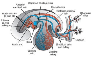
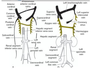
2. Introduction
• In the 5th
week, 3pairs of major veins can be
distinguished
1.vitelline veins, or omphalomesenteric veins,
carrying blood from the yolk sac to the sinus
venosus
2.umbilical veins, originating in the chorionic villi
and carrying oxygenated blood to the embryo
3.cardinal veins, draining the body of the embryo
proper
3.
4. Contd..
• Initially the cardinal veins form the main venous
drainage system of the embryo. This system
consists of the:
• anterior cardinal veins, which drain the cephalic
part of the embryo.
• posterior cardinal veins, which drain the rest of
the embryo.
• The anterior and posterior veins join before entering
the sinus horn and form the short common
cardinal veins.
5. Contd..
• During the fifth to the seventh week a number of
additional veins are formed:
1.subcardinal veins, which mainly drain the kidneys
2.sacrocardinal veins, which drain the lower
extremities
3.supracardinalveins, which drain the body wall by
way of the intercostal veins, taking over the
functions of the posterior cardinal veins
6. Contd..
• The anastomosis between the anterior cardinal
veins develops into the left brachiocephalic vein
• The terminal portion of the left posterior cardinal
vein entering into the left brachiocephalic vein is
retained as a small vessel, the left superior
intercostal vein. This vessel receives blood from
the 2nd and 3rd intercostal spaces.
• The superior vena cava is formed by the right
common cardinal vein and the proximal portion of
the right anterior cardinal vein.
7.
8. • With obliteration of the major portion of the
posterior cardinal veins, the supracardinal veins
assume a greater role in draining the body wall.
• The 4th
to 11th right intercostal veins empty into the
right supracardinal vein, which together with a
portion of the posterior cardinal vein forms the
azygos vein.
9. Contd …
• On the left the 4th to 7th intercostal veins enter into
the left supracardinal vein, and the left
supracardinal vein, then known as the hemiazygos
vein, empties into the azygos vein