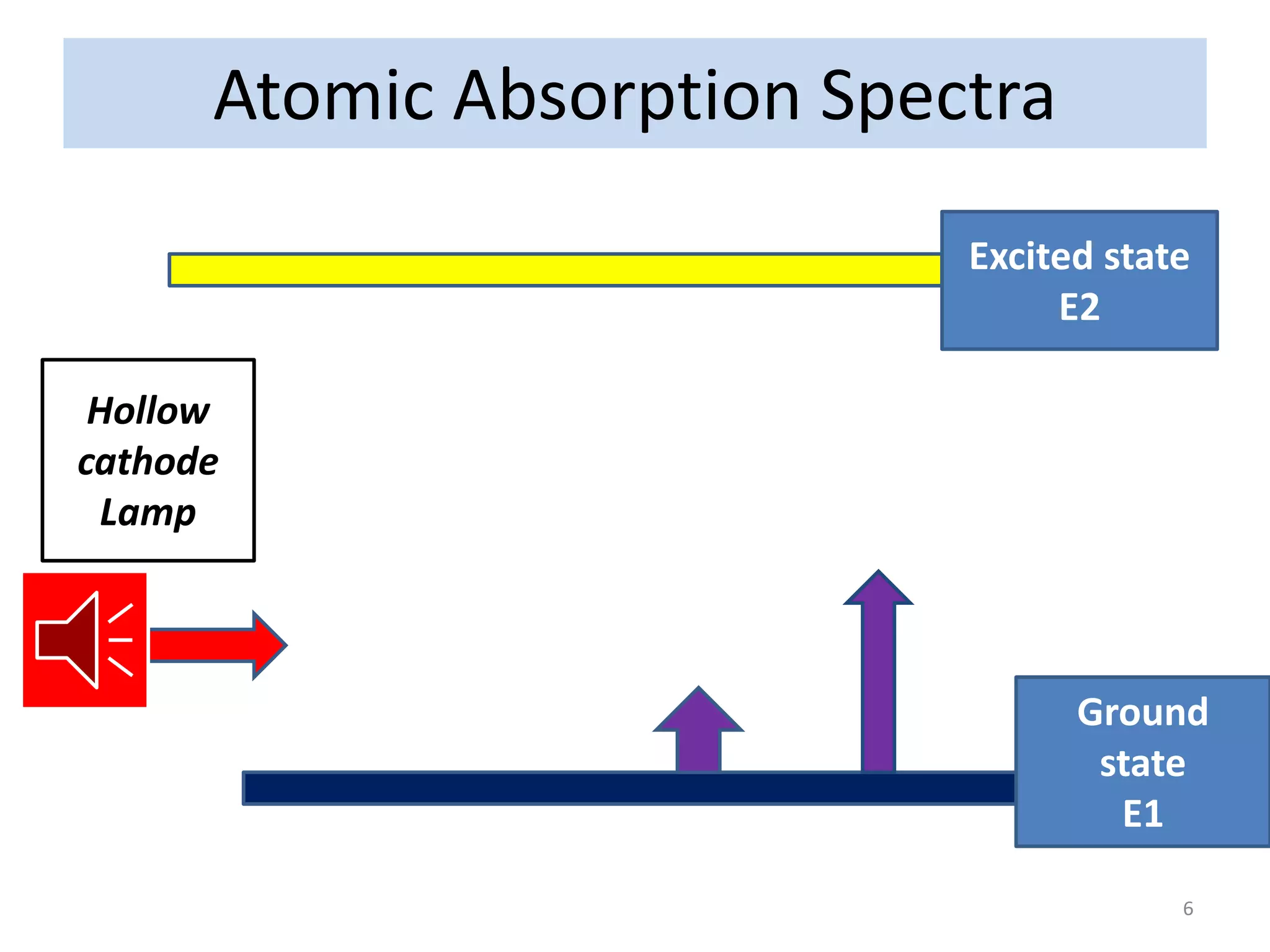
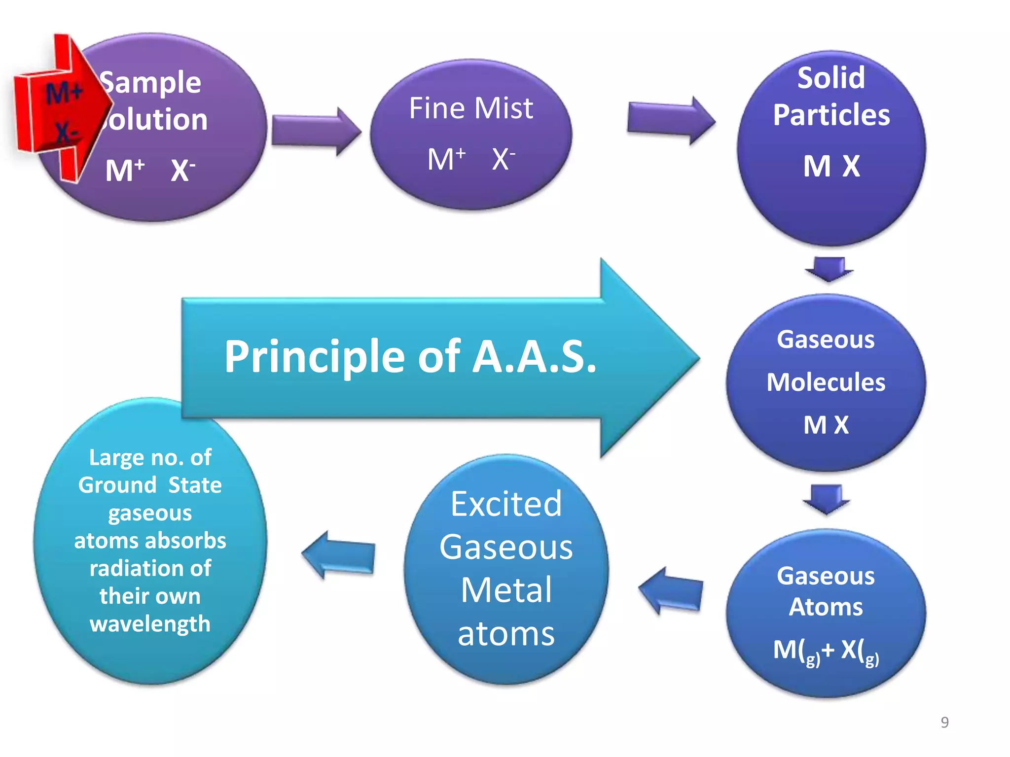
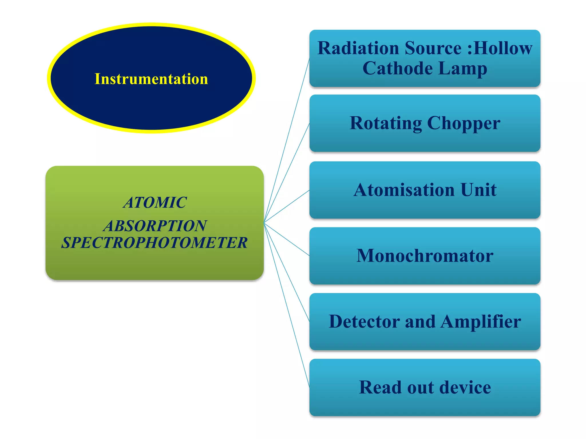
Satish Pradhan
Dnyanasadhana College, Thane
Department of Chemistry
T.Y.B.Sc. Analytical Chemistry
Paper-IV Sem-V
Atomic Absorption Spectroscopy
1
UNIT III: OPTICAL METHODS(6 UNITS)
Atomic Spectroscopy: Flame Emission spectroscopy(FES) and
Atomic Absorption Spectroscopy(AAS)
• 3.1.1 Introduction, Energy level diagrams, Atomic spectra,
Absorption and Emission Spectra
• 3.1.2 Flame Photometry – Principle, Instrumentation
(Flame atomizers, types of Burners, Wavelength selectors,
Detectors)
• 3.1.3 Atomic Absorption Spectroscopy – Principle,
Instrumentation (Source, Chopper, Flame and
Electrothermal Atomiser)
• 3.1.4 Quantification methods of FES and AAS – Calibration
curve method, Standard addition method and Internal
standard method. 3.1.5 Comparison between FES and AAS
• 3.1.6 Applications, Advantages and Limitations
Contents
• 3.1.4 Atomic Absorption Spectroscopy – Principle, Instrumentation
• (Source, Chopper, Flame and Electrothermal Atomizer)
• 3.1.5 Quantitative applications of Flame Photometry and Atomic
• Absorption Spectroscopy – Calibration curve method,
• Standard addition method and Internal standard method.
• 3.1.6 Comparison of Flame Photometry and Atomic Absorption
• Spectroscopy
• 3.1.7 Applications and Limitations of Flame Photometry and Atomic
• Absorption Spectroscopy.
Atomic Absorption
Spectroscopy
UNIT-IV 4
Introduction
• In Analytical chemistry Atomic
absorption spectroscopy is a technique
for determining the concentration of a
particular metal element in a sample.
Atomic absorption spectroscopy can be
used to analyze the concentration of over
62 different metals in a solution.
5
Atomic Absorption Spectra
Excited state
E2
Ground
state
E1
Hollow
cathode
Lamp
6
Technique
• The technique typically makes use of
a flame to atomize the sample, but
other atomizers such as a graphite
furnace are also used.
• Three steps are involved in turning
a liquid sample into an atomic gas:
7
Three steps In
AAS
Desolvation – the liquid
solvent is evaporated and
the dry sample remains
Vaporization – the solid
sample vaporizes to a gas
Volatilization – the
compounds making up the
sample are broken into free
atoms
Principle of F.E.S
Sample
Solution
M+ X-
Fine Mist
M+ X-
Solid
Particles
M X
Gaseous
Molecules
M X
Gaseous
Atoms
M(g)+ X(g)
Excited
Gaseous
Metal
atoms
Large no. of
Ground State
gaseous
atoms absorbs
radiation of
their own
wavelength
Principle of A.A.S.
9
ATOMIC
ABSORPTION
SPECTROPHOTOMETER
Radiation Source :Hollow
Cathode Lamp
Rotating Chopper
Atomisation Unit
Monochromator
Detector and Amplifier
Read out device
Instrumentation
Grating
Read OutAmplifier
Sample SolutionPower
Supply
Rotating Chopper
Flame
Hollow
Cathode
Lamp
P.M.T.Detector
Atomic Absorption Spectrophotometer
11
Inert Gas
Anode
+
Cathode
----
Quartz Window
_______________________________________
Glass Shield
Hallow Cathode Lamp
+
M
+
M
Hallow Cathode Cup
Radiation Source
12
13
14
15
Radiation Source :
• Radiation Source :Hollow Cathode Lamp
• HCL is the most common radiation source in atomic
absorption spectroscopy. The lamp consist of glass tube.
The Lamp is filled with argon or neon gas. The cathode is a
cylindrical metal cathode containing the metal for excitation,
and an anode. When a high voltage is applied across the anode
and cathode, gas particles are ionized. As voltage is increased,
gaseous ions acquire enough energy to eject metal atoms from
the cathode. Some of these atoms are in excited states and emit
light with the frequency characteristic to the metal. Many
modern hollow cathode lamps are selective for several metals.
16
Events in Hollow cathode Lamp:
Reactions in the hollow-cathode lamp
• ionization of filler gas: Ar + e- ® Ar+ + 2 sputtering
• of cathode atoms: M(s) + Ar+ ® M(g) + Ar
• excitation of metal atoms: M(g) + Ar+ ® M*(g) + Ar
• light emission: M*(g) ® M(g) + hV.
• The cathode contains the element that is analysed.
Light emitted by hallow-cathode lamp has the same
wavelength as the light absorbed by the analyte
element. Different lamp required for each element
(some are multielement)
17
• The type of hollow cathode tube depends on
the metal being analyzed. For analyzing the
concentration of copper in an ore, a copper
cathode tube would be used, and likewise for
any other metal being analyzed. The electrons
of the atoms in the flame can be promoted to
higher orbitals for an instant by absorbing a set
quantity of energy (a quantum).
18
2.Rotating Chopper:
Resonance radiation given by hollow cathode lamp as
well as sample atoms in flame due to thermal excitation
are detected by detector which creates an interference in
absorbance measurement.
To eliminate the radiation coming from atom in flame by
excitation of atoms. Rotating chopper is used.
Rotating chopper is arrange in between flame and Hollow
cathode lamp.
It is circular disc divided into four quarters, out of
which two are mirrored and two are opened.
When disc rotates at high speed when the mirror
quartered in front of the lamp it reflects the radiation.
The second moment it opens in front of lamp and
radiation passes to sample being absorbed by it and
reaches the detector in pulses.
19
Atomization
Unit
Flame atomizer
1) Total Consumption
Burner
2) Laminar flow
burner
Electro thermal
Atomizer
Disadvantages of Flame atomizer:
1) A large portion of sample flows down the drain
2) The residence time atoms in optical path is very
short less than 10 -4 seconds.
20
Inflation
Light
Electrical
Connection
Gas
Detachable Window
Water
Sample
Graphite Tube
Metal Jacket
Water
ELECTRO THERMAL ATOMIZER (Graphite furnace).
PMT
DETECTOR
21
ELECTRO THERMAL ATOMIZER (Graphite furnace).
Electro Thermal Atomizer also called as Graphite furnace.
Consist of hollow graphite tube 5cm long and 1 cm internal
diameter. The tube is opened at both ends and has central
hole to introduce of sample by micropipette. This tube is
arranged in such a way that radiation from the source
passes along the axis of the tube. Two electrodes are fitted
at the end of the tube. The inert gas like neon or argon is
allowed to flow over the tubes to avoid the oxidation of
graphite. The tube is surrounded By water cooled metal
jacket. The solution of the sample to be analysed (1-100ul)
is introduced by inserting the micro pipette through the
port in the outer jacket and gas inlet in the centre of
graphite tube. The graphite cylinder is then heated by the
passage of electric current to a temperature that is enough
to evaporate the solvent from the solutions. The current is
then increased.
22
STAGES IN ELECTRO THERMALATOMIZER
Drying stage Ashing stage Atomization
stage
Temperature
Range
Temperature of the
furnace is maintained
above the boiling point
of the solvent.
For aq. Solution
furnace temperature is
adjusted to 383K for 30
Sec
Temperature of the
furnace is maintained
at 600K -1500K for
period of 45 sec.
Temperature of the
furnace is
maintained at
2300K -3300K for
period of 5 sec.
Function Solvent undergoes
evaporation
Organic substance are
volatilized and other
chemical matrix of the
sample is destroyed
Gaseous atoms of
metal are formed
23
ADVANTAGES ELECTRO THERMALATOMIZER
1
• Temperature that is enough to ash the sample and to produce the
metal atoms.
2
• The sample volume is small
3
• No need for fuel -oxidant mixture.
4
• High sensitivity
5
• No flame noise.
6
• Solid sample can be used directly.
7
• Heat distribution is uniform and temperature is steady.
• Monochromator
• Prism or grating monochromator
are used in AAS. The function of
the monochromator is to select a
given absorbing line from spectral
lines by emitting hallow cathode
lamp.
Grating
Read OutAmplifier
Sample SolutionPower
Supply
Rotating Chopper
Flame
Hollow
Cathode
Lamp
P.M.T.Detector
Atomic Absorption Spectrophotometer
26
27
APPLICATION OF
ATOMIC
ABSORPTION
SPECTROPHOTOMETERY
Water analysis (e.g. Ca, Mg, Fe,
Si, Al, Ba content)
Food analysis
Analysis of animal Feedstuffs
(e.g. Mn, Fe, Cu, Cr, Se, Zn)
Analysis of additives in
lubricating oils and greases (Ba,
Ca, Na, Li, Zn, Mg)
Analysis of soils
Clinical analysis (blood samples:
whole blood, plasma, serum; Ca,
Mg, Li, Na, K, Fe)
ADVANTAGES OF
AAS OVER
FES
Solutions, slurries and solid samples
can be analysed.
Much More Efficient Atomization
Greater sensitivity
Smaller quantities of sample
(typically 5 – 50 uL)
provides a reducing environment for
easily oxidized elements
DISADVANTAGES OF
AAS
OVER
FES
Expensive
Low precision
Low sample throughput
Requires high level of operator
skill
Distinguish between FES and AAS
• AAS
 Solutions, slurries and
solid samples can be
analysed.
 Much More Efficient
Atomization
 Greater sensitivity
 Smaller quantities of
sample (typically 5 – 50 uL)
 Alkali metal and
transition metals can be
determined(64 elements)
• FES
 Solutions, can be analysed.
 Much More Efficient
Atomization
 Lesser sensitivity
 Larger quantities of
sample.
 Alkali metal and alkaline
earth metals can be
determined
31
 AAS
 Different hollow cathode lamps
are required as source of
radiation
 Chopper is used to eliminate
radiation emitted from flame.
 The quality of monochromator
should no be of very high degree .
 AC. Amplifier is used to amplify
only AC Current.
 The absorption intensity depends
upon unexcited atoms.
 signal is obtained due difference
of energies between excited and
ground state atom.
 High degree operator is
required
 FES
 Flame serves as source for
different element.
 Chopper is not used .
 The quality of monochromator
should be of very high degree
 Amplifier is used to increase the
output signal of detector.
 The emission intensity depends
upon excited atoms.
 Signal is obtained from
wavelength emitted by atoms.
 High degree operator is not
required
Distinguish between FES and AAS
32
 AAS
 The technique is expensive
(cost 15- 25 Lakh).
 Solutions, slurries and solid
samples can be analysed.
 Much More Efficient
Atomization.
 Greater sensitivity.
 Smaller quantities of sample
(typically 5 – 50 uL)
 Alkali metal and transition
metals can be determined(64
elements)
• FES
 The technique is not so
expensive (cost 60-70
thousands only)
 Only Solutions, can be
analysed.
 Less Efficient Atomization
 Lesser sensitivity
 Larger quantities of sample.
 Alkali metal and alkaline
earth metals can be
determined.
Distinguish between FES and AAS
33
Applications of Atomic Absorption
Spectroscopy
 Water analysis (e.g. Ca,
Mg, Fe, Si, Al, Ba content)
 Food analysis
 Analysis of animal
Feedstuffs (e.g. Mn, Fe,
Cu, Cr, Se, Zn)
 Analysis of additives in
lubricating oils and
greases (Ba, Ca, Na, Li,
Zn, Mg)
 Analysis of additives
in lubricating oils and
greases
 (Ba , Ca, Na, Li, Zn, Mg)
 Analysis of soils
 Clinical analysis (blood
samples: whole blood,
plasma, serum; Ca, Mg,
Li, Na, K, Fe)
34
Quantitative
Analysis
Calibration
Curve method
Standard
addition method
Internal
Standard
addition method
Calibration Curve method
•Absorbance--
Concentration-----------
A is Directly proportional to concentration
36
• In this method the absorbance of unknown (X) is
first found out by aspirating into flame against
blank.Then a series of standards having definite
amount of unknown (X) plus varying amount of
standard are prepared and diluted to same volume in
each case .Their absorbance are then obtained . A
graph of absorbance (A) against concentrations of
standard (S) gives a linear curve. The concentration
of the unknown can be determined by extrapolation
of line which cuts to X axis.
37
Standard addition method
Concentration-----
Absorbance---
Concentration of Unknown.
Standard addition method
38
Ax
-----
At
Concentration-----
Internal Standard addition method
39
•Thanks a lot

Atomic Absorption Spectroscopy

  • 1.
    Satish Pradhan Dnyanasadhana College,Thane Department of Chemistry T.Y.B.Sc. Analytical Chemistry Paper-IV Sem-V Atomic Absorption Spectroscopy 1
  • 2.
    UNIT III: OPTICALMETHODS(6 UNITS) Atomic Spectroscopy: Flame Emission spectroscopy(FES) and Atomic Absorption Spectroscopy(AAS) • 3.1.1 Introduction, Energy level diagrams, Atomic spectra, Absorption and Emission Spectra • 3.1.2 Flame Photometry – Principle, Instrumentation (Flame atomizers, types of Burners, Wavelength selectors, Detectors) • 3.1.3 Atomic Absorption Spectroscopy – Principle, Instrumentation (Source, Chopper, Flame and Electrothermal Atomiser) • 3.1.4 Quantification methods of FES and AAS – Calibration curve method, Standard addition method and Internal standard method. 3.1.5 Comparison between FES and AAS • 3.1.6 Applications, Advantages and Limitations
  • 3.
    Contents • 3.1.4 AtomicAbsorption Spectroscopy – Principle, Instrumentation • (Source, Chopper, Flame and Electrothermal Atomizer) • 3.1.5 Quantitative applications of Flame Photometry and Atomic • Absorption Spectroscopy – Calibration curve method, • Standard addition method and Internal standard method. • 3.1.6 Comparison of Flame Photometry and Atomic Absorption • Spectroscopy • 3.1.7 Applications and Limitations of Flame Photometry and Atomic • Absorption Spectroscopy.
  • 4.
  • 5.
    Introduction • In Analyticalchemistry Atomic absorption spectroscopy is a technique for determining the concentration of a particular metal element in a sample. Atomic absorption spectroscopy can be used to analyze the concentration of over 62 different metals in a solution. 5
  • 6.
    Atomic Absorption Spectra Excitedstate E2 Ground state E1 Hollow cathode Lamp 6
  • 7.
    Technique • The techniquetypically makes use of a flame to atomize the sample, but other atomizers such as a graphite furnace are also used. • Three steps are involved in turning a liquid sample into an atomic gas: 7
  • 8.
    Three steps In AAS Desolvation– the liquid solvent is evaporated and the dry sample remains Vaporization – the solid sample vaporizes to a gas Volatilization – the compounds making up the sample are broken into free atoms
  • 9.
    Principle of F.E.S Sample Solution M+X- Fine Mist M+ X- Solid Particles M X Gaseous Molecules M X Gaseous Atoms M(g)+ X(g) Excited Gaseous Metal atoms Large no. of Ground State gaseous atoms absorbs radiation of their own wavelength Principle of A.A.S. 9
  • 10.
    ATOMIC ABSORPTION SPECTROPHOTOMETER Radiation Source :Hollow CathodeLamp Rotating Chopper Atomisation Unit Monochromator Detector and Amplifier Read out device Instrumentation
  • 11.
    Grating Read OutAmplifier Sample SolutionPower Supply RotatingChopper Flame Hollow Cathode Lamp P.M.T.Detector Atomic Absorption Spectrophotometer 11
  • 12.
    Inert Gas Anode + Cathode ---- Quartz Window _______________________________________ GlassShield Hallow Cathode Lamp + M + M Hallow Cathode Cup Radiation Source 12
  • 13.
  • 14.
  • 15.
  • 16.
    Radiation Source : •Radiation Source :Hollow Cathode Lamp • HCL is the most common radiation source in atomic absorption spectroscopy. The lamp consist of glass tube. The Lamp is filled with argon or neon gas. The cathode is a cylindrical metal cathode containing the metal for excitation, and an anode. When a high voltage is applied across the anode and cathode, gas particles are ionized. As voltage is increased, gaseous ions acquire enough energy to eject metal atoms from the cathode. Some of these atoms are in excited states and emit light with the frequency characteristic to the metal. Many modern hollow cathode lamps are selective for several metals. 16
  • 17.
    Events in Hollowcathode Lamp: Reactions in the hollow-cathode lamp • ionization of filler gas: Ar + e- ® Ar+ + 2 sputtering • of cathode atoms: M(s) + Ar+ ® M(g) + Ar • excitation of metal atoms: M(g) + Ar+ ® M*(g) + Ar • light emission: M*(g) ® M(g) + hV. • The cathode contains the element that is analysed. Light emitted by hallow-cathode lamp has the same wavelength as the light absorbed by the analyte element. Different lamp required for each element (some are multielement) 17
  • 18.
    • The typeof hollow cathode tube depends on the metal being analyzed. For analyzing the concentration of copper in an ore, a copper cathode tube would be used, and likewise for any other metal being analyzed. The electrons of the atoms in the flame can be promoted to higher orbitals for an instant by absorbing a set quantity of energy (a quantum). 18
  • 19.
    2.Rotating Chopper: Resonance radiationgiven by hollow cathode lamp as well as sample atoms in flame due to thermal excitation are detected by detector which creates an interference in absorbance measurement. To eliminate the radiation coming from atom in flame by excitation of atoms. Rotating chopper is used. Rotating chopper is arrange in between flame and Hollow cathode lamp. It is circular disc divided into four quarters, out of which two are mirrored and two are opened. When disc rotates at high speed when the mirror quartered in front of the lamp it reflects the radiation. The second moment it opens in front of lamp and radiation passes to sample being absorbed by it and reaches the detector in pulses. 19
  • 20.
    Atomization Unit Flame atomizer 1) TotalConsumption Burner 2) Laminar flow burner Electro thermal Atomizer Disadvantages of Flame atomizer: 1) A large portion of sample flows down the drain 2) The residence time atoms in optical path is very short less than 10 -4 seconds. 20
  • 21.
    Inflation Light Electrical Connection Gas Detachable Window Water Sample Graphite Tube MetalJacket Water ELECTRO THERMAL ATOMIZER (Graphite furnace). PMT DETECTOR 21
  • 22.
    ELECTRO THERMAL ATOMIZER(Graphite furnace). Electro Thermal Atomizer also called as Graphite furnace. Consist of hollow graphite tube 5cm long and 1 cm internal diameter. The tube is opened at both ends and has central hole to introduce of sample by micropipette. This tube is arranged in such a way that radiation from the source passes along the axis of the tube. Two electrodes are fitted at the end of the tube. The inert gas like neon or argon is allowed to flow over the tubes to avoid the oxidation of graphite. The tube is surrounded By water cooled metal jacket. The solution of the sample to be analysed (1-100ul) is introduced by inserting the micro pipette through the port in the outer jacket and gas inlet in the centre of graphite tube. The graphite cylinder is then heated by the passage of electric current to a temperature that is enough to evaporate the solvent from the solutions. The current is then increased. 22
  • 23.
    STAGES IN ELECTROTHERMALATOMIZER Drying stage Ashing stage Atomization stage Temperature Range Temperature of the furnace is maintained above the boiling point of the solvent. For aq. Solution furnace temperature is adjusted to 383K for 30 Sec Temperature of the furnace is maintained at 600K -1500K for period of 45 sec. Temperature of the furnace is maintained at 2300K -3300K for period of 5 sec. Function Solvent undergoes evaporation Organic substance are volatilized and other chemical matrix of the sample is destroyed Gaseous atoms of metal are formed 23
  • 24.
    ADVANTAGES ELECTRO THERMALATOMIZER 1 •Temperature that is enough to ash the sample and to produce the metal atoms. 2 • The sample volume is small 3 • No need for fuel -oxidant mixture. 4 • High sensitivity 5 • No flame noise. 6 • Solid sample can be used directly. 7 • Heat distribution is uniform and temperature is steady.
  • 25.
    • Monochromator • Prismor grating monochromator are used in AAS. The function of the monochromator is to select a given absorbing line from spectral lines by emitting hallow cathode lamp.
  • 26.
    Grating Read OutAmplifier Sample SolutionPower Supply RotatingChopper Flame Hollow Cathode Lamp P.M.T.Detector Atomic Absorption Spectrophotometer 26
  • 27.
  • 28.
    APPLICATION OF ATOMIC ABSORPTION SPECTROPHOTOMETERY Water analysis(e.g. Ca, Mg, Fe, Si, Al, Ba content) Food analysis Analysis of animal Feedstuffs (e.g. Mn, Fe, Cu, Cr, Se, Zn) Analysis of additives in lubricating oils and greases (Ba, Ca, Na, Li, Zn, Mg) Analysis of soils Clinical analysis (blood samples: whole blood, plasma, serum; Ca, Mg, Li, Na, K, Fe)
  • 29.
    ADVANTAGES OF AAS OVER FES Solutions,slurries and solid samples can be analysed. Much More Efficient Atomization Greater sensitivity Smaller quantities of sample (typically 5 – 50 uL) provides a reducing environment for easily oxidized elements
  • 30.
    DISADVANTAGES OF AAS OVER FES Expensive Low precision Lowsample throughput Requires high level of operator skill
  • 31.
    Distinguish between FESand AAS • AAS  Solutions, slurries and solid samples can be analysed.  Much More Efficient Atomization  Greater sensitivity  Smaller quantities of sample (typically 5 – 50 uL)  Alkali metal and transition metals can be determined(64 elements) • FES  Solutions, can be analysed.  Much More Efficient Atomization  Lesser sensitivity  Larger quantities of sample.  Alkali metal and alkaline earth metals can be determined 31
  • 32.
     AAS  Differenthollow cathode lamps are required as source of radiation  Chopper is used to eliminate radiation emitted from flame.  The quality of monochromator should no be of very high degree .  AC. Amplifier is used to amplify only AC Current.  The absorption intensity depends upon unexcited atoms.  signal is obtained due difference of energies between excited and ground state atom.  High degree operator is required  FES  Flame serves as source for different element.  Chopper is not used .  The quality of monochromator should be of very high degree  Amplifier is used to increase the output signal of detector.  The emission intensity depends upon excited atoms.  Signal is obtained from wavelength emitted by atoms.  High degree operator is not required Distinguish between FES and AAS 32
  • 33.
     AAS  Thetechnique is expensive (cost 15- 25 Lakh).  Solutions, slurries and solid samples can be analysed.  Much More Efficient Atomization.  Greater sensitivity.  Smaller quantities of sample (typically 5 – 50 uL)  Alkali metal and transition metals can be determined(64 elements) • FES  The technique is not so expensive (cost 60-70 thousands only)  Only Solutions, can be analysed.  Less Efficient Atomization  Lesser sensitivity  Larger quantities of sample.  Alkali metal and alkaline earth metals can be determined. Distinguish between FES and AAS 33
  • 34.
    Applications of AtomicAbsorption Spectroscopy  Water analysis (e.g. Ca, Mg, Fe, Si, Al, Ba content)  Food analysis  Analysis of animal Feedstuffs (e.g. Mn, Fe, Cu, Cr, Se, Zn)  Analysis of additives in lubricating oils and greases (Ba, Ca, Na, Li, Zn, Mg)  Analysis of additives in lubricating oils and greases  (Ba , Ca, Na, Li, Zn, Mg)  Analysis of soils  Clinical analysis (blood samples: whole blood, plasma, serum; Ca, Mg, Li, Na, K, Fe) 34
  • 35.
  • 36.
  • 37.
    • In thismethod the absorbance of unknown (X) is first found out by aspirating into flame against blank.Then a series of standards having definite amount of unknown (X) plus varying amount of standard are prepared and diluted to same volume in each case .Their absorbance are then obtained . A graph of absorbance (A) against concentrations of standard (S) gives a linear curve. The concentration of the unknown can be determined by extrapolation of line which cuts to X axis. 37 Standard addition method
  • 38.
  • 39.
  • 40.