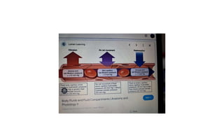
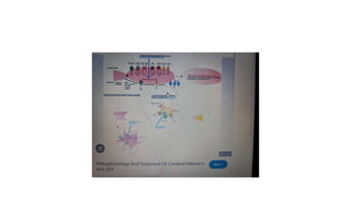
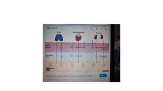
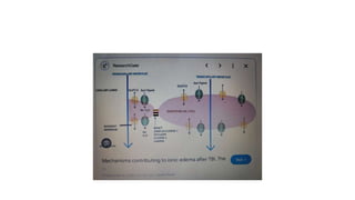
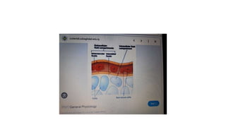
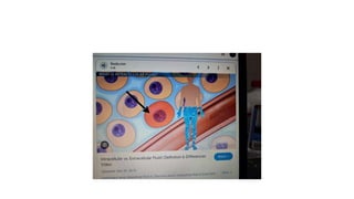
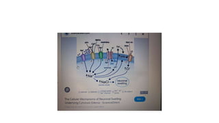
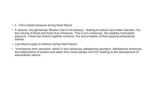
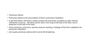
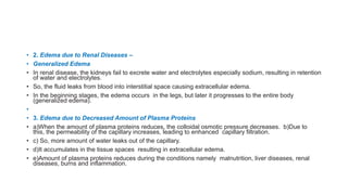
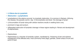
APPLIED PHYSIOLOGY – EDEMA
Dr. E. Muralinath, Kalyan C. R. Gnanalahari
• DEFINITION
• Edema is defined as the swelling happened by excessive accumulation of fluid particul;arly in the
tissues.
• It may be generalized or local. Edema that involves the entire body is termed as generalized
edema.
• Local edema is the one that happens is specific areas of the body namely abdomen, lungs and
extremities like feet, ankles and legs.
• Accumulation of fluid may occur either inside or outside the cell.
• 
TYPES OF EDEMA
• Edema is categorized into two types, depending upon the body fluid compartment where
accumulation of excess fluid happens:
• 1. Intracellular edema
• 2. Extracellular edema.
• INTRACELLULAR EDEMA
• Intracellular edema is the accumulation of fluid inside the cell. It happens due to three reasons:
• 1. Malnutrition
• 2. Poor metabolism
• 3. Inflammation of the tissues.
• 1. Edema due to Malnutrition
• a)Malnutrition happens due to poor consumpotion of food or poor circulatory system, through
which the nutritive substances are supplied.
• b) Due to the lack of nutrition, the ionic pumps of the cell membrane are depressed resulting in
poor exchange of ions.
• c) Sp[ecifically, , the sodium ions leaking into the cells cannot be pumped out. Excess sodium
inside the cells leads to endosmosis, resulting in intracellular edema.
• 2. Edema due to Poor Metabolism
• Poor metabolism is happened by poor blood supply. Poor blood supply results in lack of oxygen. It
leads to poor function of cell membrane and edema,
• 3. Edema due to Inflammation of Tissues
• a)During inflammation of the tissues, normally the permeability of cell membrane enhances. b)This
causes the movement of many ions, along with sodium into the cells leading to endosmosis and
intracellular edema.
• EXTRACELLULAR EDEMA
• Extracellular edema is defined as the accumulation of fluid outside the cell.
• Causes for extracellular edema
• 1. Abnormal leakage of fluid from capillaries into interstitial space.
• 2. Obstruction of lymphatic vessels that obstructs the fluid return from interstitium to blood.
• Conditions which lresult inextracellular edema
• 1. Heart failure.
• 2. Renal disease.
• 3. Decreased amount of plasma proteins.
• 4. Lymphatic obstruction.
• 5. Increased endothelial permeability.
• 1. Edema due to Heart Failure
• Edema happens in heart failuredue to various reasons namely :
• Failure of heart to pump blood:
• Failure of the heart to pump blood from veins to arteries enhances venous pressure and
capillary pressure. This results in enhanced capillary permeability and leakage of fluid from blood
into interstitial fluid, causing extracellular edema.
• Ii.. Fall in blood pressure during heart failure:
• It reduces the glomerular filtration rate in the kidneys, leading to sodium and water retention. So,
the volume of blood and body fluid enhances. This in turn enhances the capillary hydrostatic
pressure. These two factors together enhance the accumulation of fluid causing extracellular
edema.
• Low blood supply to kidneys during heart failure:
• It enhances renin secretion, which in turn enhances aldosterone secretion. Aldosterone enhances
the reabsorption of sodium and water from renal tubules into ECF leading to the development of
extracellular edema.
• Pulmonary Edema
• Pulmonary edema is the accumulation of fluid in pulmonary interstitium.
• In left heart failure, the blood is easily pumped into pulmonary circulation by right ventricle.
c)Whatever it may be, , the blood cannot return from lungs to left side of the heart due to
weakness of this side of the heart.
• d)This enhances pulmonary vascular pressure resulting in leakage of fluid from capillaries into
pulmonary interstitium.
• e)It causes pulmonary edema which can be life threatening.
• 2. Edema due to Renal Diseases –
• Generalized Edema
• In renal disease, the kidneys fail to excrete water and electrolytes especially sodium, resulting in retention
of water and electrolytes.
• So, the fluid leaks from blood into interstitial space causing extracellular edema.
• In the beginning stages, the edema occurs in the legs, but later it progresses to the entire body
(generalized edema).
•
• 3. Edema due to Decreased Amount of Plasma Proteins
• a)When the amount of plasma proteins reduces, the colloidal osmotic pressure decreases. b)Due to
this, the permeability of the capillary increases, leading to enhanced capillary filtration.
• c) So, more amount of water leaks out of the capillary.
• d)It accumulates in the tissue spaces resulting in extracellular edema.
• e)Amount of plasma proteins reduces during the conditions namely malnutrition, liver diseases, renal
diseases, burns and inflammation.
• 4. Edema due to Lymphatic
• Obstruction – Lymphedema
• Lymphedema is the edema occurred by lymphatic obstruction. It is common in filariasis. b)During
this disease, the parasitic worms stay in the lymphatics and block the drainage of lymph.
• c) Accumulation of lymph along with cellular reactions results in swelling that is very
• prominent in legs and scrotum.
• d)Repeated obstruction of lymphatic drainage in these regions leading to fibrosis and development
of elephantiasis.
•
• Elephantiasis
• Elephantiasis is a disorder of lymphatic system, manifested by thickening of skin and extreme
enlargement of the affected area, most commonly limbs (legs), genitals, certain areas of trunk and
parts of head.
• 5. Edema due to Increased Endothelial Permeability
• The permeability of the capillary endothelium enhances in conditions namely burns, inflammation,
trauma, allergic reactions and immunologic reactions, which result in oozing out of fluid. This fluid
accumulates resulting in development of edema.
• PITTING AND NON-PITTING EDEMA
• Interstitial fluid is observed in the form of a gel that is almost like a semisolid substance.
• It is because the interstitial fluid is not present as fluid but is bound in a proteoglycan meshwork.
• It does not permit any free space for the fluid movement except for a diameter of about a few
hundredths of a micron.
• Normal volume of interstitial fluid is 12 L and it exerts a negative pressure of about 3 mm Hg.
• It applies a slight suction effect and results in holding of the tissues together.
• Whatever it may be, in abnormal conditions, where the interstitial fluid volume enhances
enormously, the pressure becomes positive.
• Most of the fluid becomes free fluid that is not bound to proteoglycan meshwork.
• It flows freely through tissue spaces, producing a swelling termed as edema.
• This type of edema is called pitting edema because, when this area is pressed with the finger,
displacement of fluid happens producing a depression or pit.
• When the finger is removed, the pit remains for few seconds, sometimes as long as one minute, till
the fluid flows back into that area.
• Edema also develops because of swelling of the cells or clotting of interstitial fluid in the presence
of fibrinogen.
• This is termed as non-pitting edema because, it is hard and a pit is not formed by pressing.
• References
• 1.
• Cho S, Atwood JE. Peripheral edema. Am J Med. 2002 Nov;113(7):580-6. [PubMed]
• 2.
• Miserocchi G, Negrini D, Passi A, De Luca G. Development of lung edema: interstitial fluid
dynamics and molecular structure. News Physiol Sci. 2001 Apr;16:66-71. [PubMed]
• 3.
• Bhave G, Neilson EG. Body fluid dynamics: back to the future. J Am Soc Nephrol. 2011
Dec;22(12):2166-81. [PMC free article] [PubMed]
• 4.
• Levick JR, Michel CC. Microvascular fluid exchange and the revised Starling principle.
Cardiovasc Res. 2010 Jul 15;87(2):198-210. [PubMed]
• 5.
• Reed RK, Rubin K. Transcapillary exchange: role and importance of the interstitial fluid
pressure and the extracellular matrix. Cardiovasc Res. 2010 Jul 15;87(2):211-7. [PubMed]
• 6.
• Woodcock TE, Woodcock TM. Revised Starling equation and the glycocalyx model of transvascular fluid
exchange: an improved paradigm for prescribing intravenous fluid therapy. Br J Anaesth. 2012
Mar;108(3):384-94. [PubMed]
• 7.
• Wiig H, Schröder A, Neuhofer W, Jantsch J, Kopp C, Karlsen TV, Boschmann M, Goss J, Bry M, Rakova N,
Dahlmann A, Brenner S, Tenstad O, Nurmi H, Mervaala E, Wagner H, Beck FX, Müller DN, Kerjaschki D, Luft
FC, Harrison DG, Alitalo K, Titze J. Immune cells control skin lymphatic electrolyte homeostasis and blood
pressure. J Clin Invest. 2013 Jul;123(7):2803-15. [PMC free article] [PubMed]
• 8.
• Renkin EM. B. W. Zweifach Award lecture. Regulation of the microcirculation. Microvasc Res. 1985
Nov;30(3):251-63. [PubMed]
• 9.
• Taylor AE. Capillary fluid filtration. Starling forces and lymph flow. Circ Res. 1981 Sep;49(3):557-75.
[PubMed]
• 10.
• Crandall ED, Staub NC, Goldberg HS, Effros RM. Recent developments in pulmonary edema. Ann Intern Med.
1983 Dec;99(6):808-22. [PubMed]
• 11.
• Watkins L, Burton JA, Haber E, Cant JR, Smith FW, Barger AC. The renin-angiotensin-aldosterone system in
congestive failure in conscious dogs. J Clin Invest. 1976 Jun;57(6):1606-17. [PMC free article] [PubMed]
• 12.
• Dzau VJ, Colucci WS, Hollenberg NK, Williams GH. Relation of the renin-angiotensin-aldosterone system to clinical
state in congestive heart failure. Circulation. 1981 Mar;63(3):645-51. [PubMed]
• 13.
• Deitch EA. The management of burns. N Engl J Med. 1990 Nov 01;323(18):1249-53. [PubMed]
• 14.
• Ohlsson K, Björk P, Bergenfeldt M, Hageman R, Thompson RC. Interleukin-1 receptor antagonist reduces mortality
from endotoxin shock. Nature. 1990 Dec 06;348(6301):550-2. [PubMed]
• 15.
• Colletti LM, Remick DG, Burtch GD, Kunkel SL, Strieter RM, Campbell DA. Role of tumor necrosis factor-alpha in
the pathophysiologic alterations after hepatic ischemia/reperfusion injury in the rat. J Clin Invest. 1990
Jun;85(6):1936-43. [PMC free article] [PubMed]
• 16.
• Hommel E, Mathiesen ER, Aukland K, Parving HH. Pathophysiological aspects of edema formation in diabetic
nephropathy. Kidney Int. 1990 Dec;38(6):1187-92. [PubMed]
Oedema_types_causes_pathophysiology.pptx

Oedema_types_causes_pathophysiology.pptx

  • 1.
    APPLIED PHYSIOLOGY –EDEMA Dr. E. Muralinath, Kalyan C. R. Gnanalahari
  • 3.
    • DEFINITION • Edemais defined as the swelling happened by excessive accumulation of fluid particul;arly in the tissues. • It may be generalized or local. Edema that involves the entire body is termed as generalized edema. • Local edema is the one that happens is specific areas of the body namely abdomen, lungs and extremities like feet, ankles and legs. • Accumulation of fluid may occur either inside or outside the cell. •  TYPES OF EDEMA • Edema is categorized into two types, depending upon the body fluid compartment where accumulation of excess fluid happens: • 1. Intracellular edema • 2. Extracellular edema.
  • 4.
    • INTRACELLULAR EDEMA •Intracellular edema is the accumulation of fluid inside the cell. It happens due to three reasons: • 1. Malnutrition • 2. Poor metabolism • 3. Inflammation of the tissues.
  • 5.
    • 1. Edemadue to Malnutrition • a)Malnutrition happens due to poor consumpotion of food or poor circulatory system, through which the nutritive substances are supplied. • b) Due to the lack of nutrition, the ionic pumps of the cell membrane are depressed resulting in poor exchange of ions. • c) Sp[ecifically, , the sodium ions leaking into the cells cannot be pumped out. Excess sodium inside the cells leads to endosmosis, resulting in intracellular edema.
  • 6.
    • 2. Edemadue to Poor Metabolism • Poor metabolism is happened by poor blood supply. Poor blood supply results in lack of oxygen. It leads to poor function of cell membrane and edema, • 3. Edema due to Inflammation of Tissues • a)During inflammation of the tissues, normally the permeability of cell membrane enhances. b)This causes the movement of many ions, along with sodium into the cells leading to endosmosis and intracellular edema.
  • 7.
    • EXTRACELLULAR EDEMA •Extracellular edema is defined as the accumulation of fluid outside the cell. • Causes for extracellular edema • 1. Abnormal leakage of fluid from capillaries into interstitial space. • 2. Obstruction of lymphatic vessels that obstructs the fluid return from interstitium to blood.
  • 8.
    • Conditions whichlresult inextracellular edema • 1. Heart failure. • 2. Renal disease. • 3. Decreased amount of plasma proteins. • 4. Lymphatic obstruction. • 5. Increased endothelial permeability. • 1. Edema due to Heart Failure • Edema happens in heart failuredue to various reasons namely : • Failure of heart to pump blood: • Failure of the heart to pump blood from veins to arteries enhances venous pressure and capillary pressure. This results in enhanced capillary permeability and leakage of fluid from blood into interstitial fluid, causing extracellular edema.
  • 9.
    • Ii.. Fallin blood pressure during heart failure: • It reduces the glomerular filtration rate in the kidneys, leading to sodium and water retention. So, the volume of blood and body fluid enhances. This in turn enhances the capillary hydrostatic pressure. These two factors together enhance the accumulation of fluid causing extracellular edema. • Low blood supply to kidneys during heart failure: • It enhances renin secretion, which in turn enhances aldosterone secretion. Aldosterone enhances the reabsorption of sodium and water from renal tubules into ECF leading to the development of extracellular edema.
  • 10.
    • Pulmonary Edema •Pulmonary edema is the accumulation of fluid in pulmonary interstitium. • In left heart failure, the blood is easily pumped into pulmonary circulation by right ventricle. c)Whatever it may be, , the blood cannot return from lungs to left side of the heart due to weakness of this side of the heart. • d)This enhances pulmonary vascular pressure resulting in leakage of fluid from capillaries into pulmonary interstitium. • e)It causes pulmonary edema which can be life threatening.
  • 11.
    • 2. Edemadue to Renal Diseases – • Generalized Edema • In renal disease, the kidneys fail to excrete water and electrolytes especially sodium, resulting in retention of water and electrolytes. • So, the fluid leaks from blood into interstitial space causing extracellular edema. • In the beginning stages, the edema occurs in the legs, but later it progresses to the entire body (generalized edema). • • 3. Edema due to Decreased Amount of Plasma Proteins • a)When the amount of plasma proteins reduces, the colloidal osmotic pressure decreases. b)Due to this, the permeability of the capillary increases, leading to enhanced capillary filtration. • c) So, more amount of water leaks out of the capillary. • d)It accumulates in the tissue spaces resulting in extracellular edema. • e)Amount of plasma proteins reduces during the conditions namely malnutrition, liver diseases, renal diseases, burns and inflammation.
  • 12.
    • 4. Edemadue to Lymphatic • Obstruction – Lymphedema • Lymphedema is the edema occurred by lymphatic obstruction. It is common in filariasis. b)During this disease, the parasitic worms stay in the lymphatics and block the drainage of lymph. • c) Accumulation of lymph along with cellular reactions results in swelling that is very • prominent in legs and scrotum. • d)Repeated obstruction of lymphatic drainage in these regions leading to fibrosis and development of elephantiasis. • • Elephantiasis • Elephantiasis is a disorder of lymphatic system, manifested by thickening of skin and extreme enlargement of the affected area, most commonly limbs (legs), genitals, certain areas of trunk and parts of head.
  • 13.
    • 5. Edemadue to Increased Endothelial Permeability • The permeability of the capillary endothelium enhances in conditions namely burns, inflammation, trauma, allergic reactions and immunologic reactions, which result in oozing out of fluid. This fluid accumulates resulting in development of edema.
  • 14.
    • PITTING ANDNON-PITTING EDEMA • Interstitial fluid is observed in the form of a gel that is almost like a semisolid substance. • It is because the interstitial fluid is not present as fluid but is bound in a proteoglycan meshwork. • It does not permit any free space for the fluid movement except for a diameter of about a few hundredths of a micron. • Normal volume of interstitial fluid is 12 L and it exerts a negative pressure of about 3 mm Hg. • It applies a slight suction effect and results in holding of the tissues together. • Whatever it may be, in abnormal conditions, where the interstitial fluid volume enhances enormously, the pressure becomes positive. • Most of the fluid becomes free fluid that is not bound to proteoglycan meshwork. • It flows freely through tissue spaces, producing a swelling termed as edema. • This type of edema is called pitting edema because, when this area is pressed with the finger, displacement of fluid happens producing a depression or pit. • When the finger is removed, the pit remains for few seconds, sometimes as long as one minute, till the fluid flows back into that area. • Edema also develops because of swelling of the cells or clotting of interstitial fluid in the presence of fibrinogen. • This is termed as non-pitting edema because, it is hard and a pit is not formed by pressing.
  • 15.
    • References • 1. •Cho S, Atwood JE. Peripheral edema. Am J Med. 2002 Nov;113(7):580-6. [PubMed] • 2. • Miserocchi G, Negrini D, Passi A, De Luca G. Development of lung edema: interstitial fluid dynamics and molecular structure. News Physiol Sci. 2001 Apr;16:66-71. [PubMed] • 3. • Bhave G, Neilson EG. Body fluid dynamics: back to the future. J Am Soc Nephrol. 2011 Dec;22(12):2166-81. [PMC free article] [PubMed] • 4. • Levick JR, Michel CC. Microvascular fluid exchange and the revised Starling principle. Cardiovasc Res. 2010 Jul 15;87(2):198-210. [PubMed] • 5. • Reed RK, Rubin K. Transcapillary exchange: role and importance of the interstitial fluid pressure and the extracellular matrix. Cardiovasc Res. 2010 Jul 15;87(2):211-7. [PubMed]
  • 16.
    • 6. • WoodcockTE, Woodcock TM. Revised Starling equation and the glycocalyx model of transvascular fluid exchange: an improved paradigm for prescribing intravenous fluid therapy. Br J Anaesth. 2012 Mar;108(3):384-94. [PubMed] • 7. • Wiig H, Schröder A, Neuhofer W, Jantsch J, Kopp C, Karlsen TV, Boschmann M, Goss J, Bry M, Rakova N, Dahlmann A, Brenner S, Tenstad O, Nurmi H, Mervaala E, Wagner H, Beck FX, Müller DN, Kerjaschki D, Luft FC, Harrison DG, Alitalo K, Titze J. Immune cells control skin lymphatic electrolyte homeostasis and blood pressure. J Clin Invest. 2013 Jul;123(7):2803-15. [PMC free article] [PubMed] • 8. • Renkin EM. B. W. Zweifach Award lecture. Regulation of the microcirculation. Microvasc Res. 1985 Nov;30(3):251-63. [PubMed] • 9. • Taylor AE. Capillary fluid filtration. Starling forces and lymph flow. Circ Res. 1981 Sep;49(3):557-75. [PubMed] • 10. • Crandall ED, Staub NC, Goldberg HS, Effros RM. Recent developments in pulmonary edema. Ann Intern Med. 1983 Dec;99(6):808-22. [PubMed]
  • 17.
    • 11. • WatkinsL, Burton JA, Haber E, Cant JR, Smith FW, Barger AC. The renin-angiotensin-aldosterone system in congestive failure in conscious dogs. J Clin Invest. 1976 Jun;57(6):1606-17. [PMC free article] [PubMed] • 12. • Dzau VJ, Colucci WS, Hollenberg NK, Williams GH. Relation of the renin-angiotensin-aldosterone system to clinical state in congestive heart failure. Circulation. 1981 Mar;63(3):645-51. [PubMed] • 13. • Deitch EA. The management of burns. N Engl J Med. 1990 Nov 01;323(18):1249-53. [PubMed] • 14. • Ohlsson K, Björk P, Bergenfeldt M, Hageman R, Thompson RC. Interleukin-1 receptor antagonist reduces mortality from endotoxin shock. Nature. 1990 Dec 06;348(6301):550-2. [PubMed] • 15. • Colletti LM, Remick DG, Burtch GD, Kunkel SL, Strieter RM, Campbell DA. Role of tumor necrosis factor-alpha in the pathophysiologic alterations after hepatic ischemia/reperfusion injury in the rat. J Clin Invest. 1990 Jun;85(6):1936-43. [PMC free article] [PubMed] • 16. • Hommel E, Mathiesen ER, Aukland K, Parving HH. Pathophysiological aspects of edema formation in diabetic nephropathy. Kidney Int. 1990 Dec;38(6):1187-92. [PubMed]