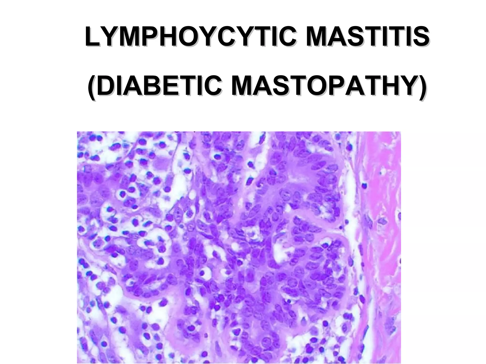
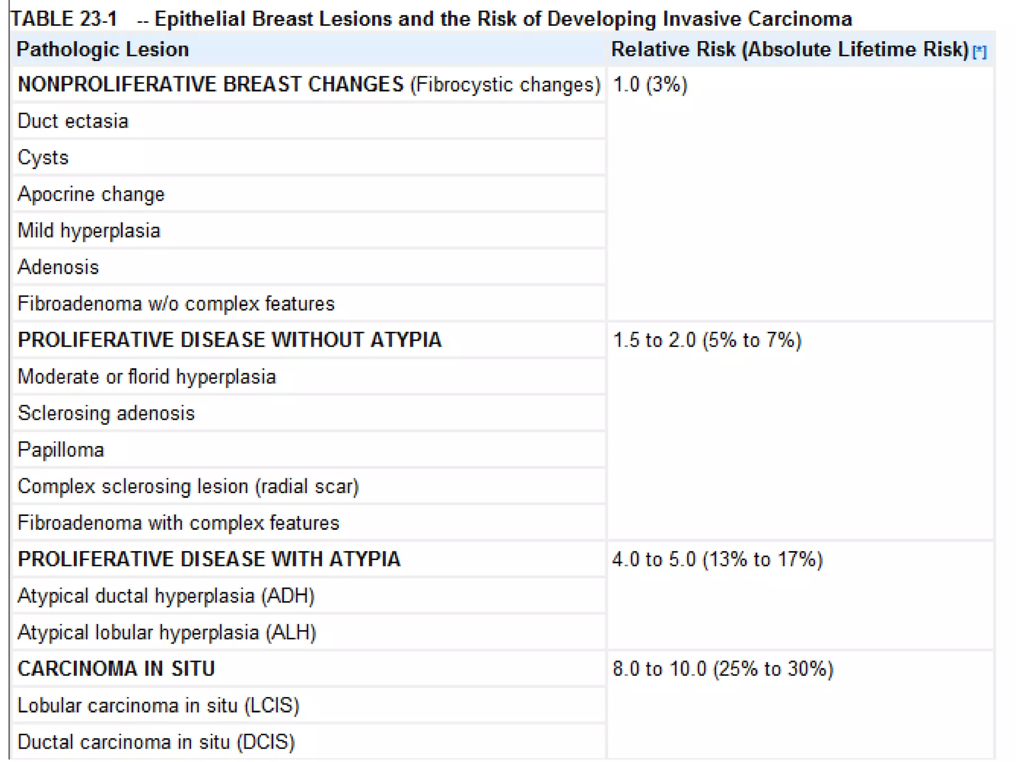
NON-NEOPLASTIC LESIONS
OF BREAST
Dr. Usman Shams
HISTOLOGY
• LOBE: (10 in whole breast)
• LOBULE: (many per lobe)
• ACINUS/I, aka ALVEOLUS/I: (many per
lobule)
• DUCT(S): INTRA- or INTER- LOB(UL)AR,
leading to the lactiferous ducts in the nipple
L
O
B
E
LOBULE
DEVELOPMENTAL
• MILKLINE REMNANTS
• ACCESSORY (axillary) BREAST
TISSUE
• NIPPLE INVERSION
• MACROMASTIA
ACCESSORY
(axillary)
BREAST
TISSUE
1) CONGENITAL
2) ACQUIRED: CARCINOMA
3) ACQUIRED: PIERCING
NIPPLE
INVERSION
INFLAMMATION
• ACUTE: staph most common
• PERIDUCTAL
• DUCT-ECTASIA
• FAT NECROSIS: usually trauma
• LYMPHOCYTIC: diabetic
• GRANULOMATOUS: sarcoidosis, TB,
etc., but mostly idiopathic
ACUTE MASTITIS
PERIDUCTAL INFLAMMATION
DUCT ECTASIA
FAT NECROSIS
FAT NECROSIS
LYMPHOYCYTIC MASTITISLYMPHOYCYTIC MASTITIS
(DIABETIC MASTOPATHY)(DIABETIC MASTOPATHY)
GRANULOMATOUS MASTITIS
CYST
CYST, GROSS
CYST,
MICROSCOPIC
ADENOSIS
↑ acini/lobule
FIBROSIS + CYSTS = FIBROCYSTIC
DISEASE
CUBOIDAL
COLUMNAR
RED
COLUMNAR
i.e. “APOCRINE”
DUCTAL
HYPERPLASIA
“SCLEROSING” ADENOSIS
“COMPLEX” SCLEROSING ADENOSIS
(RADIAL SCAR)
“MYOEPITHELIAL CELLS”
PAPILLOMA
PAPILLOMA
PAPILLOMA
FEATURES OF “ATYPIA”
• LOSS OF STROMA BETWEEN ACINI
• “SWISS CHEESE” HYPERPLASIA*
• CRIBRIFORMING**
• CELLULAR PLEOMORPHISM
• CELLULAR HYPERCHROMASIA
• INCREASED/ABNORMAL MITOSES*
• “ROMAN” BRIDGES***
• NECROSIS*** (“COMEDO-carcinoma”)
NORMAL
DUCT
NORMAL
ACINUS
ATYPICAL HYPERPLASIA
of DUCT
ATYPICAL
HYPERPLASIA, LOBULE
Non neoplastic breast diseases

Non neoplastic breast diseases

Editor's Notes

  • #3 Confusion between lobe, lobule, acini, alveolus, and duct is rampant in clinical medicine, but should never be confusing for you.
  • #4 Ther are an average of about 10 LOBES per breast. The suspensory ligament separates lobes.
  • #5 A lobule is part of a lobe composed of many acini. Lobules are separated from each other by bands of connective tissue.
  • #6 Acini are also known as alveoli.
  • #11 Nipple lines extend from the axilla to the pubic regions, these are also called milklines.
  • #12 Go home and see if you hav any “moles” on your milk lines, and if you do, they may be accessory nipples.
  • #13 Breast tissue ALWAYS extends to the axilla, and when it does form an actual protuberance, it can be called an accessory breast. Breasts are modified apocrine sweat glands embryologically.
  • #14 Nipple retraction can be congenital or acquired, when acquired, it represents suspicion for underlying fibrosis due to neoplasm or inflammation.
  • #16 All 4 of the classical signs of inflammation, heat redness, swelling, pain. What the the fifth?
  • #17 Intraductal and periductal inflammatory cells, mostly neutrophils in acute mastitis.
  • #18 Pap smear of nipple exudate in acute mastitis. What are most of these cells?
  • #19 Most of the inflammation here is PERI- ductal rather than INTRA- ductal. Acute or chronic? Why? Ans: Lymphocytes.
  • #20 Zuska Disease
  • #21 Ductesia means dilated ducts.
  • #22 Chronic inflammation and fibrosis surround an ectatic duct filled with inspissated debris. The fibrotic response can produce a firm irregular mass that mimics invasive carcinoma on palpation or mammogram.
  • #23 Classic cheesy appearance of fat in fat necrosis. Fat necrosis is usually due to mechanical trauma, surgical or otherwise.
  • #24 Giant cells and hemosiderin are usually easily found in fat necrosis.
  • #25 What is the principal inflammatory cell here? Ans: Lymphocyte. Because of this, would you like to call it “chronic” mastitis? Be my guest.
  • #26 Sclerosing lymphocytic mastitis in a diabetic woman. Some of the lymphocytes infiltrate the epithelium
  • #27 The appearance is 100% exemplary of the diagnosis.
  • #30 Breast cyst, filled with fluid, in the pathology lab.
  • #31 This image speaks for itself.
  • #32 Adenosis is defined as an increased number of acini per lobule.
  • #33 Hence the name, “fibrocystic” disease.
  • #35 “Benign” hyperplasia is characterized by, NO necrosis, the presence of MYOEPITHELIAL cells, and NO ATYPIA.
  • #37 Sclerosing adenosis. The involved terminal duct lobular unit is enlarged, and the acini are compressed and distorted by dense stroma. Calcifications are present within some of the lumens. Unlike carcinomas, the acini are arranged in a swirling pattern, and the outer border is well circumscribed.
  • #38 Sclerosing adenosis is often confused with malignancy. Why? Ans: the “sclerosis” can be mistaken for desmoplasia.
  • #40 VERy very very scary, but 100% benign, lesion.
  • #41 Note the myoepithelial cell. The presence of myoepithelial cells, means, BENIGN!!!
  • #42 Our old friend the papillopma, i.e., a fingerlike proliferation of epithelium, growing over a fibrovascular core.
  • #44 Number 1 commandment in pathology: NEVER diagnosis a malignant papilloma on a frozen section!!!!! NEVER.
  • #45 The asterisked items, are more suspicious than the non-asterisked items. Intraductal NECROSIS is the most suspicious feature of all.