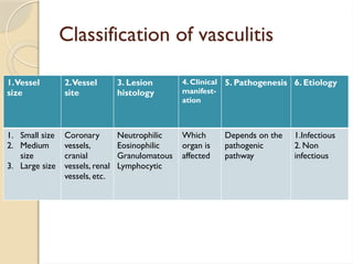
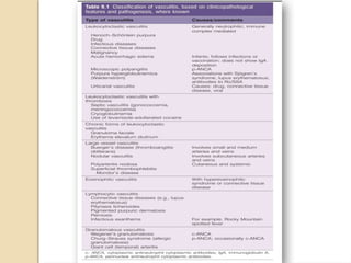
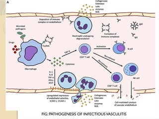
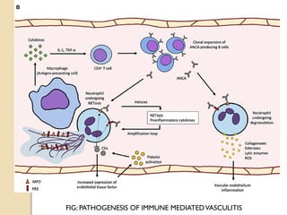
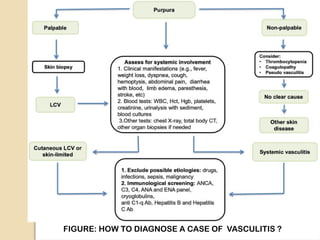
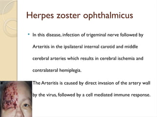
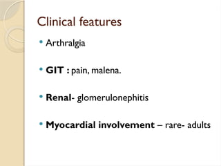
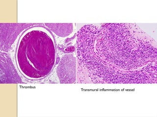
VASCULITIS
Moderator: Dr. Smita Chandra
Presenter : Dr. Saloni Garg
Basic structure of a vessel
Vessel walls are organized into three layers:
 Tunica Intima – made up of three components:
1. Endothelium: Simple squamous epithelium.
2. Basal lamina: Basement membrane of lining epithelium.
3. Sub-endothelial layer: Loose connective tissue beneath the
endothelium
Tunica Intima is limited by a fenestrated layer of elastic tissue called
internal elastic lamina.
 Tunica Media – This layer consists of concentrically arranged smooth muscle
cells, elastic fibers, reticular fibers. It is separated from tunica adventitia by
fenestrated layer of elastic tissue called external elastic lamina.
 Tunica Adventitia – It is the outer most layer of a vessel wall. It consists
of longitudinally arranged collagen and reticular fibers, which gradually
merges with the connective tissue of the surroundings.
This layer contains network of blood vessels providing nutrition to them
called VasaVasorum and network of autonomic nerves controlling their
lumen diameter called Nervi vasorum.
VASCULITIS
 Vasculitis is a general term for vessel
inflammation with protean manifestation.
 Inflammation of vessels is a primary feature
of many immune complex mediated diseases.
 It can be confined to single organ or can
affect multiple organ system.
 The endothelial lining of the blood vessels usually repels white
blood cells and has been called TEFLON like.
 This protective barrier becomes less effective when vasculitis
develops, allowing the deposition of immune complexes on
vascular endothelial surface, where they aggregate to fix
complement, which in turn attracts and activates granulocytes.
 When the neutrophils pass through the wall of the vessel and have
disintegrated. This is referred to as Leukocytoclastic vasculitis.
 Vasculitis share common pathogenic pathways that lead to
the activation of leukocytes in vessel wall, with resultant
injury.
 Inflammatory mediators act on both circulating leukocytes
and endothelial cells.
 It is characterized by inflammatory injury to vessel walls
that results from the activation of leukocytes and other
inflammatory mediators with in and adjacent to vessel walls.
 This is caused by the presence of pathogenic
factors that can drive leukocytes to complete
activation as they are adhering to and
penetrating vessel walls.
 Pathogenic factors include infectious pathogens,
immune complexes, and auto antibodies.
Classification of vasculitis
1.Vessel
size
2.Vessel
site
3. Lesion
histology
4. Clinical
manifest-
ation
5. Pathogenesis 6. Etiology
1. Small size
2. Medium
size
3. Large size
Coronary
vessels,
cranial
vessels, renal
vessels, etc.
Neutrophilic
Eosinophilic
Granulomatous
Lymphocytic
Which
organ is
affected
Depends on the
pathogenic
pathway
1.Infectious
2. Non
infectious
Pathogenesis
 The two mechanisms of vasculitis are:
1. Immune-mediated inflammation
2. Direct invasion of vessel wall by
infectious pathogens.
Physical and chemical injury can also cause
vasculitis (e.g. Irradiation, mechanical
trauma and toxins)
Note: Infections can also induce non-
infectious vasculitis.
FIG: PATHOGENESIS OF INFECTIOUSVASCULITIS
FIG: PATHOGENESIS OF IMMUNE MEDIATEDVASCULITIS
FIGURE: HOW TO DIAGNOSE A CASE OF VASCULITIS ?
 ANCA test is a blood test which if comes
out to be positive is confirmatory for
autoimmune vasculitis.
 Further tests can be done to differentiate
between c-ANCA and p- ANCA.
 A biopsy is then taken for confirmation.A
skin biopsy can be taken for cutaneous
vasculitis and a biopsy is taken from the
affected organ like temporal artery, skin,
kidney, lung etcetera.
Infectious vasculitis
 Infections cause vasculitis by the direct invasion of pathogens into the
vessel wall with resultant inflammatory injury as well as by inducing the
formation of pathogenic immune complex that localize in vessel wall.
 Infectious organism reach vessel walls by:
1. Direct extension
2. Septic embolization
3. Hematogenous spread
1. Direct extension
 Direct extension from adjacent
extravascular sites of infection such as the
extension of pulmonary fungal infection
into the walls of pulmonary vessels.
2. Septic embolization
 Septic embolization of thrombotic
material containing pathogens such as
emboli released from valvular vegetations
in a patient with bacterial endocarditis.
3. Hematogenous dissemination
 Hematogenous dissemination of pathogens such as occurring
in Neisserial septicemia and spotted fever rickettsial disease.
Vascular inflammation results not only from activation of
inflammatory mediators by microorganism or their
degradation products such as bacterial formyl tripeptides and
glycolipids but also from host immune response.
 Vascular inflammation caused by pyogenic bacteria
is characterized by the presence of neutrophils,
resulting in focal vascular necrosis, hemorrhage
and sometime thrombosis.
 Products of bacteria attract and activate leucocytes
especially neutrophils and also activate humoral
inflammatory mediators system such as the
complementary system.
 Rickettsial, Mycobacterial and Fungal invasion of
vessels induces inflammation that has a predominance
of mononuclear leukocytes rather than neutrophils.
Syphilitic vasculitis
 Inflammation in the tunica media with the infiltration
of:
=> lymphocytes, macrophages and plasma cells
results in destruction of elastic tissue and smooth
muscle cells which are replaced initially by highly
vascularized fibrous tissue and later by dense scar
tissue.
 The scarring often extends into the tunica
intima.
 Adventitial inflammation progresses to
fibrosis and theVasa vasorum become
severely stenoptic secondary to fibrotic
process.
Bacterial vasculitis
 Bacterial infection causes vasculitis by means of several
processes:
a. The invasion of vessels adjacent to and within localized sites
of infection.
b. The hematogenous spread of infected emboli
c. The systemic infection of small vessels secondary to
bacteremia.
 Neisseria meningitis and gonorrhea organism causes small vessel
vasculitis. It can be seen in the endothelial cells and neutrophils
and in Gram-stained smears of blood from the acute lesions.
 Direct invasion of vessels occurs at sites of necrotizing
inflammation, especially in pseudomonas or klebsiella pneumoniae
infection.
 Embolization of infected thrombotic material can result in the
formation of ‘mycotic aneurysm’ in any type of vessel.
Fungal vasculitis
 Caused by a variety of fungi, included
=>Aspergilosis, Mucor, Candida, Cryptococcus,
Nocardia, and Actinomyces.
It occurs in immuno-compromised hosts.
 A major pathologic feature of the disease is
Mycobacterial vasculititis
 It can also cause acute and chronic
vasculitis of central nervous system
vessels by direct invasion associated with
Tuberculous meningitis.
Viral vasculitis
 Hepatitis B and C infection cause vasculitis by
participating in immune complex formation.
 HIV virus infection is associated with development of
Polyarteritis nodosa, microscopic polyangitis and
central nervous system angitis.
 Best known example is with herpes zoster
Herpes zoster ophthalmicus
 In this disease, infection of trigeminal nerve followed by
Arteritis in the ipsilateral internal carotid and middle
cerebral arteries which results in cerebral ischemia and
contralateral hemiplegia.
 The Arteritis is caused by direct invasion of the artery wall
by the virus, followed by a cell mediated immune response.
Non infectious vasculitis
The main immunologic mechanism underlying are:
1. Immune complex formation
2. ANCA mediated
3. Anti endothelial cell mediated antibodies
4. Auto reactive T cells
Immune complex formation
 It is antigen-antibody immune complex deposition.
 Antigen can be :
Þ Viral antigen (hep-C)
Þ Auto-antigens such as immunoglobulins, proteinase 3.
Þ Drugs ( horse serum, sulphonamides antibiotics, mouse
monoclonal antibodies, cloned streptokinase)
Þ Anti DNA complex (SLE)
 The localisation of these immune complexes in the endothelial
cells causes activation of
Þ Inflammatory mediators: Neutrophils, monocytes, mast cell,
platelets, endothelial cells
Þ Humoral inflammatory mediators: Complement system (C3b,
C5a), kinin, plasmin, coagulation system.
The activation of neutrophils and monocytes results in release of
lytic enzymes and toxic oxygen metabolites that cause injury
to vessel wall.
 In many cases even the antigen-antibody deposits are
insufficient. In such instances the underlying mechanism need
to be considered such as “pauci-immune” vasculitis.
1. Drug hypersenstivity vasculitis: drug act as haptens by
binding to serum proteins(eg. Penicillin).Antibodies directed
against the drug modified proteins results in immune-
complex formation.
2. Vasculitis secondary to infections: Antibodies to
microbial constituents can form immune-complexes and
deposits in vessel wall.
ANCA mediated vasculitis
 This causes auto immune type of vasculitis.
 We have auto antibodies against normal enzymes or granules that are present in the
neutrophils.
1. c-ANCA/ anti proteinase 3 (cytoplasmic/ PR 3-ANCA): is a neutrophilic azurophilic
granule and is involved in normal inflammatory process.
2. p-ANCA/ anti myeloperoxidase ( peinuclear/ anti myelo-peroxidase): is a lysosomal
granule constituent involved in oxygen free radical generation. MPO is present in
neutrophils and monocytes.
 These auto antibodies are formed against neutrophilic granules, react with
neutrophils and undergo degranulation and release protein, enzymes and
reactive oxygen species.This will cause endothelial damage.
 Drugs or cross reactive microbial antigens induce ANCA formation.
 Infection, endotoxin exposure or inflammatory stimulus elicits cytokines
such as tumor necrosis factor (TNF) that up-regulate the surface
expression of PR 3 and MPO on neutrophils.
 ANCA’s react with these cytokines activated cells causing direct injury to
endothelial cell or further activation of neutrophils.
 ANCA’s activated neutrophils cause tissue injury by releasing granule
contents and reactive oxygen species.
ANCA’s auto-antibodies are directed against
cellular constituents and do not form
circulating immune complexes.
Vascular lesion do not contain antibody and
complement.
Thus ANCA associated vasculitides are often
described as “pauci-immune”.
Anti endothelial cell mediated antibodies
 This vasculitis will occur when we have auto antibodies which
are going to be against the cell antigen of the endothelial cell.
 Our cells have certain protein.These antigen that are
normally presented by endothelial cells.
 Auto antibodies fails to recognize them as self and they will
be developed against them and will directly or indirectly cause
cytotoxicity.
 The presence ofT-lymphocytes in the
inflammatory infiltrates of some vasculitides and
the presence of:
- Granulomatous inflammation with
multinucleated giant cell has been the major
basis for incriminating cell mediated immunity.
 The only form of vasculitis that is clearly
directly mediated by T-lymphocyte is Acute
vascular rejection of allografts.
 In this process activated T-lymphocytes attach
to endothelial cells and penetrate Intima of
arteries and in severe rejection the muscularis.
Small vessel vasculitis
A. Wegener’s granulomatosis
B. Microscopic polyangitis
C. Churg Strauss Syndrome
D. Henoch-Schonlein purpura
WEGENER GRANULOMATOSIS
Also called as “Granulomatosis with Polyangitis”
(GAP) characterized by triad of :
1. Necrotizing Granulomas – of the upper
respiratory tract or lower respiratory tract (ear,
nose, sinuses, throat, lungs)
2. Necrotizing Vasculitis – affecting small to medium-
sized vessels mostly in the lungs and upper
respiratory tract
3. Necrotizing Glomerulonephritis
Pathogenesis
 GPA represents a form of T-cell mediated
hypersensitivity response to inhaled
microbial or other environmental antigens.
 PR 3- ANCA’s are also present and they are
a useful marker of disease activity.
Classic presentations :
 Middle aged men
 Average age 40 years
 Mainly affect upper and lower respiratory tract,
kidney, lungs.
1. Bilateral pneumonitis with nodules and cavitary
lesions (95%),
2. Chronic sinusitis (90%),
3. Mucosal ulcerations of the nasopharynx (75%)
4. Renal disease (80%)
Rashes, myalgias, neuritis and fever can also occur.
If left untreated the disease is usually fatal with 80%
mortality with in 1 year.
Morphology
 Upper respiratory tract lesions- granulomatous
sinusitis to ulcerative lesions of the nose, palate, or
pharynx;
 Lung findings - diffuse parenchymal infiltrates to
granulomatous nodules.
 Renal findings: Multifocal necrotizing granulomatous
Microscopic polyangiitis
Microscopic polyangitis
 It is also called hypersensitivity vasculitis or
Leukocytoclastic vasculitis.
 It is necrotizing vasculitis that affects
capillaries as well as arterioles and venules.
 The granulomatous inflammation is absent.
Pathogenesis
 Antibodies responses to antigens such as:
Drugs (e.g., penicillin),
Microorganisms (e.g., streptococci),
Heterologous proteins
Tumor proteins
 These reaction can either lead to immune
complex deposition or trigger secondary
immune response (eg. Development of
ANCA’s) (MPO-ANCA)
Clinical features
Most common:
1. Necrotizing glomerulonephritis (90)%
2. Pulmonary capillaritis
 The skin, mucous membranes, lungs,
brain, heart, gastro- intestinal tract,
kidneys, and muscle
Depending upon the vascular bed involved,
major clinical features are:
 Hemoptysis
 Hematuria
 Proteinuria
 Abdominal pain or bleeding
 Muscle pain or weakness
 Palpable cutaneous purpura
Morphology
 Segmental fibrinoid necrosis of the
media with focal transmural
necrotizing lesions
 In some areas (typically postcapillary
venules), only infiltrating and fragmenting
neutrophils are seen giving rise to the term
Leukocytoclastic vasculitis
skin biopsy samples revealing leukocytoclastic
vasculitis with extensive fibrinoid necrosis
within dermal capillaries
Fibrinoid necrosis destroying the vessel wall
with extravasation of red blood cells and nuclear
debris in the adjacent areas
).
Churg-Strauss syndrome
 Also called Allergic granulomatosis and angitis
 Small vessel necrotizing vasculitis associated with asthma,
allergic rhinitis, lung infiltrates, peripheral eosinophilia, extra
vascular necrotizing granulomas and a striking eosinophilic
infiltration of vessels and tissue.
 It is a consequence of hyper responsiveness to some allergic
stimulus.
Clinical Features
3 phases:
 Prodromal Phase: Allergic Respiratory
disease
 Eosinophilic Phase: Peripheral blood and
tissue hypereosinophilia.
 Vasculitic Phase: Cardiac/ Coronary
involvement
 Palpable purpura
 Gastrointestinal bleeding
 Renal disease- Glomerulosclerosis
 Cardiomyopathy- cytotoxicity produced by the
myocardial eosinophilic infiltrates leads to
cardiomyopathy, cardiac involvement is seen in 60% of
patients and is the major cause of morbidity and death.
MORPHOLOGY
 Extravascular necrotizing granulomas
 Infiltration of vessels and perivascular
tissues by Eosinophils.
showing intimal fibrinoid
necrosis and mural
infiltration by histiocytes
consistent with a necrotizing
granulomatous vasculitis.
There is marked
extravascular eosinophilia.
HENOCH SCHONLEIN
PURPURA
HENOCH SCHONLEIN PURPURA
 It is a IgA immune complex mediated
vasculitis.
 IgA is present on mucosal surface.
 Children are affected.
 Follows URTI
IgA immune complex deposits in mucosal
surface and results in HSP.
 Small vessel vasculitis
Clinical features
 Arthralgia
 GIT : pain, malena.
 Renal- glomerulonephitis
 Myocardial involvement – rare- adults
The left side of the picture
demonstrates glomerulus
with segmental necrosis. The right side of the picture shows two
glomeruli; one of them apparently shows focus
of early endocapillary proliferation (arrow);
right bottom edge shows glomerulus of
adequate appearance.
Medium vessel vasculitis
a. Poly arteritis nodosa
b. Kawasaki disease
c. Buerger’s disease
Polyarteritis Nodosa
 It is systemic vasculitis of small and medium sized muscular
arteries that typically affects renal and visceral vessels but spares
pulmonary circulation. So, it let’s you breathe.
 There is no association with ANCA’s.
 It is associated with chronic Hepatitis-B which lead to formation
of HbsAG-HbsAB complexes that deposits in affected vessels.
Clinical Features
 Males> females
 Rashes, ulcerations, infarcts ,ischemic
atrophy and hemorrhage
 Firm, tender subcutaneous nodules may be
the first sign.
 Palpable purpura
 Livedo reticularis
Livedo reticularis is a common skin finding
consisting of a mottled reticulated vascular
pattern that appears as a lace-like purplish
discoloration of the skin. The discoloration is
caused by swelling of the venules owing to
obstruction of capillaries by thrombi.
Clinical features
 Typical presentation involves:
1. Accelerating hypertension
2. Abdominal pain, bloody stools
3. Myalgias
4. Peripheral neuritis (motor nerves)
5. Renal, Cardiac, Liver, GIT vessels are involved in
descending order of frequency.
Morphology
 Segmental ,transmural ,necrotizing inflammation with
superimposed aneurysm and thrombosis
 2 stages
Acute Stage: Transmural inflammation of vessel with
polynuclear and mononuclear cells along with Fibrinoid
Necrosis.
Late Stage : Fibrous thickening of vessel wall
(nodular)extending into adventitia
Thrombus
Transmural inflammation of vessel
KAWASAKI DISEASE
 An acute, febrile self-limiting illness
 Large to medium-sized vessels.
 Infancy and childhood
 Leading cause of Acquired heart disease in children
 Coronary arteries – results in Aneurysms that rupture or
thrombose, resulting in myocardial infarction.
Etiology
 Infectious agents (mostly viral)
 Delayed-type hypersensitivity response
directed against cross-reactive or newly uncovered
vascular antigens.
 Cytokine production and polyclonal B cell activation
result in autoantibodies to endothelial cells and
Clinical Features
 Oral cavity: Oral erythema , cracked lips, blisters ,
STRAWBERRYTONGUE
 Eyes: Conjunctival hemorrhage
 Hands and feet: Erythema of the palms and soles.
 Cervical lymph node: Enlargement
 It is also known as Mucocutaneous lymph node
syndrome
The epicardial vein contains
blood and shows mild
thickening
of the wall
The coronary artery shows
almost complete occlusion by
luminal myofibroblastic
proliferation with a fine slit-
like lumen.
Thromboangitis Obliterans
Buerger disease
 it is characterized by segmental, thrombosing, acute and
chronic inflammation of medium and small sized vessels.
 It occurs in heavy cigarette smokers usually before age of 35
years.
 A disorder that results in severe vascular insufficiency and
gangrene of the extremities.
 Focal acute and chronic inflammation of medium and small
arteries- Tibial and Radial.
Pathogenesis
 Associated with thrombosis
 Secondary extension into adjacent veins
and nerves.
TOBACCO
Direct endothelial cell toxicity
Modification of host vascular wall
proteins
CLINICAL FEATURES
Cold-induced Raynaud phenomenon
 Nodular phlebitis
 Extremity ulceration
 Gangrene
MORPHOLOGY
 Sharply segmental acute and chronic
transmural vasculitis of medium- sized and
small arteries, predominantly those of the
extremities.
 In early stages, mixed inflammatory infiltrates with
luminal thrombosis;
 Small micro abscesses rimmed by granulomatous
inflammation
Large vessel vasculitis
a. Temporal arteritis
b. Takayasu arteritis
Giant cell (temporal) arteritis
 A chronic granulomatous inflammatory disorder
 Old individuals>50yrs
 It affects arteries of the head. It is the most
common form of vasculitis.
 Arteries: large to small size arteries, mainly
“Temporal arteries”
 Other arteries: Vertebral and ophthalmic
arteries
Pathogenesis
 T-cell mediated immune response
 Cytokines :TNF
 Anti endothelial cell and anti smooth
muscle cell antibodies
Clinical Features
 Age : after 50.
 Raised ESR
 Fever, fatigue, weight loss
 Temporal artery :Facial pain or headache
 Ophthalmic: Diplopia to complete vision loss.
Morphology
 Intimal thickening and luminal narrowing.
 Granulomatous inflammation -
Fragmentation of the internal elastic lamina
 Focally distributed inflammatory lesions.
 Infiltrate of T cells (CD4+ > CD8+) and
macrophages.
 Multinucleated giant cells are seen.
Takayasu Arteritis
 Granulomatous vasculitis
 Medium and larger arteries
 Characterized by ocular disturbances, marked
weakening of pulse in the upper extrimeties.
Hence the name “pulseless disease”
Clinical Features
 Autoimmune etiology.
 Vascular features- Reduced upper
extremity blood pressure and pulse
strength.
 Neurologic deficits
 Ocular disturbances -Visual field defects,
retinal hemorrhages, total blindness.
 Classically affects the Aortic arch
and arch vessels causing dilation and
aortic valve insufficiency.
 Pulmonary arteries, renal and
coronary arteries
 High ESR
 Distal aorta disease : leg claudication
 Pulmonary artery :pulmonary
hypertension.
 Coronary artery: myocardial infarction
 Renal arteries : systemic hypertension
Morphology
 Transmural mononuclear cells
infiltration
 Perivascular infiltration of vasa vasorum.
 Intima : Marked thickening and luminal
narrowing.
 Media : Granulomatous change with
central necrosis , Langhan’s giant cells and
mononuclear inflammatory cells.
 Adventitia : mononuclear cell infiltration
A. Intimal fibrosis
B. Fibrosis of media and infiltration of inflammatory cells around vasa vasorum.
C. and D. Inflammatory cell infiltrate consists of predominantly mononuclear lymphocytes.
NEUTROPHILIC DERMATOSES
 Includes many entities characterized
histopathologicaly by a heavy dermal
infiltrate of neutrophils and variable
lymphocytoclasis.
 It includes the following diseases:
- Periodic fever syndromes (autoinflammatory diseases)
- Amicrobial pustulosis of the folds
- Sweet’s syndrome
- Bowel associated dermatosis- arthritis syndrome
- Rheumatoid neutrophilic dermatosis
- Acute generalized pustulosis
- Behcet’s syndrome
- Abscess- forming neutrophilic dermatosis
EosinophilicVasculitis
 It is an eosinophil predominant
necrotizing vasculitis affecting small
dermal vessels.
 It presents as either pruritic,
erythematous or purpuric papules and
plaques.
 Causes:
- Connective tissue diseases associated.
- Hypereosinophilic syndrome.
- Exposure to drugs like propylthiouracil,
hydralazine, minocycline, penicillamine.
- Idiopathic.
Reference
 Robbins and cotran; pathologic basic of
disease (10th
edition)
 Anderson’s pathology (10th
edition)
 Weedons’s skin pathology (5th
edition)
THANKYOU

VASCULITIS.pptx different types of vasculitis

  • 1.
    VASCULITIS Moderator: Dr. SmitaChandra Presenter : Dr. Saloni Garg
  • 2.
    Basic structure ofa vessel Vessel walls are organized into three layers:  Tunica Intima – made up of three components: 1. Endothelium: Simple squamous epithelium. 2. Basal lamina: Basement membrane of lining epithelium. 3. Sub-endothelial layer: Loose connective tissue beneath the endothelium Tunica Intima is limited by a fenestrated layer of elastic tissue called internal elastic lamina.
  • 3.
     Tunica Media– This layer consists of concentrically arranged smooth muscle cells, elastic fibers, reticular fibers. It is separated from tunica adventitia by fenestrated layer of elastic tissue called external elastic lamina.  Tunica Adventitia – It is the outer most layer of a vessel wall. It consists of longitudinally arranged collagen and reticular fibers, which gradually merges with the connective tissue of the surroundings. This layer contains network of blood vessels providing nutrition to them called VasaVasorum and network of autonomic nerves controlling their lumen diameter called Nervi vasorum.
  • 5.
    VASCULITIS  Vasculitis isa general term for vessel inflammation with protean manifestation.  Inflammation of vessels is a primary feature of many immune complex mediated diseases.  It can be confined to single organ or can affect multiple organ system.
  • 6.
     The endotheliallining of the blood vessels usually repels white blood cells and has been called TEFLON like.  This protective barrier becomes less effective when vasculitis develops, allowing the deposition of immune complexes on vascular endothelial surface, where they aggregate to fix complement, which in turn attracts and activates granulocytes.  When the neutrophils pass through the wall of the vessel and have disintegrated. This is referred to as Leukocytoclastic vasculitis.
  • 7.
     Vasculitis sharecommon pathogenic pathways that lead to the activation of leukocytes in vessel wall, with resultant injury.  Inflammatory mediators act on both circulating leukocytes and endothelial cells.  It is characterized by inflammatory injury to vessel walls that results from the activation of leukocytes and other inflammatory mediators with in and adjacent to vessel walls.
  • 8.
     This iscaused by the presence of pathogenic factors that can drive leukocytes to complete activation as they are adhering to and penetrating vessel walls.  Pathogenic factors include infectious pathogens, immune complexes, and auto antibodies.
  • 9.
    Classification of vasculitis 1.Vessel size 2.Vessel site 3.Lesion histology 4. Clinical manifest- ation 5. Pathogenesis 6. Etiology 1. Small size 2. Medium size 3. Large size Coronary vessels, cranial vessels, renal vessels, etc. Neutrophilic Eosinophilic Granulomatous Lymphocytic Which organ is affected Depends on the pathogenic pathway 1.Infectious 2. Non infectious
  • 11.
    Pathogenesis  The twomechanisms of vasculitis are: 1. Immune-mediated inflammation 2. Direct invasion of vessel wall by infectious pathogens. Physical and chemical injury can also cause vasculitis (e.g. Irradiation, mechanical trauma and toxins) Note: Infections can also induce non- infectious vasculitis.
  • 12.
    FIG: PATHOGENESIS OFINFECTIOUSVASCULITIS
  • 13.
    FIG: PATHOGENESIS OFIMMUNE MEDIATEDVASCULITIS
  • 14.
    FIGURE: HOW TODIAGNOSE A CASE OF VASCULITIS ?
  • 15.
     ANCA testis a blood test which if comes out to be positive is confirmatory for autoimmune vasculitis.  Further tests can be done to differentiate between c-ANCA and p- ANCA.  A biopsy is then taken for confirmation.A skin biopsy can be taken for cutaneous vasculitis and a biopsy is taken from the affected organ like temporal artery, skin, kidney, lung etcetera.
  • 16.
    Infectious vasculitis  Infectionscause vasculitis by the direct invasion of pathogens into the vessel wall with resultant inflammatory injury as well as by inducing the formation of pathogenic immune complex that localize in vessel wall.  Infectious organism reach vessel walls by: 1. Direct extension 2. Septic embolization 3. Hematogenous spread
  • 17.
    1. Direct extension Direct extension from adjacent extravascular sites of infection such as the extension of pulmonary fungal infection into the walls of pulmonary vessels.
  • 18.
    2. Septic embolization Septic embolization of thrombotic material containing pathogens such as emboli released from valvular vegetations in a patient with bacterial endocarditis.
  • 19.
    3. Hematogenous dissemination Hematogenous dissemination of pathogens such as occurring in Neisserial septicemia and spotted fever rickettsial disease. Vascular inflammation results not only from activation of inflammatory mediators by microorganism or their degradation products such as bacterial formyl tripeptides and glycolipids but also from host immune response.
  • 20.
     Vascular inflammationcaused by pyogenic bacteria is characterized by the presence of neutrophils, resulting in focal vascular necrosis, hemorrhage and sometime thrombosis.
  • 21.
     Products ofbacteria attract and activate leucocytes especially neutrophils and also activate humoral inflammatory mediators system such as the complementary system.  Rickettsial, Mycobacterial and Fungal invasion of vessels induces inflammation that has a predominance of mononuclear leukocytes rather than neutrophils.
  • 22.
    Syphilitic vasculitis  Inflammationin the tunica media with the infiltration of: => lymphocytes, macrophages and plasma cells results in destruction of elastic tissue and smooth muscle cells which are replaced initially by highly vascularized fibrous tissue and later by dense scar tissue.
  • 23.
     The scarringoften extends into the tunica intima.  Adventitial inflammation progresses to fibrosis and theVasa vasorum become severely stenoptic secondary to fibrotic process.
  • 24.
    Bacterial vasculitis  Bacterialinfection causes vasculitis by means of several processes: a. The invasion of vessels adjacent to and within localized sites of infection. b. The hematogenous spread of infected emboli c. The systemic infection of small vessels secondary to bacteremia.
  • 25.
     Neisseria meningitisand gonorrhea organism causes small vessel vasculitis. It can be seen in the endothelial cells and neutrophils and in Gram-stained smears of blood from the acute lesions.  Direct invasion of vessels occurs at sites of necrotizing inflammation, especially in pseudomonas or klebsiella pneumoniae infection.  Embolization of infected thrombotic material can result in the formation of ‘mycotic aneurysm’ in any type of vessel.
  • 26.
    Fungal vasculitis  Causedby a variety of fungi, included =>Aspergilosis, Mucor, Candida, Cryptococcus, Nocardia, and Actinomyces. It occurs in immuno-compromised hosts.  A major pathologic feature of the disease is
  • 27.
    Mycobacterial vasculititis  Itcan also cause acute and chronic vasculitis of central nervous system vessels by direct invasion associated with Tuberculous meningitis.
  • 28.
    Viral vasculitis  HepatitisB and C infection cause vasculitis by participating in immune complex formation.  HIV virus infection is associated with development of Polyarteritis nodosa, microscopic polyangitis and central nervous system angitis.  Best known example is with herpes zoster
  • 29.
    Herpes zoster ophthalmicus In this disease, infection of trigeminal nerve followed by Arteritis in the ipsilateral internal carotid and middle cerebral arteries which results in cerebral ischemia and contralateral hemiplegia.  The Arteritis is caused by direct invasion of the artery wall by the virus, followed by a cell mediated immune response.
  • 30.
    Non infectious vasculitis Themain immunologic mechanism underlying are: 1. Immune complex formation 2. ANCA mediated 3. Anti endothelial cell mediated antibodies 4. Auto reactive T cells
  • 31.
    Immune complex formation It is antigen-antibody immune complex deposition.  Antigen can be : Þ Viral antigen (hep-C) Þ Auto-antigens such as immunoglobulins, proteinase 3. Þ Drugs ( horse serum, sulphonamides antibiotics, mouse monoclonal antibodies, cloned streptokinase) Þ Anti DNA complex (SLE)
  • 32.
     The localisationof these immune complexes in the endothelial cells causes activation of Þ Inflammatory mediators: Neutrophils, monocytes, mast cell, platelets, endothelial cells Þ Humoral inflammatory mediators: Complement system (C3b, C5a), kinin, plasmin, coagulation system. The activation of neutrophils and monocytes results in release of lytic enzymes and toxic oxygen metabolites that cause injury to vessel wall.
  • 33.
     In manycases even the antigen-antibody deposits are insufficient. In such instances the underlying mechanism need to be considered such as “pauci-immune” vasculitis. 1. Drug hypersenstivity vasculitis: drug act as haptens by binding to serum proteins(eg. Penicillin).Antibodies directed against the drug modified proteins results in immune- complex formation. 2. Vasculitis secondary to infections: Antibodies to microbial constituents can form immune-complexes and deposits in vessel wall.
  • 34.
    ANCA mediated vasculitis This causes auto immune type of vasculitis.  We have auto antibodies against normal enzymes or granules that are present in the neutrophils. 1. c-ANCA/ anti proteinase 3 (cytoplasmic/ PR 3-ANCA): is a neutrophilic azurophilic granule and is involved in normal inflammatory process. 2. p-ANCA/ anti myeloperoxidase ( peinuclear/ anti myelo-peroxidase): is a lysosomal granule constituent involved in oxygen free radical generation. MPO is present in neutrophils and monocytes.
  • 35.
     These autoantibodies are formed against neutrophilic granules, react with neutrophils and undergo degranulation and release protein, enzymes and reactive oxygen species.This will cause endothelial damage.  Drugs or cross reactive microbial antigens induce ANCA formation.  Infection, endotoxin exposure or inflammatory stimulus elicits cytokines such as tumor necrosis factor (TNF) that up-regulate the surface expression of PR 3 and MPO on neutrophils.  ANCA’s react with these cytokines activated cells causing direct injury to endothelial cell or further activation of neutrophils.  ANCA’s activated neutrophils cause tissue injury by releasing granule contents and reactive oxygen species.
  • 36.
    ANCA’s auto-antibodies aredirected against cellular constituents and do not form circulating immune complexes. Vascular lesion do not contain antibody and complement. Thus ANCA associated vasculitides are often described as “pauci-immune”.
  • 38.
    Anti endothelial cellmediated antibodies  This vasculitis will occur when we have auto antibodies which are going to be against the cell antigen of the endothelial cell.  Our cells have certain protein.These antigen that are normally presented by endothelial cells.  Auto antibodies fails to recognize them as self and they will be developed against them and will directly or indirectly cause cytotoxicity.
  • 39.
     The presenceofT-lymphocytes in the inflammatory infiltrates of some vasculitides and the presence of: - Granulomatous inflammation with multinucleated giant cell has been the major basis for incriminating cell mediated immunity.
  • 40.
     The onlyform of vasculitis that is clearly directly mediated by T-lymphocyte is Acute vascular rejection of allografts.  In this process activated T-lymphocytes attach to endothelial cells and penetrate Intima of arteries and in severe rejection the muscularis.
  • 41.
    Small vessel vasculitis A.Wegener’s granulomatosis B. Microscopic polyangitis C. Churg Strauss Syndrome D. Henoch-Schonlein purpura
  • 42.
    WEGENER GRANULOMATOSIS Also calledas “Granulomatosis with Polyangitis” (GAP) characterized by triad of : 1. Necrotizing Granulomas – of the upper respiratory tract or lower respiratory tract (ear, nose, sinuses, throat, lungs) 2. Necrotizing Vasculitis – affecting small to medium- sized vessels mostly in the lungs and upper respiratory tract 3. Necrotizing Glomerulonephritis
  • 43.
    Pathogenesis  GPA representsa form of T-cell mediated hypersensitivity response to inhaled microbial or other environmental antigens.  PR 3- ANCA’s are also present and they are a useful marker of disease activity.
  • 44.
    Classic presentations : Middle aged men  Average age 40 years  Mainly affect upper and lower respiratory tract, kidney, lungs. 1. Bilateral pneumonitis with nodules and cavitary lesions (95%), 2. Chronic sinusitis (90%), 3. Mucosal ulcerations of the nasopharynx (75%) 4. Renal disease (80%) Rashes, myalgias, neuritis and fever can also occur. If left untreated the disease is usually fatal with 80% mortality with in 1 year.
  • 45.
    Morphology  Upper respiratorytract lesions- granulomatous sinusitis to ulcerative lesions of the nose, palate, or pharynx;  Lung findings - diffuse parenchymal infiltrates to granulomatous nodules.  Renal findings: Multifocal necrotizing granulomatous
  • 47.
  • 48.
    Microscopic polyangitis  Itis also called hypersensitivity vasculitis or Leukocytoclastic vasculitis.  It is necrotizing vasculitis that affects capillaries as well as arterioles and venules.  The granulomatous inflammation is absent.
  • 49.
    Pathogenesis  Antibodies responsesto antigens such as: Drugs (e.g., penicillin), Microorganisms (e.g., streptococci), Heterologous proteins Tumor proteins  These reaction can either lead to immune complex deposition or trigger secondary immune response (eg. Development of ANCA’s) (MPO-ANCA)
  • 50.
    Clinical features Most common: 1.Necrotizing glomerulonephritis (90)% 2. Pulmonary capillaritis  The skin, mucous membranes, lungs, brain, heart, gastro- intestinal tract, kidneys, and muscle
  • 51.
    Depending upon thevascular bed involved, major clinical features are:  Hemoptysis  Hematuria  Proteinuria  Abdominal pain or bleeding  Muscle pain or weakness  Palpable cutaneous purpura
  • 52.
    Morphology  Segmental fibrinoidnecrosis of the media with focal transmural necrotizing lesions  In some areas (typically postcapillary venules), only infiltrating and fragmenting neutrophils are seen giving rise to the term Leukocytoclastic vasculitis
  • 53.
    skin biopsy samplesrevealing leukocytoclastic vasculitis with extensive fibrinoid necrosis within dermal capillaries Fibrinoid necrosis destroying the vessel wall with extravasation of red blood cells and nuclear debris in the adjacent areas
  • 54.
  • 55.
    Churg-Strauss syndrome  Alsocalled Allergic granulomatosis and angitis  Small vessel necrotizing vasculitis associated with asthma, allergic rhinitis, lung infiltrates, peripheral eosinophilia, extra vascular necrotizing granulomas and a striking eosinophilic infiltration of vessels and tissue.  It is a consequence of hyper responsiveness to some allergic stimulus.
  • 56.
    Clinical Features 3 phases: Prodromal Phase: Allergic Respiratory disease  Eosinophilic Phase: Peripheral blood and tissue hypereosinophilia.  Vasculitic Phase: Cardiac/ Coronary involvement
  • 57.
     Palpable purpura Gastrointestinal bleeding  Renal disease- Glomerulosclerosis  Cardiomyopathy- cytotoxicity produced by the myocardial eosinophilic infiltrates leads to cardiomyopathy, cardiac involvement is seen in 60% of patients and is the major cause of morbidity and death.
  • 58.
    MORPHOLOGY  Extravascular necrotizinggranulomas  Infiltration of vessels and perivascular tissues by Eosinophils.
  • 59.
    showing intimal fibrinoid necrosisand mural infiltration by histiocytes consistent with a necrotizing granulomatous vasculitis. There is marked extravascular eosinophilia.
  • 60.
  • 61.
    HENOCH SCHONLEIN PURPURA It is a IgA immune complex mediated vasculitis.  IgA is present on mucosal surface.  Children are affected.  Follows URTI IgA immune complex deposits in mucosal surface and results in HSP.  Small vessel vasculitis
  • 62.
    Clinical features  Arthralgia GIT : pain, malena.  Renal- glomerulonephitis  Myocardial involvement – rare- adults
  • 63.
    The left sideof the picture demonstrates glomerulus with segmental necrosis. The right side of the picture shows two glomeruli; one of them apparently shows focus of early endocapillary proliferation (arrow); right bottom edge shows glomerulus of adequate appearance.
  • 64.
    Medium vessel vasculitis a.Poly arteritis nodosa b. Kawasaki disease c. Buerger’s disease
  • 65.
    Polyarteritis Nodosa  Itis systemic vasculitis of small and medium sized muscular arteries that typically affects renal and visceral vessels but spares pulmonary circulation. So, it let’s you breathe.  There is no association with ANCA’s.  It is associated with chronic Hepatitis-B which lead to formation of HbsAG-HbsAB complexes that deposits in affected vessels.
  • 66.
    Clinical Features  Males>females  Rashes, ulcerations, infarcts ,ischemic atrophy and hemorrhage  Firm, tender subcutaneous nodules may be the first sign.  Palpable purpura  Livedo reticularis
  • 67.
    Livedo reticularis isa common skin finding consisting of a mottled reticulated vascular pattern that appears as a lace-like purplish discoloration of the skin. The discoloration is caused by swelling of the venules owing to obstruction of capillaries by thrombi.
  • 68.
    Clinical features  Typicalpresentation involves: 1. Accelerating hypertension 2. Abdominal pain, bloody stools 3. Myalgias 4. Peripheral neuritis (motor nerves) 5. Renal, Cardiac, Liver, GIT vessels are involved in descending order of frequency.
  • 69.
    Morphology  Segmental ,transmural,necrotizing inflammation with superimposed aneurysm and thrombosis  2 stages Acute Stage: Transmural inflammation of vessel with polynuclear and mononuclear cells along with Fibrinoid Necrosis. Late Stage : Fibrous thickening of vessel wall (nodular)extending into adventitia
  • 70.
  • 71.
    KAWASAKI DISEASE  Anacute, febrile self-limiting illness  Large to medium-sized vessels.  Infancy and childhood  Leading cause of Acquired heart disease in children  Coronary arteries – results in Aneurysms that rupture or thrombose, resulting in myocardial infarction.
  • 72.
    Etiology  Infectious agents(mostly viral)  Delayed-type hypersensitivity response directed against cross-reactive or newly uncovered vascular antigens.  Cytokine production and polyclonal B cell activation result in autoantibodies to endothelial cells and
  • 73.
    Clinical Features  Oralcavity: Oral erythema , cracked lips, blisters , STRAWBERRYTONGUE  Eyes: Conjunctival hemorrhage  Hands and feet: Erythema of the palms and soles.  Cervical lymph node: Enlargement  It is also known as Mucocutaneous lymph node syndrome
  • 76.
    The epicardial veincontains blood and shows mild thickening of the wall The coronary artery shows almost complete occlusion by luminal myofibroblastic proliferation with a fine slit- like lumen.
  • 77.
    Thromboangitis Obliterans Buerger disease it is characterized by segmental, thrombosing, acute and chronic inflammation of medium and small sized vessels.  It occurs in heavy cigarette smokers usually before age of 35 years.  A disorder that results in severe vascular insufficiency and gangrene of the extremities.  Focal acute and chronic inflammation of medium and small arteries- Tibial and Radial.
  • 78.
    Pathogenesis  Associated withthrombosis  Secondary extension into adjacent veins and nerves. TOBACCO Direct endothelial cell toxicity Modification of host vascular wall proteins
  • 79.
    CLINICAL FEATURES Cold-induced Raynaudphenomenon  Nodular phlebitis  Extremity ulceration  Gangrene
  • 80.
    MORPHOLOGY  Sharply segmentalacute and chronic transmural vasculitis of medium- sized and small arteries, predominantly those of the extremities.  In early stages, mixed inflammatory infiltrates with luminal thrombosis;  Small micro abscesses rimmed by granulomatous inflammation
  • 82.
    Large vessel vasculitis a.Temporal arteritis b. Takayasu arteritis
  • 83.
    Giant cell (temporal)arteritis  A chronic granulomatous inflammatory disorder  Old individuals>50yrs  It affects arteries of the head. It is the most common form of vasculitis.  Arteries: large to small size arteries, mainly “Temporal arteries”  Other arteries: Vertebral and ophthalmic arteries
  • 84.
    Pathogenesis  T-cell mediatedimmune response  Cytokines :TNF  Anti endothelial cell and anti smooth muscle cell antibodies
  • 85.
    Clinical Features  Age: after 50.  Raised ESR  Fever, fatigue, weight loss  Temporal artery :Facial pain or headache  Ophthalmic: Diplopia to complete vision loss.
  • 86.
    Morphology  Intimal thickeningand luminal narrowing.  Granulomatous inflammation - Fragmentation of the internal elastic lamina  Focally distributed inflammatory lesions.  Infiltrate of T cells (CD4+ > CD8+) and macrophages.  Multinucleated giant cells are seen.
  • 88.
    Takayasu Arteritis  Granulomatousvasculitis  Medium and larger arteries  Characterized by ocular disturbances, marked weakening of pulse in the upper extrimeties. Hence the name “pulseless disease”
  • 89.
    Clinical Features  Autoimmuneetiology.  Vascular features- Reduced upper extremity blood pressure and pulse strength.  Neurologic deficits  Ocular disturbances -Visual field defects, retinal hemorrhages, total blindness.
  • 90.
     Classically affectsthe Aortic arch and arch vessels causing dilation and aortic valve insufficiency.  Pulmonary arteries, renal and coronary arteries  High ESR
  • 91.
     Distal aortadisease : leg claudication  Pulmonary artery :pulmonary hypertension.  Coronary artery: myocardial infarction  Renal arteries : systemic hypertension
  • 92.
    Morphology  Transmural mononuclearcells infiltration  Perivascular infiltration of vasa vasorum.  Intima : Marked thickening and luminal narrowing.  Media : Granulomatous change with central necrosis , Langhan’s giant cells and mononuclear inflammatory cells.  Adventitia : mononuclear cell infiltration
  • 93.
    A. Intimal fibrosis B.Fibrosis of media and infiltration of inflammatory cells around vasa vasorum. C. and D. Inflammatory cell infiltrate consists of predominantly mononuclear lymphocytes.
  • 95.
    NEUTROPHILIC DERMATOSES  Includesmany entities characterized histopathologicaly by a heavy dermal infiltrate of neutrophils and variable lymphocytoclasis.
  • 96.
     It includesthe following diseases: - Periodic fever syndromes (autoinflammatory diseases) - Amicrobial pustulosis of the folds - Sweet’s syndrome - Bowel associated dermatosis- arthritis syndrome - Rheumatoid neutrophilic dermatosis - Acute generalized pustulosis - Behcet’s syndrome - Abscess- forming neutrophilic dermatosis
  • 98.
    EosinophilicVasculitis  It isan eosinophil predominant necrotizing vasculitis affecting small dermal vessels.  It presents as either pruritic, erythematous or purpuric papules and plaques.
  • 99.
     Causes: - Connectivetissue diseases associated. - Hypereosinophilic syndrome. - Exposure to drugs like propylthiouracil, hydralazine, minocycline, penicillamine. - Idiopathic.
  • 100.
    Reference  Robbins andcotran; pathologic basic of disease (10th edition)  Anderson’s pathology (10th edition)  Weedons’s skin pathology (5th edition)
  • 101.

Editor's Notes

  • #42 Limited” respiratory tract a widespread eyes, skin, heart;
  • #44 HEMATURIA,PROTEINUEIA AND RAPIDLY PROGRESSIVE RF
  • #46 EPITHELOID CELLS GIANT CELLS
  • #47 purpura
  • #49 Pan same age
  • #54 NEUTRO FRAGMENTATION artery shows transmural infiltration by leukocytes including numerous neutrophils. There is extensive leukocytoclasia characterized by nuclear karyorrhectic fragments, which are admixed with the intact leukocytes. Note the fibrinoid necrosis (arrows
  • #55 The vascular lesions differ from those of polyarteritis nodosa or microscopic polyangiitis by virtue of the presence of granu- lomas and eosinophils
  • #57 HEART-- EOSINOPHILS
  • #58 The vas- cular lesions differ from those of polyarteritis nodosa or microscopic polyangiitis by virtue of the presence of granu- lomas and eosinophils.
  • #59 EOSINOPHILLIC INFILTRATEES
  • #61 4-11 YEARS
  • #62 IDSOLATED HEMATURUIA WITH RED BLOOD CELL CASTS-----ARF---CRF
  • #65 Descending order HBS AG- AB 30% PULM AR SPARED
  • #66 . It can be caused by any condition that makes venules swell.
  • #69 Suggesting on going and recurrent insults ANEURYSMS AND WALL RUPTURE
  • #79 undoubtedly from the neural involvement
  • #81 LUMEN OCCLUDED BY THROMBUS
  • #87 ARROW GIAANT CELLS AT INTERNAL ELASTIC LAMINA ELASTIC TISSUE STAIN SHOWS FOCAL DESTRUCTION OF INT ELASTIC LAMINA AND INTIMAL THICKENING
  • #88 FEMALES
  • #89 VASCULAR BRUITS HTN SCLAVIAN AR
  • #90 Transmural thickening of aortic arch: luminal narrowing of major branch vessels.
  • #92 Transmural- mainly adv and media
  • #94 Media gone Media giant clls and mononuclear cells