From Microscope toAlgorithm:
The Impact of AI on
Histopathology Practice
Dr. Muhammad Usman Shams
MBBS, M.Phil, FCPS (Histopathology)
Diploma in Healthcare Management
2.
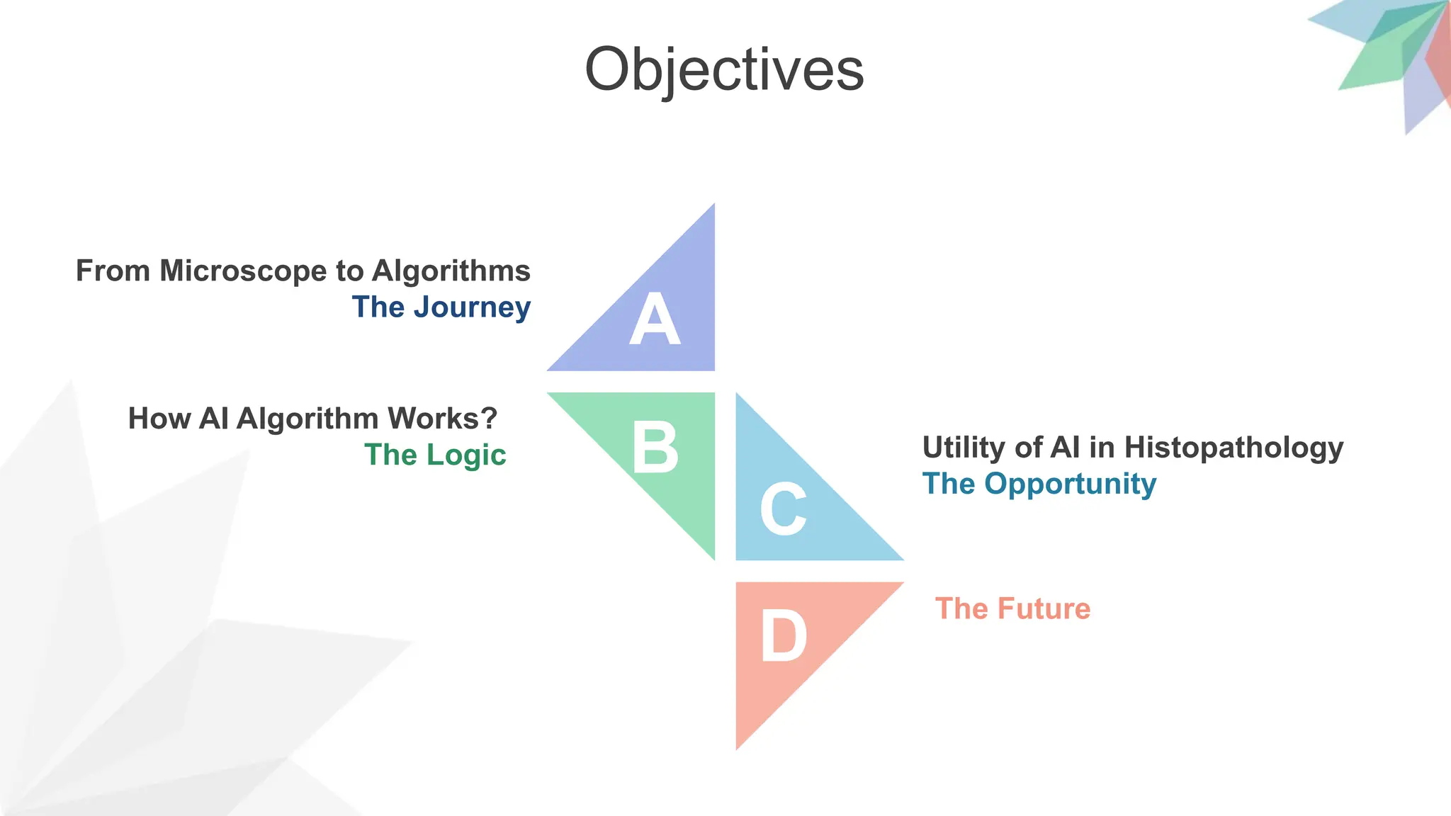
Objectives
From Microscope toAlgorithms
The Journey
The Future
Utility of AI in Histopathology
The Opportunity
How AI Algorithm Works?
The Logic B
D
C
A
3.
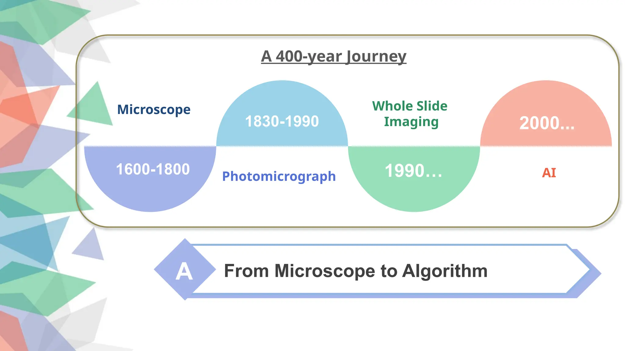
A From Microscopeto Algorithm
1600-1800
1830-1990 2000...
1990…
Microscope
Photomicrograph
Whole Slide
Imaging
AI
A 400-year Journey
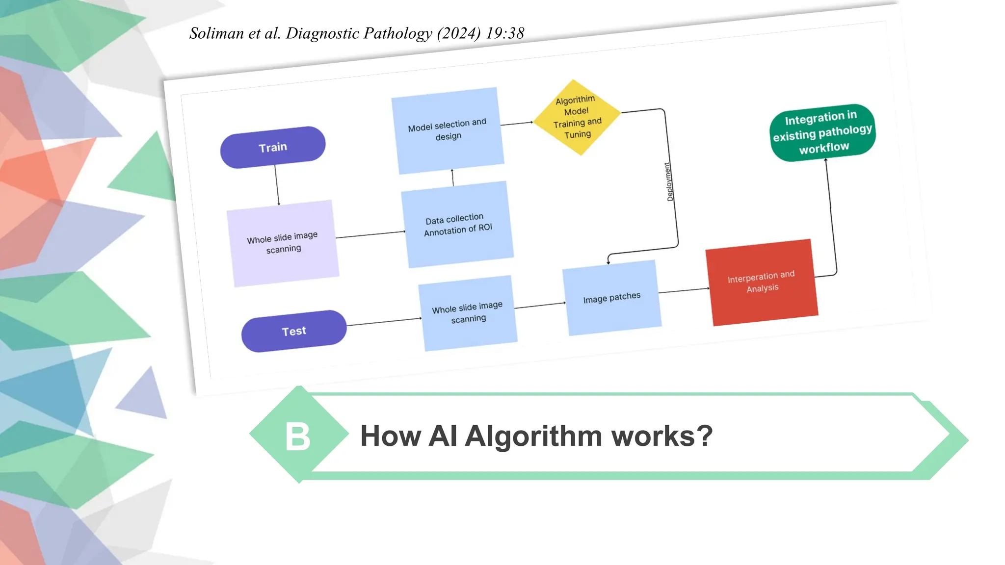
How AI Algorithmworks?
B
Soliman et al. Diagnostic Pathology (2024) 19:38
9.
Computational Pathology
Five DifferentTypes of Annotations
Computational pathology: A survey review and the way forward. Journal of Pathology Informatics (2024)
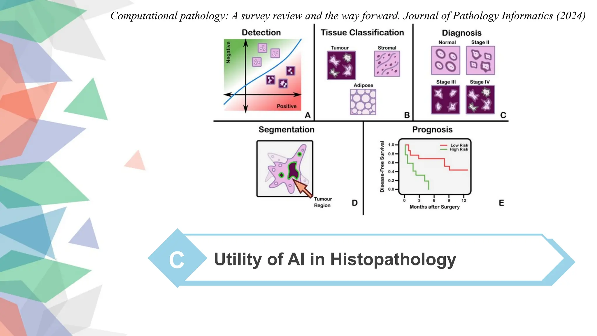
C Utility ofAI in Histopathology
Computational pathology: A survey review and the way forward. Journal of Pathology Informatics (2024)
14.
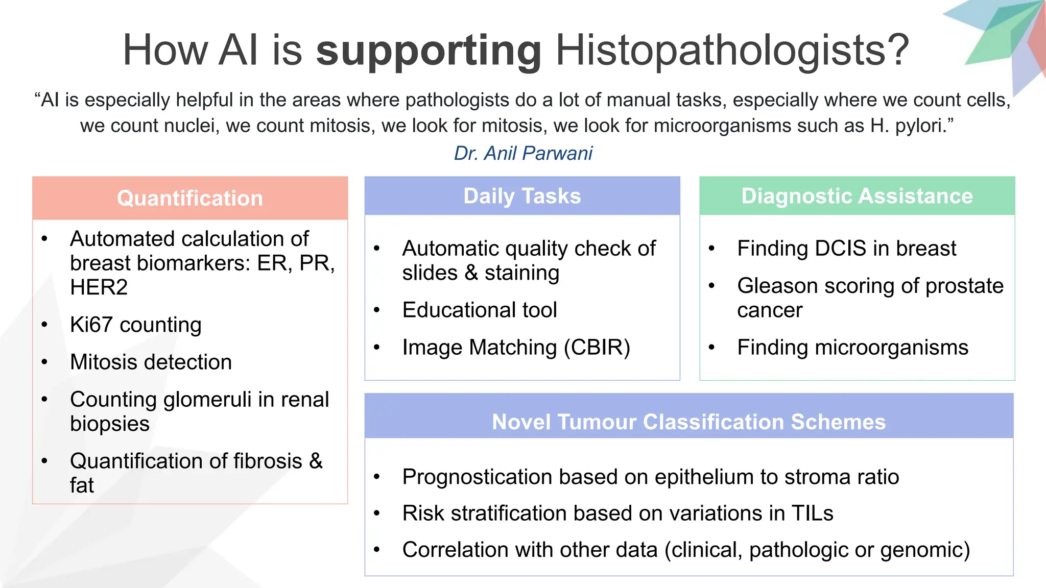
How AI issupporting Histopathologists?
“AI is especially helpful in the areas where pathologists do a lot of manual tasks, especially where we count cells,
we count nuclei, we count mitosis, we look for mitosis, we look for microorganisms such as H. pylori.”
Dr. Anil Parwani
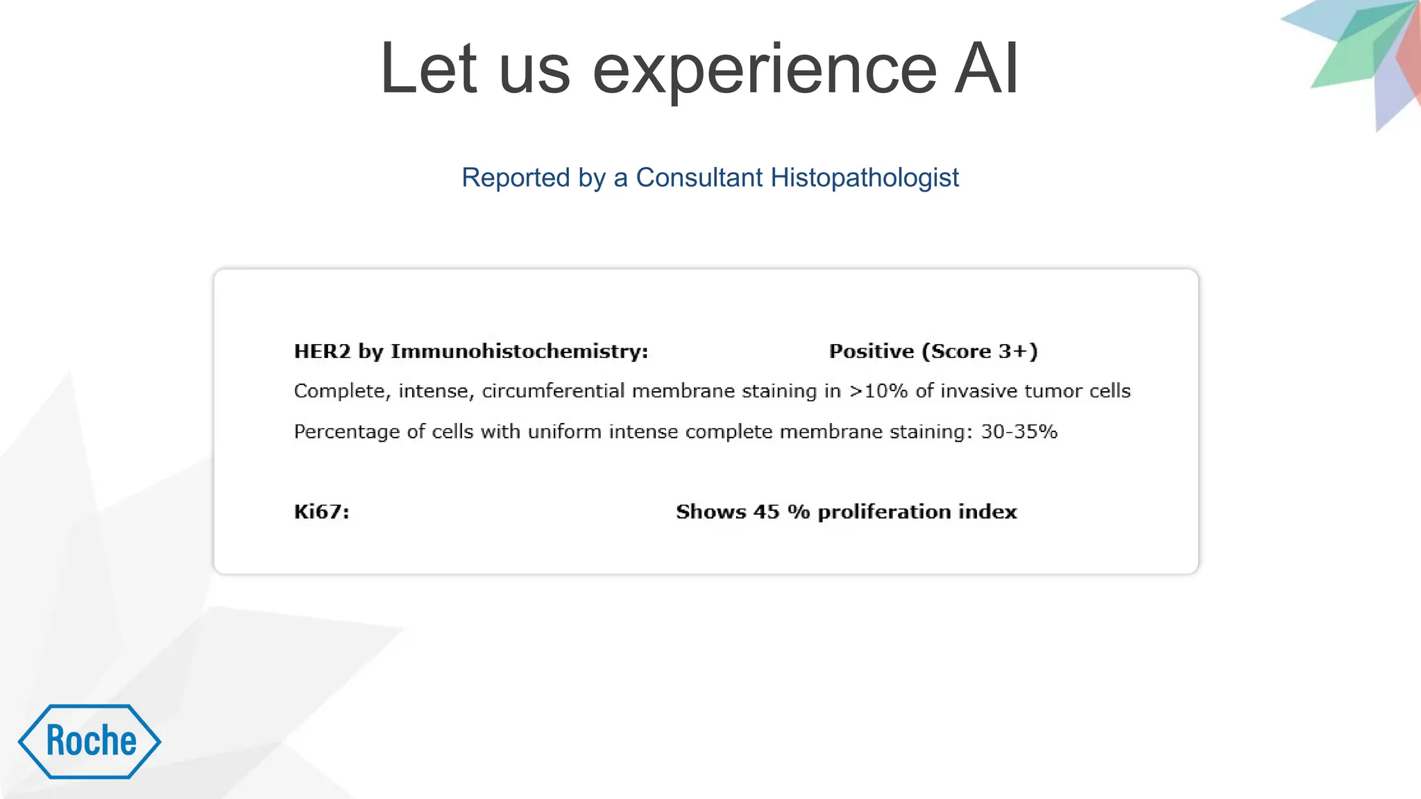
Quantification
• Automated calculation of
breast biomarkers: ER, PR,
HER2
• Ki67 counting
• Mitosis detection
• Counting glomeruli in renal
biopsies
• Quantification of fibrosis &
fat
Diagnostic Assistance
• Finding DCIS in breast
• Gleason scoring of prostate
cancer
• Finding microorganisms
Daily Tasks
• Automatic quality check of
slides & staining
• Educational tool
• Image Matching (CBIR)
Novel Tumour Classification Schemes
• Prognostication based on epithelium to stroma ratio
• Risk stratification based on variations in TILs
• Correlation with other data (clinical, pathologic or genomic)
15.
400+ CPath DiagnosticTasks from 2018 to 2022
Journal of Pathology Informatics 15 (2024) 100357
16.
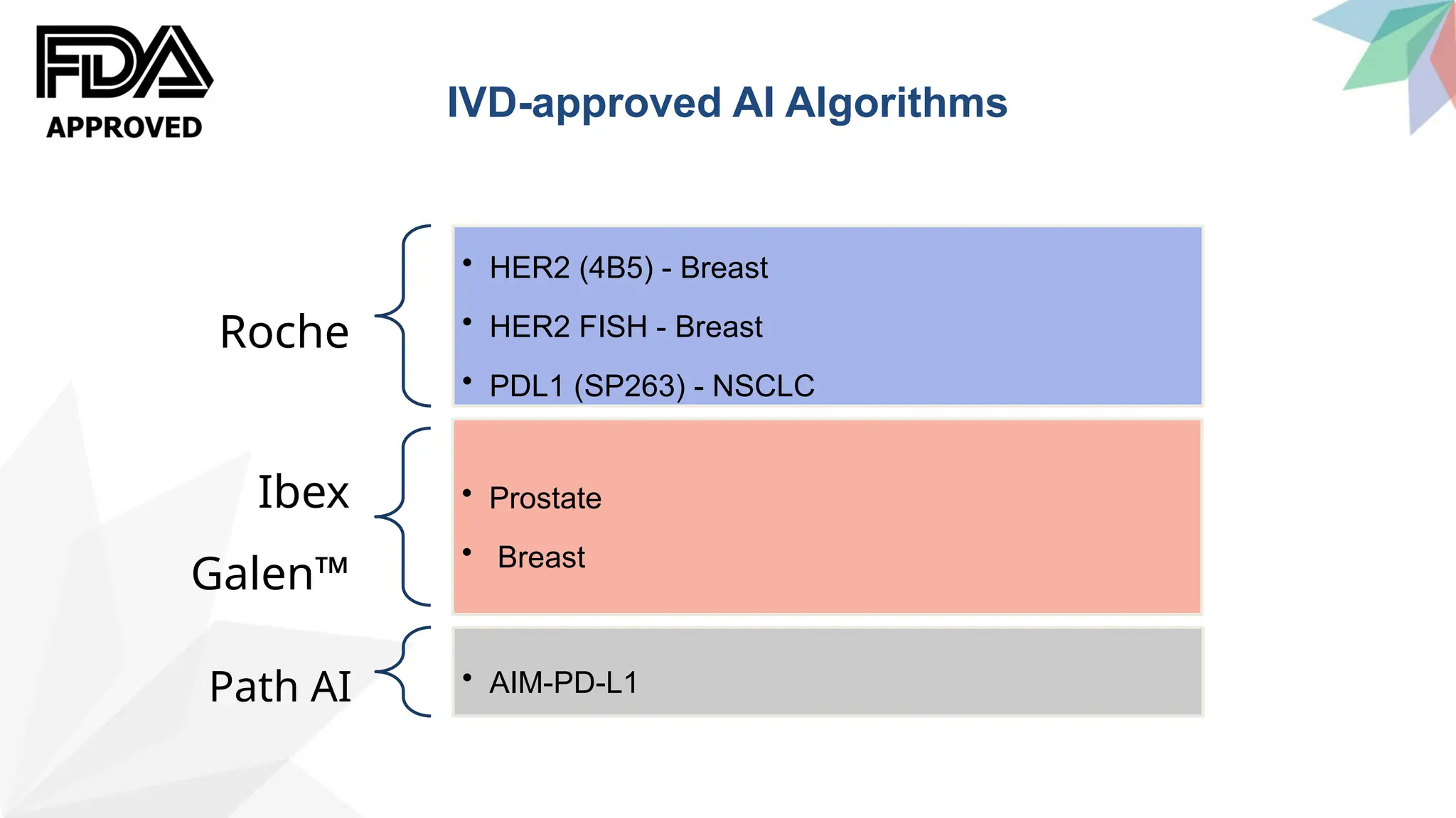
Roche
• HER2 (4B5)- Breast
• HER2 FISH - Breast
• PDL1 (SP263) - NSCLC
Ibex
Galen™
• Prostate
• Breast
Path AI • AIM-PD-L1
IVD-approved AI Algorithms
17.
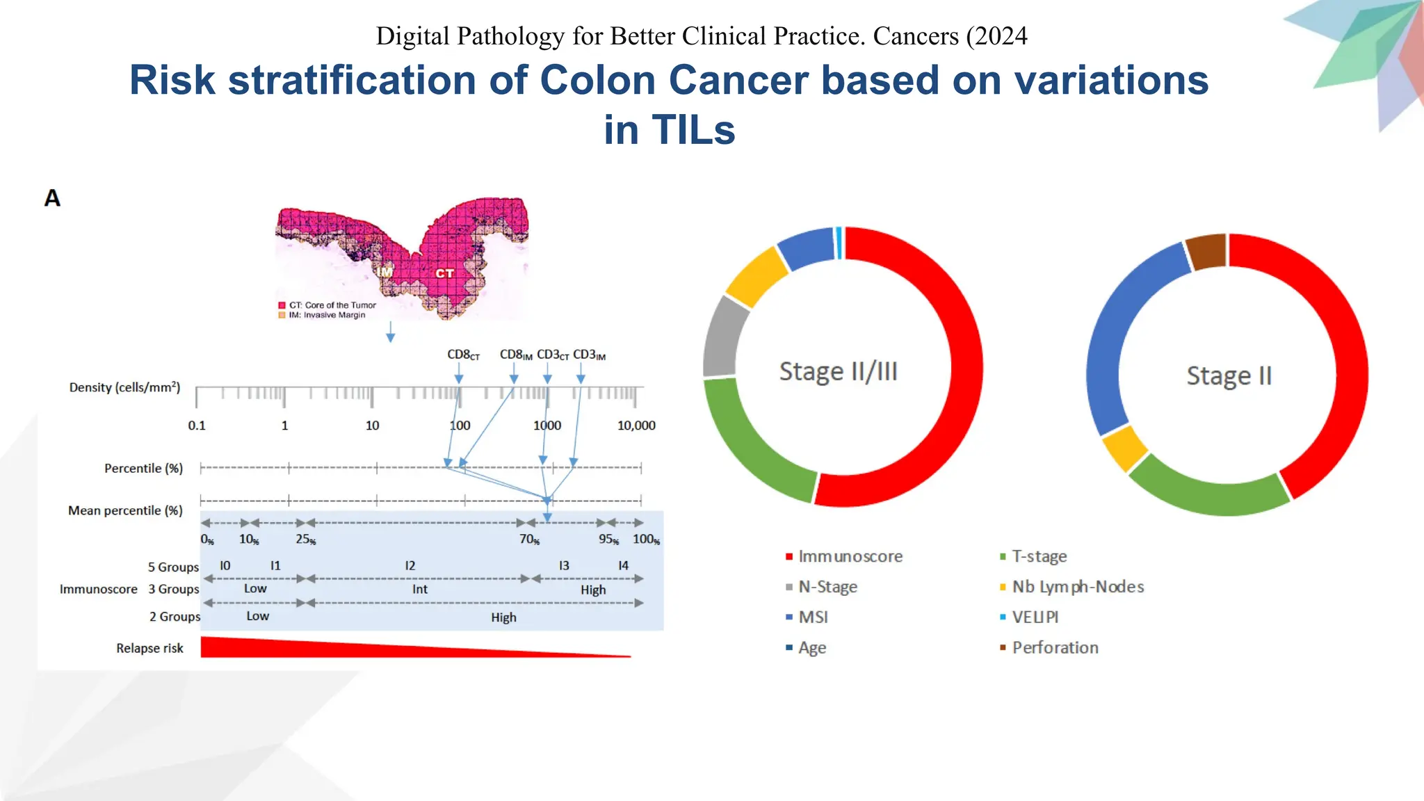
Risk stratification ofColon Cancer based on variations
in TILs
Digital Pathology for Better Clinical Practice. Cancers (2024
18.
How AI ischallenging Histopathologists?
• ROI
• Reimburse-
ment
Cost
• Utility in real
world
• Cost :
Benefit ratio
Acceptance
• Data &
Storage
• Overfitting
• ‘Black box’
problem
Technology
• Quality of
Data
• Quality of
Slides &
Staining
Pre-Analytical
Variables
Those who understandAI limitations and its uses, and overcome the
barriers to implementation, will be well poised to deliver the best
possible patient care now and in the future.
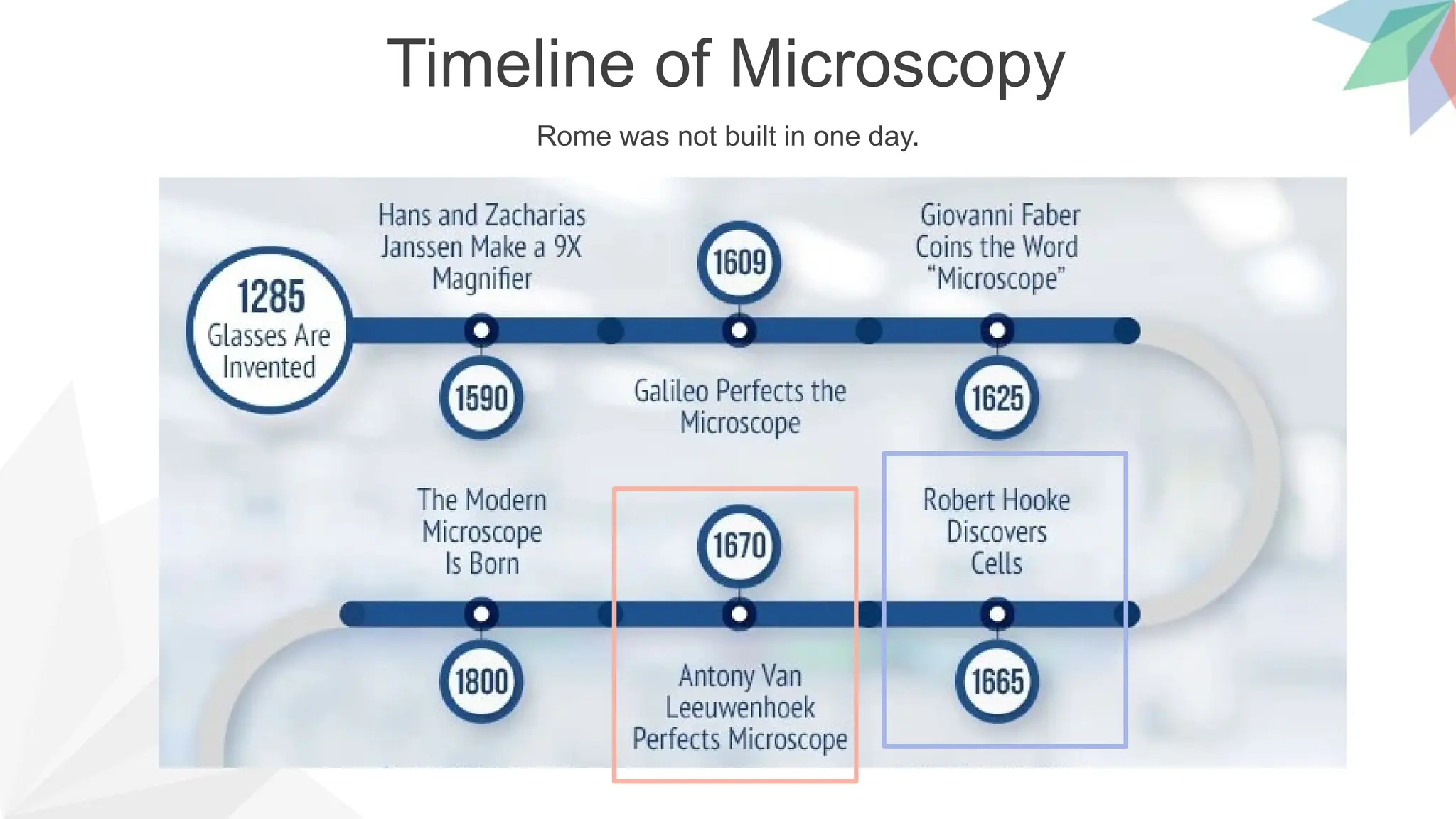
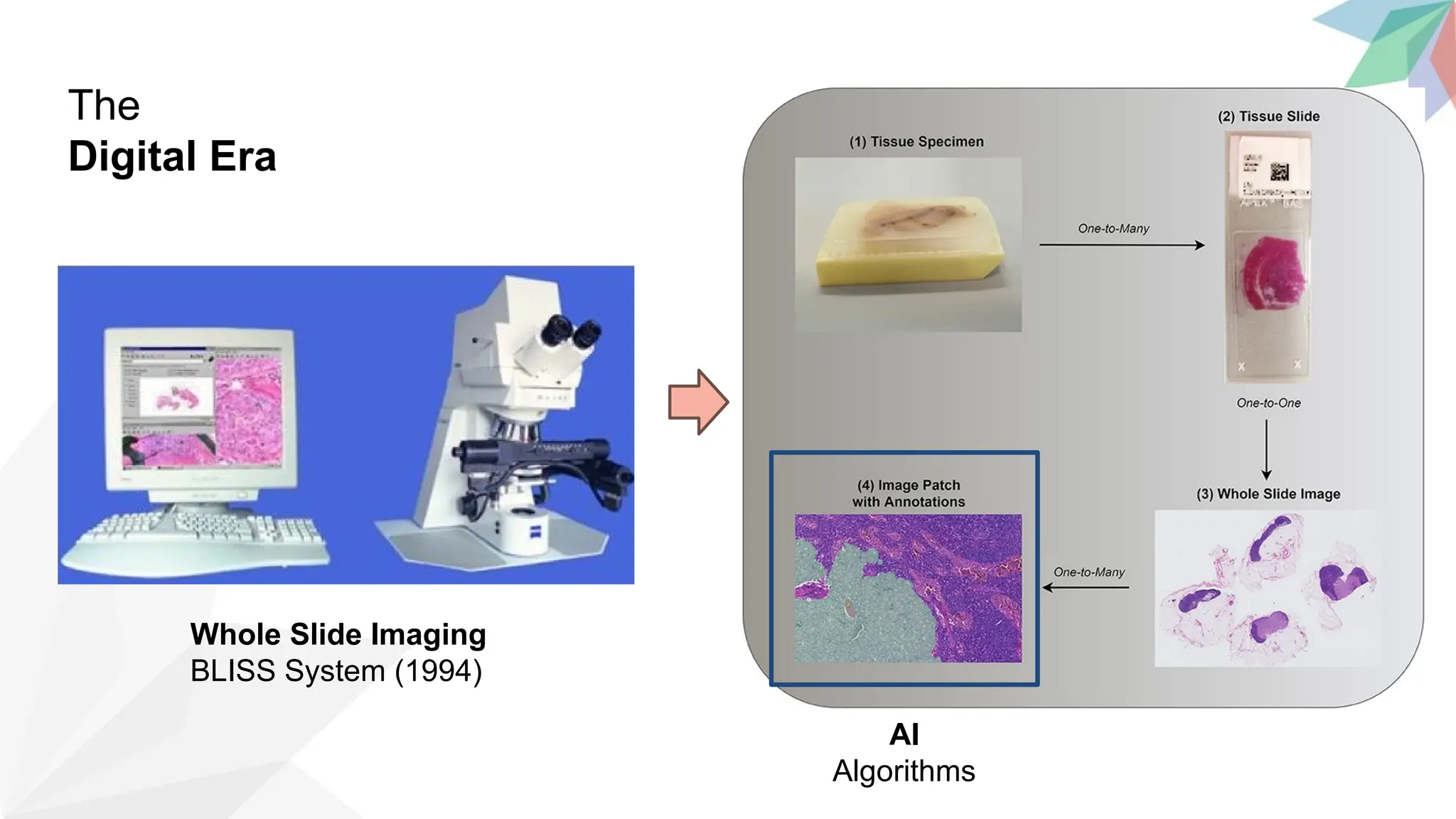
#6 First ever photomicrographs, 1845. This image was published by Alfred Donne and Leon Foucault in 1845 in French the medical textbook Cours de microscopie. Donne and Foucault took Daguerreotype photographs of specimens through a light microscope. The four images are: (fig 45) urea nitrate crystals, (fig 46) crystals of typhoid urine, (fig 47) crystals of uric acid, (fig 48) group of uric acid crystals.
#7 BLISS System (1994). The first digital microscope systems cost about $300,000 to set up and took over 24 h to scan a single slide. He worked with the American Board of Pathology to introduce virtual microscopy into board certification of pathologists. Company acquired by Olympus
#9 Five Different Types of Annotations. The most beneficial support of AI to pathology will be building up computational pathology on traditional histopathology. FOV: Field of View
Computational pathology is a brand-new discipline that aims to enhance patient care by utilizing advances in artificial intelligence and data generated from anatomic and clinical pathology.
#10 AI: Intelligence exhibited by machines, particularly computer systems
ML: Enables systems to learn from data without explicit programming
DL: Uses multiple layers to learn complex understandings, inspired by neural network in humans
CNN: Designed for image and video analysis, Spatial information & Extraction filters
#15 Distribution of diagnostic tasks in CPath for different organs from Table 9.11. This distribution includes more than 400 cited works from 2018 to 2022 inclusive. The x-axis covers different organs, the y-axis displays different diagnostic tasks, and the height of the bars along the vertical axis measures the number of works that have examined the specific task and organ
#17 Figure 2. Illustration of the DP-Immunoscore calculation method. (A) Densities of CD3+ and CD8+ at both CT and IM are converted into percentile values. The mean percentile of the four markers is calculated and represented into a five-category (IS0, IS1, IS2, IS3, IS4) or a three- (IS-Low, IS-Int, IS-High) or a two-category scoring system (IS-Low, IS-High). Based on measuring immune response at the tumor site, IS predicts the risk of relapse in localized colon cancer to identify patients who could be spared from chemotherapy. (B) Ring charts illustrating the relative contribution of each risk parameter to recurrence risk in patients with stages II and II/III colon cancer. IS (red) is the highest predictor of time to recurrence (TTR) in both subgroups.
#18 Overfitting” is when AI algorithms, trained on one dataset, have limited applicability to other datasets.
The ‘black box’ problem is the inability of deep learning algorithms to demonstrate how they arrive at their conclusions.
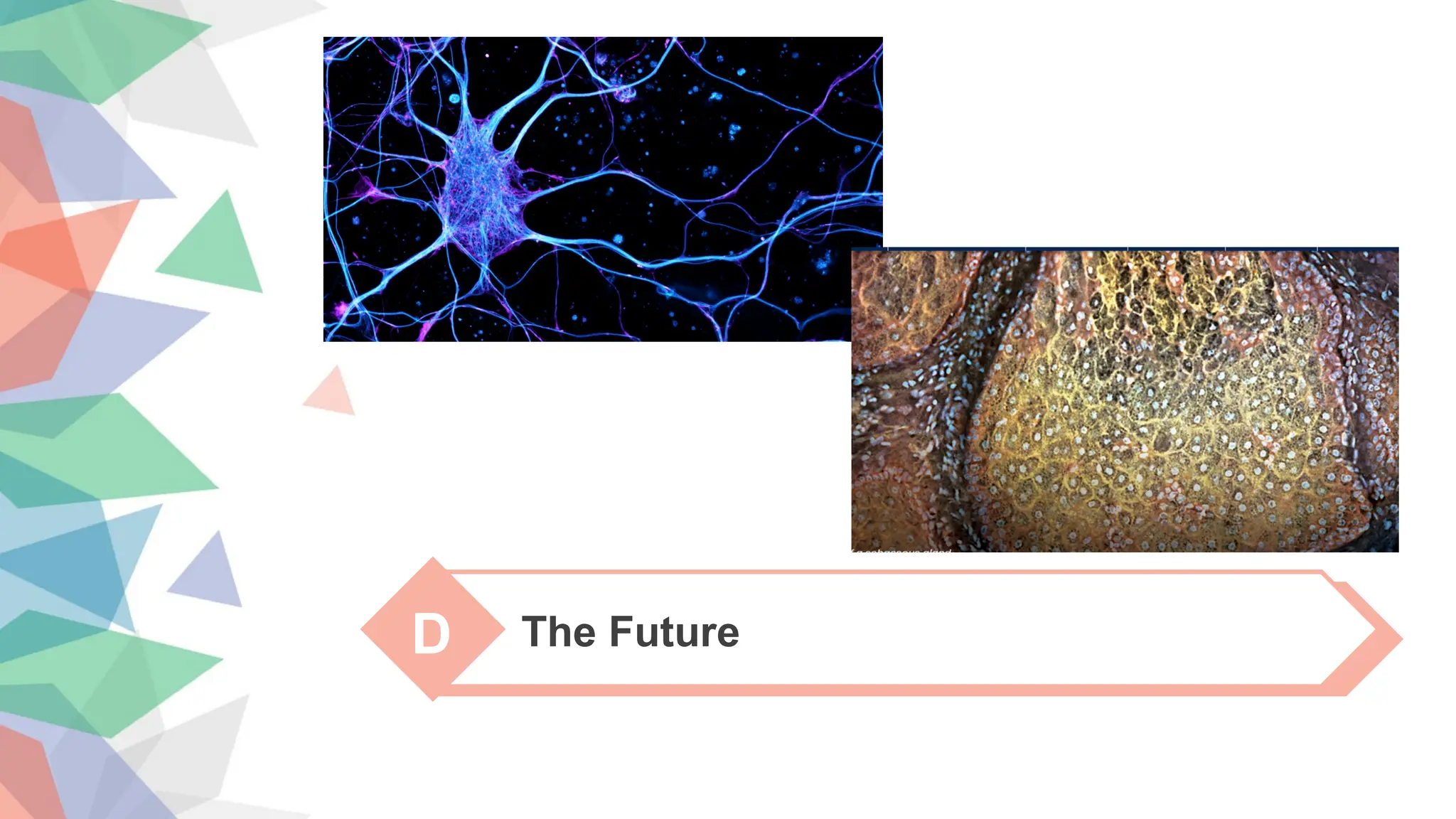
#19 A STED (stimulated emission/depletion) micrograph image revealing actin (magenta) and microtubules (cyan)
MUSE image of sebaceous gland
A STED (stimulated emission/depletion) micrograph image revealing actin (magenta) and microtubules (cyan) of a young dissociated hippocampal neuron. Image by K. Jansen and E. Katrukha, Kapitein Lab, Molecular and Cellular Biophysics, Utrecht University, The Netherlands
Where the light is focused from all sides to a common focus that is used to scan the object by 'point-by-point' excitation combined with 'point-by-point' detection
Photon-tunneling microscopy[4] as well as those that use the Pendry Superlens and near field scanning optical microscopy
“More than 80 percent of the time, patient biopsies will come back normal,” says Richard Levenson, professor and vice chair for strategic technologies in the UC Davis Department of Pathology and Laboratory Medicine. “Meanwhile, the patient is waiting for results for a potentially lethal disease. They live with a great deal of anxiety.”
To remedy this, researchers at UC Davis are working on a new way to view tissue. They’ve created the MUSE microscope, which uses ultraviolet rather than visible light. MUSE samples do not require the rigorous preparation that can slow down analysis of conventional slides.


![[대한병리학회] 의료 인공지능 101: 병리를 중심으로](https://cdn.slidesharecdn.com/ss_thumbnails/pathology-201106004112-thumbnail.jpg?width=640&height=640&fit=bounds)


![Understanding Parkinson’s Disease: Causes, Symptoms, and Treatment [2025]](https://cdn.slidesharecdn.com/ss_thumbnails/understandingparkinson-251208102525-80ba3223-thumbnail.jpg?width=640&height=640&fit=bounds)


