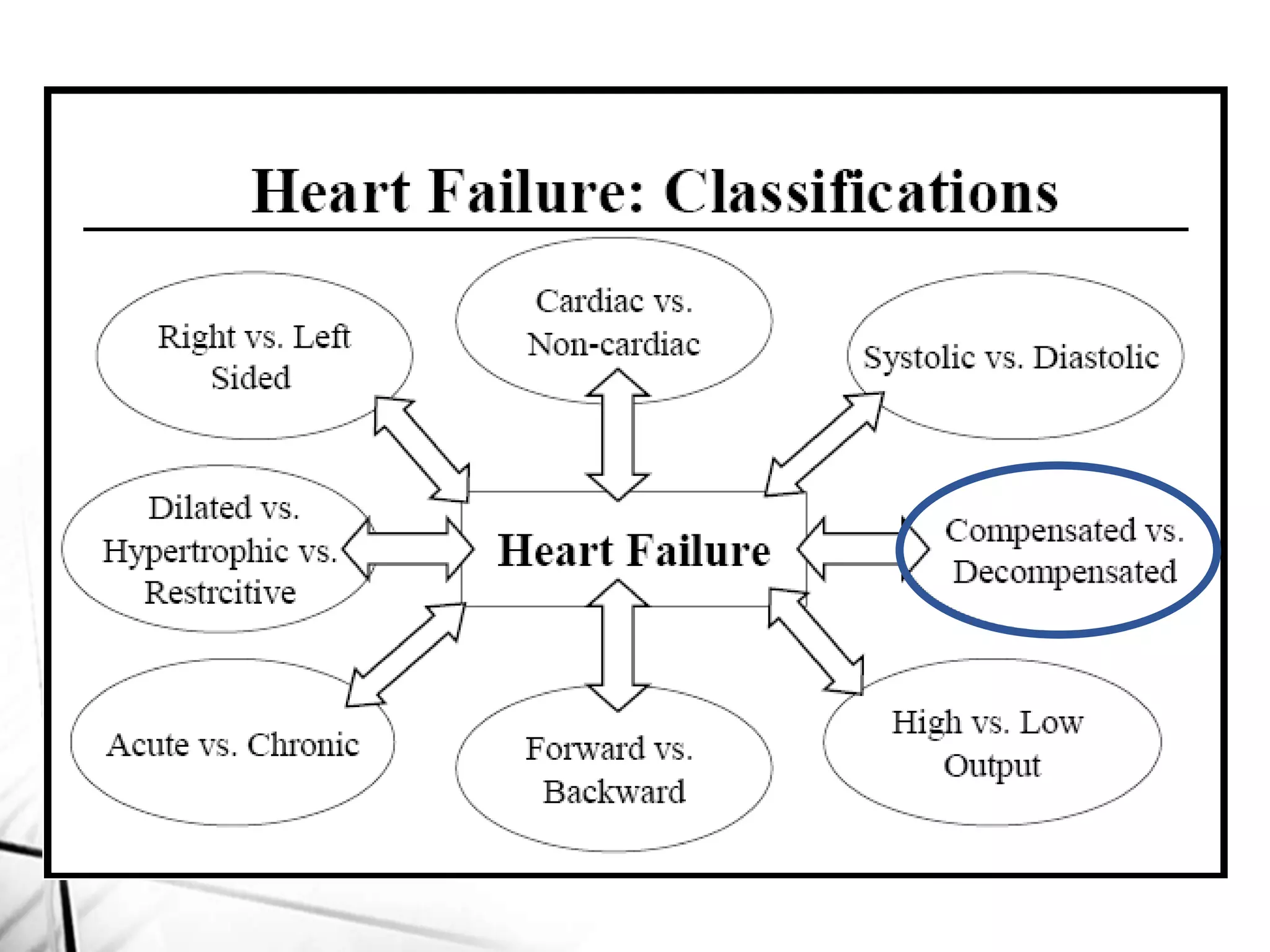
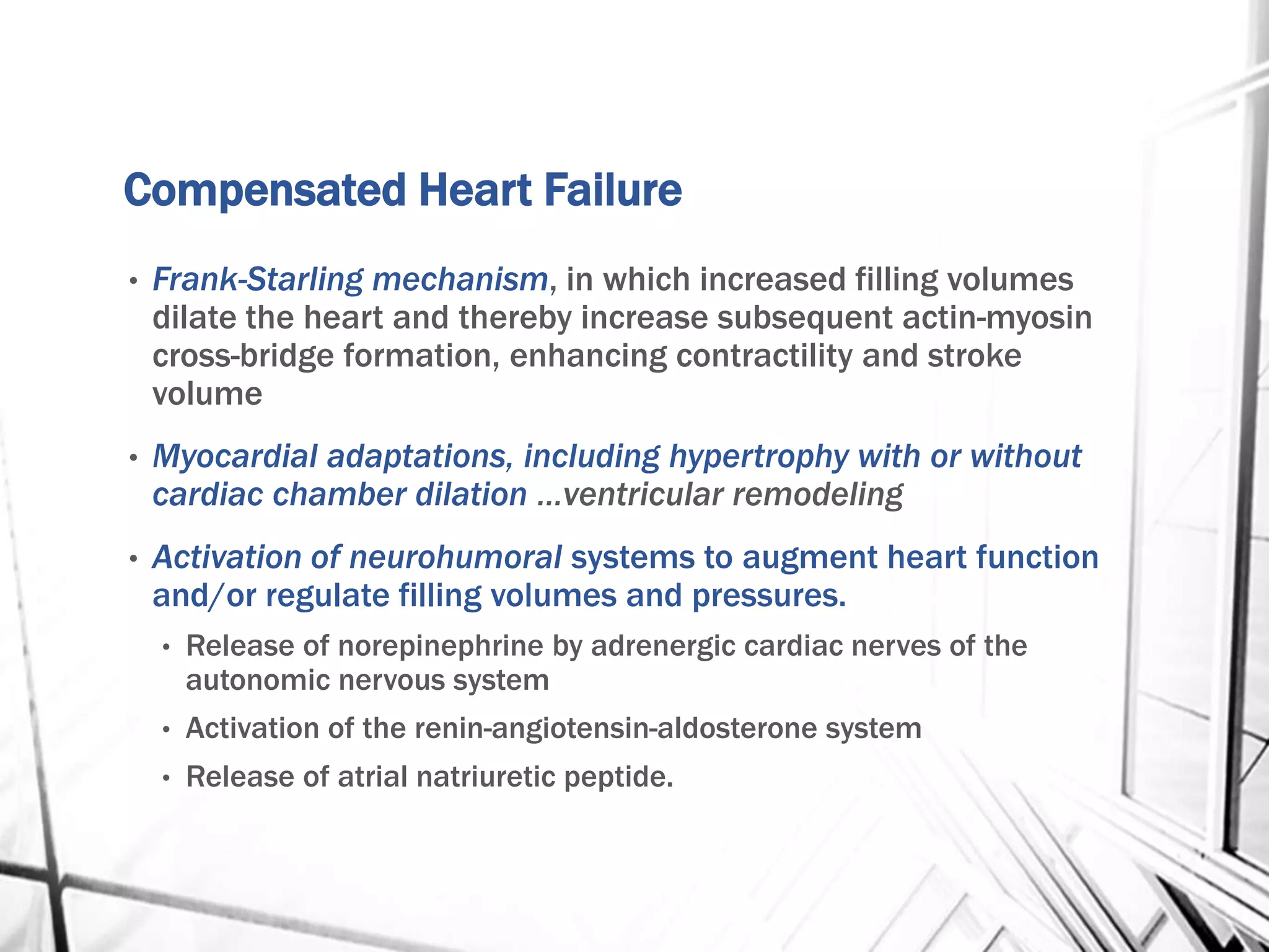
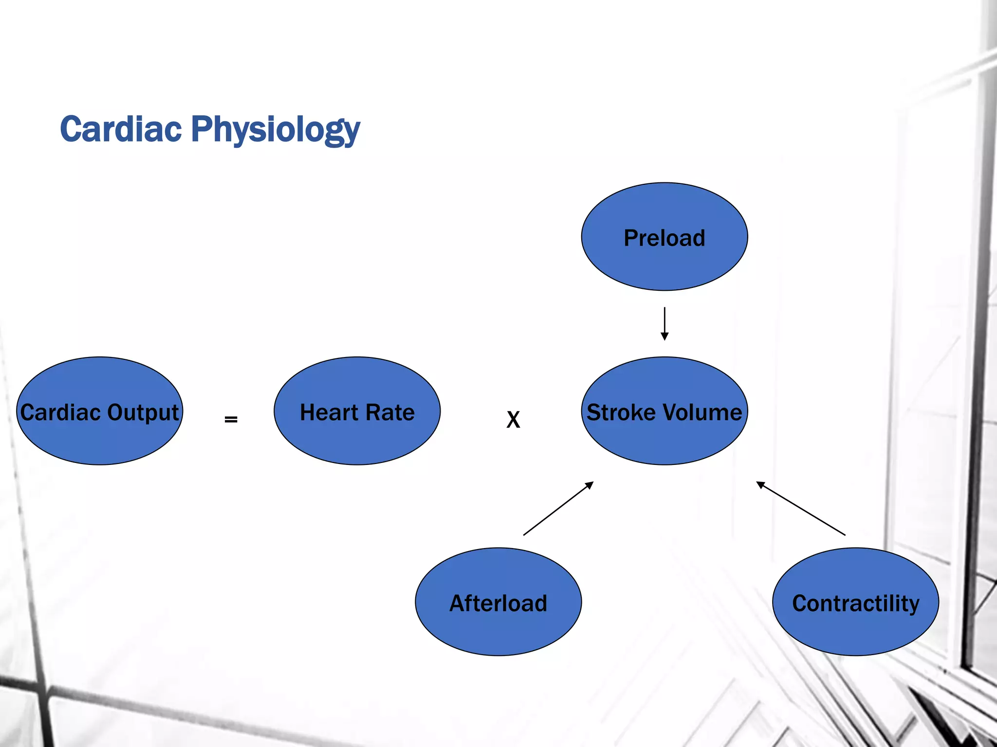
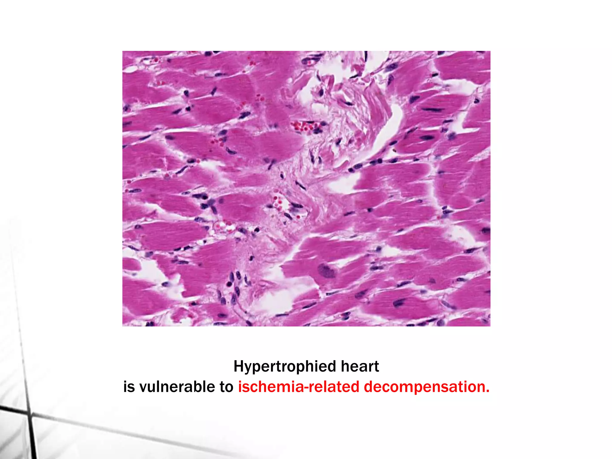
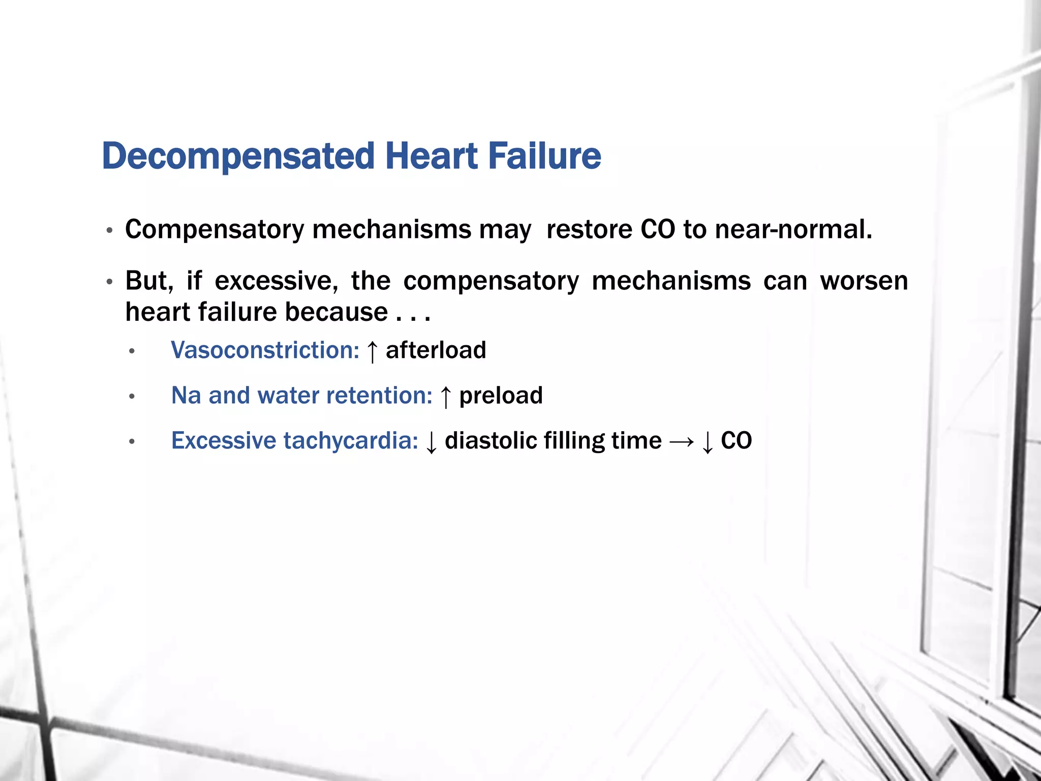
HEART FAILURE
(CHF)
Dr. Usman Shams
Normal Heart
• Weight of the heart varies with body weight & height.
• Males: 300 to 350 grams
• Females: 250 to 300 grams
• Usual thickness of
• Right Ventricle: 0.3 to 0.5 cm
• Left Ventricle: 1.3 to 1.5 cm
Hypertrophy: Greater heart weight or Ventricular thickness
Dilation: Enlarged chamber size
Cardiomegaly: Increase in cardiac weight or size owing to hypertrophy
and/or dilation
HEART MAy DIE
Congestive
Heart
Failure
(CHF)
Impaired cardiac pumping
such that heart is unable to
pump adequate amount of
blood to meet metabolic
needs.
Not a disease but a
“syndrome”
Common
Causes
HEART
MAY
DIE
• A complex clinical syndrome that can result from any
structural or functional cardiac disorder that impairs the
ability of the ventricle to fill with or eject blood.
• Four Basic Mechanisms
• Increased Blood Volume (Excessive Preload)
• Increased Resistant to Blood Flow (Excessive Afterload)
• Decreased contractility
• Decreased Filling
Increased Blood Volume
• Mitral Regurgitation
• Aortic Regurgitation
• Volume Overload
• Left to Right Shunts
• Chronic Kidney Disease
Decreased Contractility
• Ischemic Cardiomyopathy
– Myocardial Infarction
– Myocardial Ischemia
• Myocarditis
• Toxins
– Anthracycline
– Alcohol
– Cocaine
Decreased Filling
• Mitral Stenosis
• Constriction
• Restrictive
Cardiomyopathy
• Cardiac Tamponade
• Hypertrophic
Cardiomyopathy
• Infiltrative
Cardiomyopathy
Increased Afterload
• Aortic Stenosis
• Aortic Coarctation
• Hypertension
Compensated Heart Failure
• Frank-Starling mechanism, in which increased filling volumes
dilate the heart and thereby increase subsequent actin-myosin
cross-bridge formation, enhancing contractility and stroke
volume
• Myocardial adaptations, including hypertrophy with or without
cardiac chamber dilation …ventricular remodeling
• Activation of neurohumoral systems to augment heart function
and/or regulate filling volumes and pressures.
• Release of norepinephrine by adrenergic cardiac nerves of the
autonomic nervous system
• Activation of the renin-angiotensin-aldosterone system
• Release of atrial natriuretic peptide.
Cardiac Physiology
Cardiac Output
Preload
Afterload Contractility
Heart Rate Stroke Volume= X
HYPERTROPHY
• PRESSURE OVERLOAD … concentric increase in wall thickness
• VOLUME OVERLOAD … ventricular dilatation
• 2X normal weight … ischemia
• 3X normal weight … HTN
• >3X normal weight … cardiomyopathy, aortic regurgitation
Wall thickness vs. Heart weight
Hypertrophied heart
is vulnerable to ischemia-related decompensation.
Decompensated Heart Failure
• Compensatory mechanisms may restore CO to near-normal.
• But, if excessive, the compensatory mechanisms can worsen
heart failure because . . .
• Vasoconstriction: ↑ afterload
• Na and water retention: ↑ preload
• Excessive tachycardia: ↓ diastolic filling time → ↓ CO
Not types, but characteristics of CHF.
FORWARD FAILURE
Variable degrees of decreased
cardiac output and tissue
perfusion.
BACKWARD FAILURE
Pooling of blood in the venous
capacitance system
Usually CHF is the common end stage of many
forms of chronic heart disease.
Chronic Acute
Coronary Artery Disease
Hypertensive HD
Rheumatic Heart Disorders
Congenital Heart Disorders
Cor pulmonale
Cardiomyopathy
Anemia
Bacterial endocarditis
Valvular disorders
Acute MI
Arrhythmias
Pulmonary emboli
Thyrotoxicosis
Hypertensive crisis
Rupture of papillary muscle
VSD
Myocarditis
Left Sided Heart Failure
• Lungs
• pulmonary congestion and edema
• heart failure cells
• Kidneys
• pre-renal azotemia
• salt and fluid retention
• renin-aldosterone activation
• natriuretic peptides
• Brain
• Irritability, decreased attention, stupor
• coma
Right Sided Heart Failure
• Liver & Spleen
• passive congestion (nutmeg liver)
• Centrilobular necrosis
• congestive splenomegaly
• Pleura/Pericardium/Peritoneum
• Pleural and pericardial effusions
• Ascites
• Transudates
• Peripheral tissues
• Ankle and pretibial edema
• Anasarca
Edema
Kerley B lines
Heart Failure Cells
Nutmeg Liver
Two types of left heart failure.
When the abnormality
is primary in and
localized to the
myocardium, the
condition is called
CARDIOMYOPATHY.
Functional
pattern
Ejection
Fraction
Mechanism of
heart Failure
Causes Myocardial
dysfunction
Dilated <40% ↓ contractility
(systolic)
Idiopathic, Alcohol,
Peripartum, Genetic,
Myocarditis,
Hemochromatosis,
Chronic anemia,
Doxorubicin,
Sarcoidosis
Ischemic HD,
Valvular HD,
Hypertensive HD,
Congenital HD.
Hypertrophic 50-80% ↓ compliance
(diastolic)
Genetic, Friedreich
ataxia, storage
diseases, infants of
diabetic mothers.
Hypertensive HD,
Aortic stenosis
Restrictive 45-90% ↓compliance
(diastolic)
Idiopathic,
Amyloidosis,
Radiation-induced
fibrosis
Pericardial
constriction
Cardiomyopathy & Indirect Myocardial Dysfunction
Dilated
Cardiomyopathy.
Four chamber dilatation
and hypertrophy are
evident.
There is granular mural
thrombus at the apex of
the left ventricle (white
arrow).
The coronary arteries
were unobstructed.
12-32
Dilated Cardiomyopathy:
Histology demonstrating variable myocyte hypertrophy
and interstitial fibrosis. (Collagen is highlighted as
blue in this Masson trichrome stain.)
12-32 B
Arrhythmogenic Right Ventricular Cardiomyopathy
(Arrhythmogenic Right Ventricular Dysplasia)
Right ventricular dysplasia:
Adipose tissue nearly replaces the myocardium. This may
occur from uncontrolled apoptosis of myocardium.
Hypertrophic Cardiomyopathy with
asymmetric septal hypertrophy
The septal muscle bulges into the left ventricular outflow tract, and
the left atrium is enlarged.
12-
34
Banana-like
configuration
of left
ventricular
cavity due to
asymmetrical
septal
hypertrophy
12-
34
LA
LV
Hypertrophic Cardiomyopathy:
Histologic appearance demonstrating disarray, extreme
hypertrophy and characteristic branching of the myocytes as
well as interstitial fibrosis characteristic of the hypertrophic
cardiomyopathy.
12-34 B
Restrictive Cardiomyopathy
• It is a rare entity with multiple etiologies marked by a
restriction of ventricular filling leading to reduced cardiac
output.
• Interstitial myocardial fibrosis is usually present.
• Specific entities include:
• Endomyocardial fibrosis
• Loeffler endocarditis
• Endocardial fibroelastosis
• Restrictive cardiomyopathy can be idiopathic or associated
with distinct diseases that affect the myocardium (radiation
fibrosis, amyloidosis, sarcoidosis, metastatic tumors, products
of inborn error of metabolism, iron overload).

Congestive Heart Failure (CHF)

  • 1.
  • 2.
    Normal Heart • Weightof the heart varies with body weight & height. • Males: 300 to 350 grams • Females: 250 to 300 grams • Usual thickness of • Right Ventricle: 0.3 to 0.5 cm • Left Ventricle: 1.3 to 1.5 cm Hypertrophy: Greater heart weight or Ventricular thickness Dilation: Enlarged chamber size Cardiomegaly: Increase in cardiac weight or size owing to hypertrophy and/or dilation
  • 3.
    HEART MAy DIE Congestive Heart Failure (CHF) Impairedcardiac pumping such that heart is unable to pump adequate amount of blood to meet metabolic needs. Not a disease but a “syndrome”
  • 4.
  • 5.
    • A complexclinical syndrome that can result from any structural or functional cardiac disorder that impairs the ability of the ventricle to fill with or eject blood. • Four Basic Mechanisms • Increased Blood Volume (Excessive Preload) • Increased Resistant to Blood Flow (Excessive Afterload) • Decreased contractility • Decreased Filling
  • 6.
    Increased Blood Volume •Mitral Regurgitation • Aortic Regurgitation • Volume Overload • Left to Right Shunts • Chronic Kidney Disease Decreased Contractility • Ischemic Cardiomyopathy – Myocardial Infarction – Myocardial Ischemia • Myocarditis • Toxins – Anthracycline – Alcohol – Cocaine Decreased Filling • Mitral Stenosis • Constriction • Restrictive Cardiomyopathy • Cardiac Tamponade • Hypertrophic Cardiomyopathy • Infiltrative Cardiomyopathy Increased Afterload • Aortic Stenosis • Aortic Coarctation • Hypertension
  • 8.
    Compensated Heart Failure •Frank-Starling mechanism, in which increased filling volumes dilate the heart and thereby increase subsequent actin-myosin cross-bridge formation, enhancing contractility and stroke volume • Myocardial adaptations, including hypertrophy with or without cardiac chamber dilation …ventricular remodeling • Activation of neurohumoral systems to augment heart function and/or regulate filling volumes and pressures. • Release of norepinephrine by adrenergic cardiac nerves of the autonomic nervous system • Activation of the renin-angiotensin-aldosterone system • Release of atrial natriuretic peptide.
  • 9.
    Cardiac Physiology Cardiac Output Preload AfterloadContractility Heart Rate Stroke Volume= X
  • 10.
    HYPERTROPHY • PRESSURE OVERLOAD… concentric increase in wall thickness • VOLUME OVERLOAD … ventricular dilatation • 2X normal weight … ischemia • 3X normal weight … HTN • >3X normal weight … cardiomyopathy, aortic regurgitation
  • 11.
    Wall thickness vs.Heart weight
  • 13.
    Hypertrophied heart is vulnerableto ischemia-related decompensation.
  • 14.
    Decompensated Heart Failure •Compensatory mechanisms may restore CO to near-normal. • But, if excessive, the compensatory mechanisms can worsen heart failure because . . . • Vasoconstriction: ↑ afterload • Na and water retention: ↑ preload • Excessive tachycardia: ↓ diastolic filling time → ↓ CO
  • 16.
    Not types, butcharacteristics of CHF. FORWARD FAILURE Variable degrees of decreased cardiac output and tissue perfusion. BACKWARD FAILURE Pooling of blood in the venous capacitance system
  • 18.
    Usually CHF isthe common end stage of many forms of chronic heart disease. Chronic Acute Coronary Artery Disease Hypertensive HD Rheumatic Heart Disorders Congenital Heart Disorders Cor pulmonale Cardiomyopathy Anemia Bacterial endocarditis Valvular disorders Acute MI Arrhythmias Pulmonary emboli Thyrotoxicosis Hypertensive crisis Rupture of papillary muscle VSD Myocarditis
  • 22.
    Left Sided HeartFailure • Lungs • pulmonary congestion and edema • heart failure cells • Kidneys • pre-renal azotemia • salt and fluid retention • renin-aldosterone activation • natriuretic peptides • Brain • Irritability, decreased attention, stupor • coma
  • 25.
    Right Sided HeartFailure • Liver & Spleen • passive congestion (nutmeg liver) • Centrilobular necrosis • congestive splenomegaly • Pleura/Pericardium/Peritoneum • Pleural and pericardial effusions • Ascites • Transudates • Peripheral tissues • Ankle and pretibial edema • Anasarca
  • 28.
  • 29.
  • 30.
  • 31.
  • 33.
    Two types ofleft heart failure.
  • 39.
    When the abnormality isprimary in and localized to the myocardium, the condition is called CARDIOMYOPATHY.
  • 41.
    Functional pattern Ejection Fraction Mechanism of heart Failure CausesMyocardial dysfunction Dilated <40% ↓ contractility (systolic) Idiopathic, Alcohol, Peripartum, Genetic, Myocarditis, Hemochromatosis, Chronic anemia, Doxorubicin, Sarcoidosis Ischemic HD, Valvular HD, Hypertensive HD, Congenital HD. Hypertrophic 50-80% ↓ compliance (diastolic) Genetic, Friedreich ataxia, storage diseases, infants of diabetic mothers. Hypertensive HD, Aortic stenosis Restrictive 45-90% ↓compliance (diastolic) Idiopathic, Amyloidosis, Radiation-induced fibrosis Pericardial constriction Cardiomyopathy & Indirect Myocardial Dysfunction
  • 42.
    Dilated Cardiomyopathy. Four chamber dilatation andhypertrophy are evident. There is granular mural thrombus at the apex of the left ventricle (white arrow). The coronary arteries were unobstructed. 12-32
  • 43.
    Dilated Cardiomyopathy: Histology demonstratingvariable myocyte hypertrophy and interstitial fibrosis. (Collagen is highlighted as blue in this Masson trichrome stain.) 12-32 B
  • 44.
    Arrhythmogenic Right VentricularCardiomyopathy (Arrhythmogenic Right Ventricular Dysplasia)
  • 45.
    Right ventricular dysplasia: Adiposetissue nearly replaces the myocardium. This may occur from uncontrolled apoptosis of myocardium.
  • 46.
    Hypertrophic Cardiomyopathy with asymmetricseptal hypertrophy The septal muscle bulges into the left ventricular outflow tract, and the left atrium is enlarged. 12- 34 Banana-like configuration of left ventricular cavity due to asymmetrical septal hypertrophy 12- 34 LA LV
  • 47.
    Hypertrophic Cardiomyopathy: Histologic appearancedemonstrating disarray, extreme hypertrophy and characteristic branching of the myocytes as well as interstitial fibrosis characteristic of the hypertrophic cardiomyopathy. 12-34 B
  • 48.
    Restrictive Cardiomyopathy • Itis a rare entity with multiple etiologies marked by a restriction of ventricular filling leading to reduced cardiac output. • Interstitial myocardial fibrosis is usually present. • Specific entities include: • Endomyocardial fibrosis • Loeffler endocarditis • Endocardial fibroelastosis • Restrictive cardiomyopathy can be idiopathic or associated with distinct diseases that affect the myocardium (radiation fibrosis, amyloidosis, sarcoidosis, metastatic tumors, products of inborn error of metabolism, iron overload).