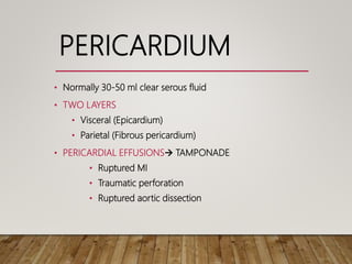
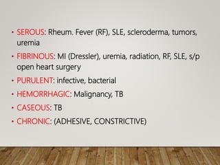
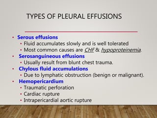
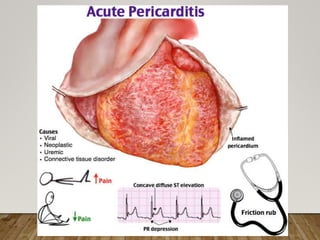
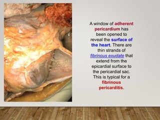
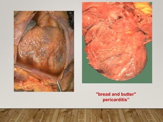
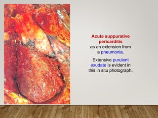
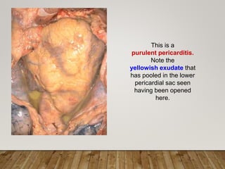
PERICARDITIS
DR. USMAN SHAMS
PERICARDIUM
• Normally 30-50 ml clear serous fluid
• TWO LAYERS
• Visceral (Epicardium)
• Parietal (Fibrous pericardium)
• PERICARDIAL EFFUSIONS TAMPONADE
• Ruptured MI
• Traumatic perforation
• Ruptured aortic dissection
• SEROUS: Rheum. Fever (RF), SLE, scleroderma, tumors,
uremia
• FIBRINOUS: MI (Dressler), uremia, radiation, RF, SLE, s/p
open heart surgery
• PURULENT: infective, bacterial
• HEMORRHAGIC: Malignancy, TB
• CASEOUS: TB
• CHRONIC: (ADHESIVE, CONSTRICTIVE)
TYPES OF PLEURAL EFFUSIONS
• Serous effusions
• Fluid accumulates slowly and is well tolerated
• Most common causes are CHF & hypoproteinemia.
• Serosanguineous effusions
• Usually result from blunt chest trauma.
• Chylous fluid accumulations
• Due to lymphatic obstruction (benign or malignant).
• Hemopericardium
• Traumatic perforation
• Cardiac rupture
• Intrapericardial aortic rupture
SEROUS PERICARDITIS
• Nonbacterial causes
• Microscopically there is scant pericardial acute and
chronic inflammatory infiltrate, mostly lymphocytes.
•
FIBRINOUS PERICARDITIS
• Most common clinical form of pericarditis.
• Associated by pericardial friction rub.
• Exudate may either be completely absorbed or can
organize leaving delicate stringy adhesions (Adhesive
pericarditis) or there may be a plaque-like thickening
of epicardium.
A window of adherent
pericardium has
been opened to
reveal the surface of
the heart. There are
thin strands of
fibrinous exudate that
extend from the
epicardial surface to
the pericardial sac.
This is typical for a
fibrinous
pericarditis.
"bread and butter"
pericarditis”
SUPPURATIVE PERICARDITIS
• Purulent pericarditis is typically composed of 400
to 500 ml of thin-to-creamy pus with erythematous,
granular serosal surfaces.
• Clinical presentation: High fever ; Rigors ; Friction
rub
• On organization: Constrictive / Mediastino-
pericarditis
Acute suppurative
pericarditis
as an extension from
a pneumonia.
Extensive purulent
exudate is evident in
this in situ photograph.
This is a
purulent pericarditis.
Note the
yellowish exudate that
has pooled in the lower
pericardial sac seen
having been opened
here.
CASEOUS
PERICARDITIS
A tuberculous
pericarditis can
produce extensive
granulomatous
inflammation with
calcification that can
severely restrict cardiac
function
HEMORRHAGIC PERICARDITIS
• The term hemorrhagic pericarditis is used when
fibrinous or suppurative pericarditis exudate is mixed
with blood oozing into the pericardial sac.
• It usually organizes with or without calcification.
CHRONIC OR HEALED
PERICARDITIS
• An acute pericarditis on healing may have:
• Complete resolution
• Pericardial fibrosis
• Thick nonadherent epicardial plaque
• Thin delicate adhesions
• Massive thick adhesions
ADHESIVE MEDIASTINO-
PERICARDITIS
• It is a clinically significant pericarditis in which the
cavity of pericardial sac is obliterated and the parietal
pericardium is adherent to mediastinal tissues.
• The heart contracts against the load of all attached
structures.
• There is subsequent hypertrophy of ventricles and
later dilation.
CONSTRICTIVE PERICARDITIS
• It is a clinically significant pericarditis with markedly
thick (up to 1 cm thick) dense fibrous obliteration of
the pericardial sac with or without calcification.
• The heart is encased with limited diastolic expansion
and restricted cardiac output.
Pericarditis

Pericarditis

  • 1.
  • 2.
    PERICARDIUM • Normally 30-50ml clear serous fluid • TWO LAYERS • Visceral (Epicardium) • Parietal (Fibrous pericardium) • PERICARDIAL EFFUSIONS TAMPONADE • Ruptured MI • Traumatic perforation • Ruptured aortic dissection
  • 3.
    • SEROUS: Rheum.Fever (RF), SLE, scleroderma, tumors, uremia • FIBRINOUS: MI (Dressler), uremia, radiation, RF, SLE, s/p open heart surgery • PURULENT: infective, bacterial • HEMORRHAGIC: Malignancy, TB • CASEOUS: TB • CHRONIC: (ADHESIVE, CONSTRICTIVE)
  • 4.
    TYPES OF PLEURALEFFUSIONS • Serous effusions • Fluid accumulates slowly and is well tolerated • Most common causes are CHF & hypoproteinemia. • Serosanguineous effusions • Usually result from blunt chest trauma. • Chylous fluid accumulations • Due to lymphatic obstruction (benign or malignant). • Hemopericardium • Traumatic perforation • Cardiac rupture • Intrapericardial aortic rupture
  • 6.
    SEROUS PERICARDITIS • Nonbacterialcauses • Microscopically there is scant pericardial acute and chronic inflammatory infiltrate, mostly lymphocytes. •
  • 7.
    FIBRINOUS PERICARDITIS • Mostcommon clinical form of pericarditis. • Associated by pericardial friction rub. • Exudate may either be completely absorbed or can organize leaving delicate stringy adhesions (Adhesive pericarditis) or there may be a plaque-like thickening of epicardium.
  • 8.
    A window ofadherent pericardium has been opened to reveal the surface of the heart. There are thin strands of fibrinous exudate that extend from the epicardial surface to the pericardial sac. This is typical for a fibrinous pericarditis.
  • 9.
  • 10.
    SUPPURATIVE PERICARDITIS • Purulentpericarditis is typically composed of 400 to 500 ml of thin-to-creamy pus with erythematous, granular serosal surfaces. • Clinical presentation: High fever ; Rigors ; Friction rub • On organization: Constrictive / Mediastino- pericarditis
  • 11.
    Acute suppurative pericarditis as anextension from a pneumonia. Extensive purulent exudate is evident in this in situ photograph.
  • 12.
    This is a purulentpericarditis. Note the yellowish exudate that has pooled in the lower pericardial sac seen having been opened here.
  • 13.
    CASEOUS PERICARDITIS A tuberculous pericarditis can produceextensive granulomatous inflammation with calcification that can severely restrict cardiac function
  • 14.
    HEMORRHAGIC PERICARDITIS • Theterm hemorrhagic pericarditis is used when fibrinous or suppurative pericarditis exudate is mixed with blood oozing into the pericardial sac. • It usually organizes with or without calcification.
  • 15.
    CHRONIC OR HEALED PERICARDITIS •An acute pericarditis on healing may have: • Complete resolution • Pericardial fibrosis • Thick nonadherent epicardial plaque • Thin delicate adhesions • Massive thick adhesions
  • 16.
    ADHESIVE MEDIASTINO- PERICARDITIS • Itis a clinically significant pericarditis in which the cavity of pericardial sac is obliterated and the parietal pericardium is adherent to mediastinal tissues. • The heart contracts against the load of all attached structures. • There is subsequent hypertrophy of ventricles and later dilation.
  • 17.
    CONSTRICTIVE PERICARDITIS • Itis a clinically significant pericarditis with markedly thick (up to 1 cm thick) dense fibrous obliteration of the pericardial sac with or without calcification. • The heart is encased with limited diastolic expansion and restricted cardiac output.