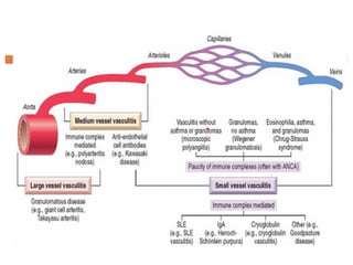
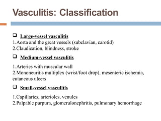
Vasculitis
- Vasculitis refers to a broad and heterogeneous disease spectrum
characterized by inflammation and damage of the blood vessel.
 Vessels of any type in virtually any organ can be affected,but most
vasculitis affect small vessels ranging in size from arterioles to capillaries
to venules.
 The two common pathogenic mechanisms of vasculitis are immune-
mediated inflammation and direct invasion of vascular walls by infectious
pathogens.
Vasculitis: Definition
- Pathologist Rheumatologist
A clinicopathologic process
characterized by inflammatory
destruction of blood vessels
that results in occlusion or
destruction of the vessel and
ischemia of the tissues supplied
by that vessel.
"Systemic vasculilides"
Inflammatory destruction of
blood vessels​
Infiltration of vessel wall with
inflammatory cells​
Leukocytoclasis​
Elastic membrane disruption​
Fibrinoid necrosis of the vessel
walls​
chemia, occlusion,
thrombosisAneurysm formation
Rupture, hemorrhage
Pathogenesis
 IgAV is a leukocytoclastic vasculitis with an IgA-
dominant
immune complex deposited within or around the small
vessels.
 IgAVN manifests similarly to IgAN, both characterized by
hematuria, proteinuria and glomerular mesangial immune
complex deposition, and its pathogenesis can be explained
by
a similar “four-hit” theory (25), that is, increased production
of
circulating galactose-deficient IgA1 (hit1) binds to specific
IgA1
autoantibodies (hit2), forming pathogenic circulating
immune
-
 This hypothesis highlights the critical role of Gd-IgA1 in
renal injury.
 First hit is provided by elevated AECA levels of IgA1
isotype (30), followed by the binding of IgA1-AECA
complexes to specific b2 glycoprotein I receptors on
vascular endothelial cells (hit2), inducing excessive
production of proinflammatory factors such as
interleukin-8 (IL-8), which in turn stimulates neutrophil
recruitment (hit3), and then neutrophils are activated by
the interaction of IgA1 and IgA1 Fc alpha receptor I
(FcaRI, also called CD89), causing extensive damage to
the vascular endothelium wall via antibody-dependent
cellular cytotoxicity (ADCC), complement-mediated
cytotoxicity (CDC), and reactive oxygen species (ROS),
ultimately leading to systemic vascular inflammation and
Vasculitis: Classification
-  Large-vessel vasculitis
1.Aorta and the great vessels (subclavian, carotid)
2.Claudication, blindness, stroke
 Medium-vessel vasculitis
1.Arteries with muscular wall
2.Mononeuritis multiplex (wrist/foot drop), mesenteric ischemia,
cutaneous ulcers
 Small-vessel vasculitis
1.Capillaries, arterioles, venules
2.Palpable purpura, glomerulonephritis, pulmonary hemorrhage
Giant Cell (Temporal) Arteritis
-
 It is a chronic inflammatory disorder of large to small-sized
arteries that principally affects arteries in the head-
especially the temporal arteries-but also the vertebral and
ophthalmic arteries. Ophthalmic arterial involvement can lead
abruptly to permanent blindness; consequently, giant cell
arteritis is a medical emergency requiring prompt recognition
and treatment.
-
 T-cell-mediated immune response against one of
handful of vessel wall antigens that drives subsequent
proinflammatory cytokine production (particularly
TNF).
 Anti-endothelial cell and anti-smooth muscle cell
antibodies can also be demonstrated in roughly two
thirds of patients, although it is unclear whether these
are causal or a consequence of other immune injury.
-  A cellular immune etiology is supported by the
characteristic granulomatous response, a correlation with
certain MHC class II haplotypes, and a prompt therapeutic
response to steroids.
MORPHOLOGY
-  Intimal thickening (with occasional thromboses) that reduces
the luminal diameter.
 Classic lesions exhibit medial granulomatous inflammation
centered on the internal elastic lamina that produce elastic
lamina fragmentation
 There is an infiltrate of T cells (CD4+ CDS+) and
macrophages. Although multinucleated giant cells are seen in
approximately 75% of adequately biopsied specimens
 Granulomas and giant cells can be rare or absent.
-
 Inflammatory lesions are only focally distributed along the
vessel and long segments of relatively normal artery may
be interposed.
 The healed stage is marked by medial attenuation and
scarring with intimal thickening, typically with residual
elastic tissue fragmentation and adventitial fibrosis.
Clinical Features.
-  Rare before age 50
 Symptoms may be only vague and constitutional-
fever,fatigue, weight loss-or there may be facial pain or
headache, most intense along the course of the superficial
temporal artery, which can be painful to palpation.
 Ocular symptoms (associated with involvement of the
ophthalmic artery) appear abruptly in about 50% of
patients.
ELASTIN STAIN
-
Doctor, Doctor, my teenage son
coughed up blood
-  15 y/o male with hx of chronic sinusitis and
congestion, here with complaints of hemoptysis and
dyspnea but stable now.
 Previously called Wegeners
 granulomatosis Differential
diagnosis
1. Sarcoid
2. TB
3. Churg Strauss
 Epidemiology of WGN
• mostly kidney and lung with
granuolomas
• adults (4th and 5th decades) >>>kids
• caucasians >>>noncaucasian
• initially with respiratory symptoms
and then renal insufficiency
 Diagnosis:
1. Clinical findings of sinusitis and pulm finding, order high
resolution CT.
2. Check ANCA: (90% of WGN have the marker) with 70%
sensitivity
 Bx:
1. lung: granulomas with geographic patterns of central necrosis
and accompanying vasculitis
2. renal: 10% with granulomas, segmental necrotizing GN
 RX:
1. rapid therapy needed
2. Steroids
3. cyclophosphamide
Showing features characteristic of granulomatosis with polyangiitis - a vasculitis and
granulomas with multi-nucleated giant cells.
-
Churg-Strauss Syndrome
 Churg-Strauss syndrome is a small-vessel necrotizing
vasculitis classically associated with asthma, allergic
rhinitis,lung infiltrates, peripheral hypereosinophilia, and
extravascular necrotizing granulomata.
 Multisystem diseases with cutaneous involvement (palpable
purpura)gastrointestinal tract bleeding, and renal disease
(primarily as focal and segmental glomerulosclerosis).
Myocardial involvement may give rise to cardiomyopathy
-  Lung and extrapulmonary
sites (skin, heart, nervous
system, GI) have
prominent eosinophilic
infiltrate, granulomatous
reaction around necrotic
foci with radially arranged
histiocytes and pallisading
giant cells near small
arteries or arterioles,
eosinophilic vasculitis
 May have fibrin-rich edema,
lymphocytes, sarcoid-like
granulomas, focal fibrosis
and eosinophilic
microabscesses
Behcet Disease
-  Behcet disease is a small- to medium-vessel neutrophilic
vasculitis that classically presents as a clinical triad of
recurrent oral aphthous ulcers, genital ulcers, and uveitis.
 Thromboangiitis obliterans or Buerger's disease is a
segmental occlusive inflammatory condition of arteries and
veins, characterized by thrombosis and recanalization of the
affected vessels.
 It is a non-atherosclerotic inflammatory disease affecting
small and medium sized arteries and veins of upper and
lower extremities.
 The etiology of thromboangiitis obliterans is unknown, but
use or exposure to tobacco is central to the initiation and
progression of the disease.
Thromboangiitisobliterans
Buerger'sDisease
-
MORPHOLOGY
-  focal acute and chronic vasculitis of small­and medium-
sized arteries.predominantly of the extremities.
 Acute and chronic inflammation, accompanied by luminal
thrombosis. The thrombus can contain small microabscesses
composed of neutrophils surrounded by granulomatous
inflammation
Clinical Features
- Early manifestations include cold induced Raynaud
phenomenon (see later), leg pain induced by exercise that
is relieved on rest (intermittent claudication), instep foot pain
induced by exercise (instep claudication), and a superficial
nodular phlebitis.
 Chronic extremity ulcerations develop, progressing over time
to frank gangrene. Smoking abstinence in the early stages of
the disease can often ameliorate further attacks; however, once
established, the vascular lesions typically do not respond to
smoking abstinence.
Infectious Vasculitis
-  Arteritis can be caused by the direct invasion of infectious
agents, usually bacteria (Pseudomonas being the classic
example) or fungi, in particular Aspergillus and Mucor
species.
 Vascular invasion can be part of a localized tissue
infection (e.g., bacterial pneumonia or adjacent to
abscesses).
-  Vascular infections can weaken arterial walls and
culminate in mycotic aneurysms (see earlier), or can
induce thrombosis and infarction.
Management
 Spectrum of Disease: Cutaneous vasculitis includes various
disease states, ranging from skin-limited to systemic
involvement.
 Therapeutic Approaches: Treatment varies based on severity
and systemic involvement, with options including topical
steroids, oral glucocorticoids, and steroid-sparing agents like
colchicine and dapsone.
 Management Challenges: Effective management is crucial due
to the significant impact on patients’ quality of life, but high-
quality data guiding treatment is limited.
 A nonsteroidal therapeutical agent that has shown
promise in treating a wide array of vascular diseases is
Pentoxifylline (Trental), a potent hemorheological
agent that modifies the plasticity of red blood cells,
allowing them to pass through narrowed vascular
spaces.
 Drugs that are targeted specifically at neutrophil
function can also be effective in cases of neutrophilic
vasculitis.
 Therapeutical success with the sulfonamide
sulfasalazine (Azulfidine), which is split at its azo linkage
by colonic bacteria into 5-aminosalicylic acid and
-  Nelson's Pediatrics
 Harrison's Internal Medicine
 Google search for pictures
 AAP Board PREP
 CMDT 2004
References

vasculitis blood vessel pathology imp pdf

  • 2.
    Vasculitis - Vasculitis refersto a broad and heterogeneous disease spectrum characterized by inflammation and damage of the blood vessel.  Vessels of any type in virtually any organ can be affected,but most vasculitis affect small vessels ranging in size from arterioles to capillaries to venules.  The two common pathogenic mechanisms of vasculitis are immune- mediated inflammation and direct invasion of vascular walls by infectious pathogens.
  • 3.
    Vasculitis: Definition - PathologistRheumatologist A clinicopathologic process characterized by inflammatory destruction of blood vessels that results in occlusion or destruction of the vessel and ischemia of the tissues supplied by that vessel. "Systemic vasculilides" Inflammatory destruction of blood vessels​ Infiltration of vessel wall with inflammatory cells​ Leukocytoclasis​ Elastic membrane disruption​ Fibrinoid necrosis of the vessel walls​ chemia, occlusion, thrombosisAneurysm formation Rupture, hemorrhage
  • 5.
    Pathogenesis  IgAV isa leukocytoclastic vasculitis with an IgA- dominant immune complex deposited within or around the small vessels.  IgAVN manifests similarly to IgAN, both characterized by hematuria, proteinuria and glomerular mesangial immune complex deposition, and its pathogenesis can be explained by a similar “four-hit” theory (25), that is, increased production of circulating galactose-deficient IgA1 (hit1) binds to specific IgA1 autoantibodies (hit2), forming pathogenic circulating immune -
  • 6.
     This hypothesishighlights the critical role of Gd-IgA1 in renal injury.  First hit is provided by elevated AECA levels of IgA1 isotype (30), followed by the binding of IgA1-AECA complexes to specific b2 glycoprotein I receptors on vascular endothelial cells (hit2), inducing excessive production of proinflammatory factors such as interleukin-8 (IL-8), which in turn stimulates neutrophil recruitment (hit3), and then neutrophils are activated by the interaction of IgA1 and IgA1 Fc alpha receptor I (FcaRI, also called CD89), causing extensive damage to the vascular endothelium wall via antibody-dependent cellular cytotoxicity (ADCC), complement-mediated cytotoxicity (CDC), and reactive oxygen species (ROS), ultimately leading to systemic vascular inflammation and
  • 8.
    Vasculitis: Classification - Large-vessel vasculitis 1.Aorta and the great vessels (subclavian, carotid) 2.Claudication, blindness, stroke  Medium-vessel vasculitis 1.Arteries with muscular wall 2.Mononeuritis multiplex (wrist/foot drop), mesenteric ischemia, cutaneous ulcers  Small-vessel vasculitis 1.Capillaries, arterioles, venules 2.Palpable purpura, glomerulonephritis, pulmonary hemorrhage
  • 9.
    Giant Cell (Temporal)Arteritis -  It is a chronic inflammatory disorder of large to small-sized arteries that principally affects arteries in the head- especially the temporal arteries-but also the vertebral and ophthalmic arteries. Ophthalmic arterial involvement can lead abruptly to permanent blindness; consequently, giant cell arteritis is a medical emergency requiring prompt recognition and treatment.
  • 10.
    -  T-cell-mediated immuneresponse against one of handful of vessel wall antigens that drives subsequent proinflammatory cytokine production (particularly TNF).  Anti-endothelial cell and anti-smooth muscle cell antibodies can also be demonstrated in roughly two thirds of patients, although it is unclear whether these are causal or a consequence of other immune injury.
  • 11.
    -  Acellular immune etiology is supported by the characteristic granulomatous response, a correlation with certain MHC class II haplotypes, and a prompt therapeutic response to steroids.
  • 12.
    MORPHOLOGY -  Intimalthickening (with occasional thromboses) that reduces the luminal diameter.  Classic lesions exhibit medial granulomatous inflammation centered on the internal elastic lamina that produce elastic lamina fragmentation  There is an infiltrate of T cells (CD4+ CDS+) and macrophages. Although multinucleated giant cells are seen in approximately 75% of adequately biopsied specimens  Granulomas and giant cells can be rare or absent.
  • 13.
    -  Inflammatory lesionsare only focally distributed along the vessel and long segments of relatively normal artery may be interposed.  The healed stage is marked by medial attenuation and scarring with intimal thickening, typically with residual elastic tissue fragmentation and adventitial fibrosis.
  • 14.
    Clinical Features. - Rare before age 50  Symptoms may be only vague and constitutional- fever,fatigue, weight loss-or there may be facial pain or headache, most intense along the course of the superficial temporal artery, which can be painful to palpation.  Ocular symptoms (associated with involvement of the ophthalmic artery) appear abruptly in about 50% of patients.
  • 16.
  • 17.
    Doctor, Doctor, myteenage son coughed up blood -  15 y/o male with hx of chronic sinusitis and congestion, here with complaints of hemoptysis and dyspnea but stable now.
  • 19.
     Previously calledWegeners  granulomatosis Differential diagnosis 1. Sarcoid 2. TB 3. Churg Strauss  Epidemiology of WGN • mostly kidney and lung with granuolomas • adults (4th and 5th decades) >>>kids • caucasians >>>noncaucasian • initially with respiratory symptoms and then renal insufficiency
  • 20.
     Diagnosis: 1. Clinicalfindings of sinusitis and pulm finding, order high resolution CT. 2. Check ANCA: (90% of WGN have the marker) with 70% sensitivity  Bx: 1. lung: granulomas with geographic patterns of central necrosis and accompanying vasculitis 2. renal: 10% with granulomas, segmental necrotizing GN  RX: 1. rapid therapy needed 2. Steroids 3. cyclophosphamide
  • 21.
    Showing features characteristicof granulomatosis with polyangiitis - a vasculitis and granulomas with multi-nucleated giant cells. -
  • 22.
    Churg-Strauss Syndrome  Churg-Strausssyndrome is a small-vessel necrotizing vasculitis classically associated with asthma, allergic rhinitis,lung infiltrates, peripheral hypereosinophilia, and extravascular necrotizing granulomata.  Multisystem diseases with cutaneous involvement (palpable purpura)gastrointestinal tract bleeding, and renal disease (primarily as focal and segmental glomerulosclerosis). Myocardial involvement may give rise to cardiomyopathy
  • 23.
    -  Lungand extrapulmonary sites (skin, heart, nervous system, GI) have prominent eosinophilic infiltrate, granulomatous reaction around necrotic foci with radially arranged histiocytes and pallisading giant cells near small arteries or arterioles, eosinophilic vasculitis  May have fibrin-rich edema, lymphocytes, sarcoid-like granulomas, focal fibrosis and eosinophilic microabscesses
  • 24.
    Behcet Disease - Behcet disease is a small- to medium-vessel neutrophilic vasculitis that classically presents as a clinical triad of recurrent oral aphthous ulcers, genital ulcers, and uveitis.
  • 25.
     Thromboangiitis obliteransor Buerger's disease is a segmental occlusive inflammatory condition of arteries and veins, characterized by thrombosis and recanalization of the affected vessels.  It is a non-atherosclerotic inflammatory disease affecting small and medium sized arteries and veins of upper and lower extremities.  The etiology of thromboangiitis obliterans is unknown, but use or exposure to tobacco is central to the initiation and progression of the disease. Thromboangiitisobliterans Buerger'sDisease -
  • 26.
    MORPHOLOGY -  focalacute and chronic vasculitis of small­and medium- sized arteries.predominantly of the extremities.  Acute and chronic inflammation, accompanied by luminal thrombosis. The thrombus can contain small microabscesses composed of neutrophils surrounded by granulomatous inflammation
  • 27.
    Clinical Features - Earlymanifestations include cold induced Raynaud phenomenon (see later), leg pain induced by exercise that is relieved on rest (intermittent claudication), instep foot pain induced by exercise (instep claudication), and a superficial nodular phlebitis.  Chronic extremity ulcerations develop, progressing over time to frank gangrene. Smoking abstinence in the early stages of the disease can often ameliorate further attacks; however, once established, the vascular lesions typically do not respond to smoking abstinence.
  • 28.
    Infectious Vasculitis - Arteritis can be caused by the direct invasion of infectious agents, usually bacteria (Pseudomonas being the classic example) or fungi, in particular Aspergillus and Mucor species.  Vascular invasion can be part of a localized tissue infection (e.g., bacterial pneumonia or adjacent to abscesses).
  • 29.
    -  Vascularinfections can weaken arterial walls and culminate in mycotic aneurysms (see earlier), or can induce thrombosis and infarction.
  • 30.
    Management  Spectrum ofDisease: Cutaneous vasculitis includes various disease states, ranging from skin-limited to systemic involvement.  Therapeutic Approaches: Treatment varies based on severity and systemic involvement, with options including topical steroids, oral glucocorticoids, and steroid-sparing agents like colchicine and dapsone.  Management Challenges: Effective management is crucial due to the significant impact on patients’ quality of life, but high- quality data guiding treatment is limited.
  • 31.
     A nonsteroidaltherapeutical agent that has shown promise in treating a wide array of vascular diseases is Pentoxifylline (Trental), a potent hemorheological agent that modifies the plasticity of red blood cells, allowing them to pass through narrowed vascular spaces.  Drugs that are targeted specifically at neutrophil function can also be effective in cases of neutrophilic vasculitis.  Therapeutical success with the sulfonamide sulfasalazine (Azulfidine), which is split at its azo linkage by colonic bacteria into 5-aminosalicylic acid and
  • 32.
    -  Nelson'sPediatrics  Harrison's Internal Medicine  Google search for pictures  AAP Board PREP  CMDT 2004 References