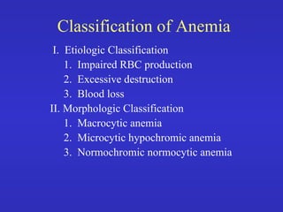
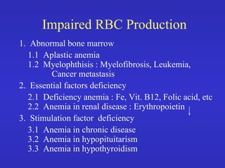
ANEMIA
By Dr Bashir Ahmed Dar
Chinki pora sopore kashmir
Associate professor of Medicine
Classification of Anemia
I. Etiologic Classification
1. Impaired RBC production
2. Excessive destruction
3. Blood loss
II. Morphologic Classification
1. Macrocytic anemia
2. Microcytic hypochromic anemia
3. Normochromic normocytic anemia
Impaired RBC Production
1. Abnormal bone marrow
1.1 Aplastic anemia
1.2 Myelophthisis : Myelofibrosis, Leukemia,
Cancer metastasis
2. Essential factors deficiency
2.1 Deficiency anemia : Fe, Vit. B12, Folic acid, etc
2.2 Anemia in renal disease : Erythropoietin
3. Stimulation factor deficiency
3.1 Anemia in chronic disease
3.2 Anemia in hypopituitarism
3.3 Anemia in hypothyroidism
Excessive Destruction of RBC(cont.)
Hemolytic anemia
1. Intracorpuscular defect
1.1 Membrane : Hereditary spherocytosis
Hereditary ovalocytosis, etc.
1.2 Enzyme : G-6PD deficiency, PK def., etc.
1.3 Hemoglobin : Thalassemia, Hemoglobino-
pathies
Excessive Destruction of RBC
2. Extracorpuscular defect
2.1 Mechanical : March hemolytic anemia
MAHA (Microangiopathic HA)
2.2 Chemical/Physical
2.3 Infection : Clostridium tetani
2.4 Antibodies : HTR, SLE
2.5 Hypersplenism
Blood Loss
1. Acute blood loss : Accident, GI bleeding
2. Chronic blood loss : Hypermenorrhea
Parasitic infestation
Macrocytic Anemia
MCV > 94
MCHC > 31
1. Megaloblastic dyspoiesis
1.1 Vit. B12 deficiency : Pernicious anemia
1.2 Folic acid deficiency : Nutritional megaloblas-
tic anemia, Sprue, Other malabsorption
1.3 Inborn errors of metabolism : Orotic aciduria,
etc.
1.4 Abnormal DNA synthesis : Chemotherapy,
Anticonvulsant, Oral contraceptives
Microcytic Hypochromic Anemia
MCV < 80
MCHC < 31
1. Fe deficiency anemia : Chronic blood loss,
Inadequate diet, Malabsorption, Increased
demand, etc.
2. Abnormal globin synthesis : Thalassemia with or
without Hemoglobinopathies
3. Abnormal porphyrin and heme synthesis :
Pyridoxine responsive anemia, etc.
4. Other abnormal Fe metabolism :
Normocytic Normochromic Anemia
MCV 82 - 92
MCHC > 30
1. Blood loss
2. Increased plasma volume : Pregnancy, Overhydration
3. Hemolytic anemia : depend on each cause
4. Hypoplastic marrow : Aplastic anemia, RBC aplasia
5. Infiltrate BM : Leukemia, Multiple myeloma,
Myelofibrosis, etc.
6. Abnormal endocrine : Hypothyroidism, Adrenal
insufficiency, etc.
7. Kidney disease / Liver disease / Cirrhosis
Hemolytic Anemia
What is Hemolysis
- Premature destruction of dead cells.
- Causes hereditary and acquired disorders.
- Hemolysis occurs at two sites:
Intravascular
- Hemolysis occurs within systemic
circulation.
- Hemoglobin is released into plasma.
- Hemoglobin is lost through kidneys or
catabolized in the liver.
Extravascular
- Trapping of red cells in spleen or liver
sinuses.
- Lyses of trapped red cells.
- Release of lysed hemoglobin and
catabolism within the sequestering organ.
Classification of Hereditary
Hemolytic Anemia
• Based on side effect:
- Metabolic defect
- Membrane defect
- Hemoglobin defect
A) Metabolic defect:
- Defect in hexose monophosphate shunt:
G-6-PD deficiency.
- Defects of glycolysis; pyrovate kinase def.,
glucose phosphate isomerase def.
- Defects in red cell nucleotide metabolism:
pyramidine-5-nucleotidase def.
B) Membrane defect:
- Heriditary spherocytosis
- Heriditary elliptocytosis
- Hereditary pyropoikilocytosis
C) Hemoglobin defect:
- Thalassemias
- Sickle cell anemia
- Hemoglobin C disease
- Hemoglobin E disease
- Unstable hemoglobin
Laboratory Findings
• Chemistry
Hyperbilirubinemia, predominantly unconjugated bilirubin
due to breakdown of heme ring by reticuloendothelial cells
in the liver.
elevated LDH: released from destroyed cells.
Hemoglobinemia: free hemoglobin level increases in
hemolysis esp. intravascular hemolysis: levels of 10-20
mg/dl gives plasma amber color and 50-100 gm/dl reddish
color.
Hemoglobinuria: red-brown color of urine due to free
hemoglobin and methamoglobin.
Decreased Heptaglobin level: it is a alpha-2-globin produced
in the liver. It binds free hemoglobin thus level is reduced in
hemolysis.
Hemosidrinuria: it reflects extensive hemolysis for a
prolonged period of time. When hemoglobin is filtered by
nephron, proximal tubular cells metabolize hemoglobin and
iron accumulate in the cells. Cells then exfoliate in the urine
and iron can be detected by Prussian blue reaction.
Drug-Induced Acute Hemolysis
• Drugs that have been linked to G6PD:
• Primaquine (an antimalarial)
• Sulphonamide antibiotics
• Sulphones (e.g. dapsone, used against leprosy)
• Other sulphur-containing drugs: glibenclamide (an anti-diabetic drug)
• Nitrofurantoin (an antibiotic often used for urinary tract infections)
• Vitamin K analogues
• Several others
• Henna can cause a hemolytic crisis in G6PD deficient infants
Acquired hemolytic anaemia
Immune haemolytic anaemias
Are caused by AB production by the body
against its own red cells.
Divided into  1) warm 37c
2) cold  4 c
Warm AIHA
• IgG alone ,Bind at 37c
Cold AIHA
• Usually IgM
• Bind to red cell at 4c
Coombs Test
Coombs Test
Antiglobulin Test
Antiglobulin Test
RBC
RBC
Red cell with
Red cell with
bound antibody to
bound antibody to
membrane antigen
membrane antigen
+
+
Anti-immunoglobulin
Anti-immunoglobulin
Coombs Test
Coombs Test
Antiglobulin Test
Antiglobulin Test
RBC
RBC RBC
RBC
Agglutination
Agglutination
Non-Immune Hemolytic Anemias
Hemolytic anaemias due to mechanisms or agents
other than antibodies +/or complement e.g.:
• Mechanical (traumatic)
Mechanical (traumatic)
• Toxins
Toxins
• Infections
Infections
• Splenomegaly (hypersplenism)
Splenomegaly (hypersplenism)
• Burn (physical)
Burn (physical)
• Renal failure and liver failure
Renal failure and liver failure
• Chemical
Chemical
Mechanical (Traumatic)
(Fragmentation)
(Fragmentation)
This is due to direct trauma (stress) to the RBCs
causing fragmentation of the RBCs & intra-
vascular hemolysis. The fragmented cells can be
seen on peripheral blood smears & are called
(schistocytes).
(schistocytes).
Due to:
Due to:
1.
1. Prosthetic valves
Prosthetic valves
2.
2. Patches
Patches
3.
3. Valvular diseasse e.g., stenosis
Valvular diseasse e.g., stenosis
Cont…
Mechanical (Traumatic)
(Fragmentation)
(cont…)
1.
1. Microangiopathic:
Microangiopathic: mechanical hemolysis due to
mechanical hemolysis due to
contact between the RBCs & the abnormal intema
of thrombosed, narrowed, necrotic small vessels or
fibrin strand formation.
Caused by many diseases e.g., DIC (disseminated
intravascular coagulation), malignant hypertension,
disseminated malignancies especially mucin
secreting adenocarcinomas, TTP
(thrombocytopenic purpura), hemolytic uremic
syndrome (HUS).
syndrome (HUS).
THE END
• THANK YOU

Anaemia classification .pdf

  • 1.
    ANEMIA By Dr BashirAhmed Dar Chinki pora sopore kashmir Associate professor of Medicine
  • 2.
    Classification of Anemia I.Etiologic Classification 1. Impaired RBC production 2. Excessive destruction 3. Blood loss II. Morphologic Classification 1. Macrocytic anemia 2. Microcytic hypochromic anemia 3. Normochromic normocytic anemia
  • 3.
    Impaired RBC Production 1.Abnormal bone marrow 1.1 Aplastic anemia 1.2 Myelophthisis : Myelofibrosis, Leukemia, Cancer metastasis 2. Essential factors deficiency 2.1 Deficiency anemia : Fe, Vit. B12, Folic acid, etc 2.2 Anemia in renal disease : Erythropoietin 3. Stimulation factor deficiency 3.1 Anemia in chronic disease 3.2 Anemia in hypopituitarism 3.3 Anemia in hypothyroidism
  • 4.
    Excessive Destruction ofRBC(cont.) Hemolytic anemia 1. Intracorpuscular defect 1.1 Membrane : Hereditary spherocytosis Hereditary ovalocytosis, etc. 1.2 Enzyme : G-6PD deficiency, PK def., etc. 1.3 Hemoglobin : Thalassemia, Hemoglobino- pathies
  • 5.
    Excessive Destruction ofRBC 2. Extracorpuscular defect 2.1 Mechanical : March hemolytic anemia MAHA (Microangiopathic HA) 2.2 Chemical/Physical 2.3 Infection : Clostridium tetani 2.4 Antibodies : HTR, SLE 2.5 Hypersplenism
  • 6.
    Blood Loss 1. Acuteblood loss : Accident, GI bleeding 2. Chronic blood loss : Hypermenorrhea Parasitic infestation
  • 7.
    Macrocytic Anemia MCV >94 MCHC > 31 1. Megaloblastic dyspoiesis 1.1 Vit. B12 deficiency : Pernicious anemia 1.2 Folic acid deficiency : Nutritional megaloblas- tic anemia, Sprue, Other malabsorption 1.3 Inborn errors of metabolism : Orotic aciduria, etc. 1.4 Abnormal DNA synthesis : Chemotherapy, Anticonvulsant, Oral contraceptives
  • 8.
    Microcytic Hypochromic Anemia MCV< 80 MCHC < 31 1. Fe deficiency anemia : Chronic blood loss, Inadequate diet, Malabsorption, Increased demand, etc. 2. Abnormal globin synthesis : Thalassemia with or without Hemoglobinopathies 3. Abnormal porphyrin and heme synthesis : Pyridoxine responsive anemia, etc. 4. Other abnormal Fe metabolism :
  • 9.
    Normocytic Normochromic Anemia MCV82 - 92 MCHC > 30 1. Blood loss 2. Increased plasma volume : Pregnancy, Overhydration 3. Hemolytic anemia : depend on each cause 4. Hypoplastic marrow : Aplastic anemia, RBC aplasia 5. Infiltrate BM : Leukemia, Multiple myeloma, Myelofibrosis, etc. 6. Abnormal endocrine : Hypothyroidism, Adrenal insufficiency, etc. 7. Kidney disease / Liver disease / Cirrhosis
  • 10.
  • 11.
    What is Hemolysis -Premature destruction of dead cells. - Causes hereditary and acquired disorders. - Hemolysis occurs at two sites:
  • 12.
    Intravascular - Hemolysis occurswithin systemic circulation. - Hemoglobin is released into plasma. - Hemoglobin is lost through kidneys or catabolized in the liver.
  • 13.
    Extravascular - Trapping ofred cells in spleen or liver sinuses. - Lyses of trapped red cells. - Release of lysed hemoglobin and catabolism within the sequestering organ.
  • 14.
    Classification of Hereditary HemolyticAnemia • Based on side effect: - Metabolic defect - Membrane defect - Hemoglobin defect
  • 15.
    A) Metabolic defect: -Defect in hexose monophosphate shunt: G-6-PD deficiency. - Defects of glycolysis; pyrovate kinase def., glucose phosphate isomerase def. - Defects in red cell nucleotide metabolism: pyramidine-5-nucleotidase def.
  • 16.
    B) Membrane defect: -Heriditary spherocytosis - Heriditary elliptocytosis - Hereditary pyropoikilocytosis
  • 17.
    C) Hemoglobin defect: -Thalassemias - Sickle cell anemia - Hemoglobin C disease - Hemoglobin E disease - Unstable hemoglobin
  • 18.
    Laboratory Findings • Chemistry Hyperbilirubinemia,predominantly unconjugated bilirubin due to breakdown of heme ring by reticuloendothelial cells in the liver. elevated LDH: released from destroyed cells. Hemoglobinemia: free hemoglobin level increases in hemolysis esp. intravascular hemolysis: levels of 10-20 mg/dl gives plasma amber color and 50-100 gm/dl reddish color.
  • 19.
    Hemoglobinuria: red-brown colorof urine due to free hemoglobin and methamoglobin. Decreased Heptaglobin level: it is a alpha-2-globin produced in the liver. It binds free hemoglobin thus level is reduced in hemolysis. Hemosidrinuria: it reflects extensive hemolysis for a prolonged period of time. When hemoglobin is filtered by nephron, proximal tubular cells metabolize hemoglobin and iron accumulate in the cells. Cells then exfoliate in the urine and iron can be detected by Prussian blue reaction.
  • 20.
    Drug-Induced Acute Hemolysis •Drugs that have been linked to G6PD: • Primaquine (an antimalarial) • Sulphonamide antibiotics • Sulphones (e.g. dapsone, used against leprosy) • Other sulphur-containing drugs: glibenclamide (an anti-diabetic drug) • Nitrofurantoin (an antibiotic often used for urinary tract infections) • Vitamin K analogues • Several others • Henna can cause a hemolytic crisis in G6PD deficient infants
  • 21.
  • 22.
    Immune haemolytic anaemias Arecaused by AB production by the body against its own red cells. Divided into  1) warm 37c 2) cold  4 c
  • 23.
    Warm AIHA • IgGalone ,Bind at 37c
  • 24.
    Cold AIHA • UsuallyIgM • Bind to red cell at 4c
  • 25.
    Coombs Test Coombs Test AntiglobulinTest Antiglobulin Test RBC RBC Red cell with Red cell with bound antibody to bound antibody to membrane antigen membrane antigen + + Anti-immunoglobulin Anti-immunoglobulin
  • 26.
    Coombs Test Coombs Test AntiglobulinTest Antiglobulin Test RBC RBC RBC RBC Agglutination Agglutination
  • 27.
    Non-Immune Hemolytic Anemias Non-ImmuneHemolytic Anemias Hemolytic anaemias due to mechanisms or agents Hemolytic anaemias due to mechanisms or agents other than antibodies +/or complement e.g.: other than antibodies +/or complement e.g.: • Mechanical (traumatic) Mechanical (traumatic) • Toxins Toxins • Infections Infections • Splenomegaly (hypersplenism) Splenomegaly (hypersplenism) • Burn (physical) Burn (physical) • Renal failure and liver failure Renal failure and liver failure • Chemical Chemical
  • 28.
    Mechanical (Traumatic) Mechanical (Traumatic) (Fragmentation) (Fragmentation) Thisis due to direct trauma (stress) to the RBCs This is due to direct trauma (stress) to the RBCs causing fragmentation of the RBCs & intra- causing fragmentation of the RBCs & intra- vascular hemolysis. The fragmented cells can be vascular hemolysis. The fragmented cells can be seen on peripheral blood smears & are called seen on peripheral blood smears & are called (schistocytes). (schistocytes). Due to: Due to: 1. 1. Prosthetic valves Prosthetic valves 2. 2. Patches Patches 3. 3. Valvular diseasse e.g., stenosis Valvular diseasse e.g., stenosis Cont…
  • 29.
    Mechanical (Traumatic) (Fragmentation) (cont…) 1. 1. Microangiopathic: Microangiopathic:mechanical hemolysis due to mechanical hemolysis due to contact between the RBCs & the abnormal intema contact between the RBCs & the abnormal intema of thrombosed, narrowed, necrotic small vessels or of thrombosed, narrowed, necrotic small vessels or fibrin strand formation. fibrin strand formation. Caused by many diseases e.g., DIC (disseminated Caused by many diseases e.g., DIC (disseminated intravascular coagulation), malignant hypertension, intravascular coagulation), malignant hypertension, disseminated malignancies especially mucin disseminated malignancies especially mucin secreting adenocarcinomas, TTP secreting adenocarcinomas, TTP (thrombocytopenic purpura), hemolytic uremic (thrombocytopenic purpura), hemolytic uremic syndrome (HUS). syndrome (HUS).
  • 30.