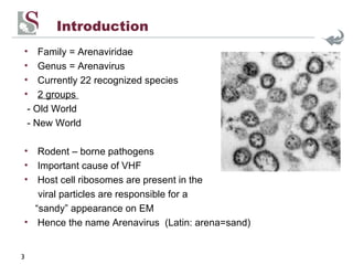
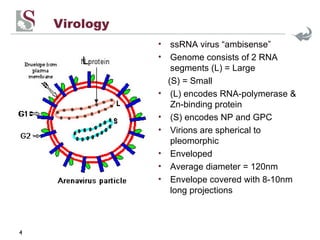
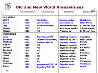
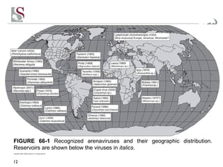
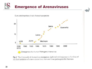
Arenaviruses Dr Preneshni R Naicker
Arenaviruses - outline Introduction Virology Classification Reservoirs Transmission Viruses and Distribution Phylogeny Clinical Presentation (Lassa Fever/LCM/S. American VHF) Laboratory Diagnosis Treatment Prevention & Control Emergence & Evolution New viruses
Introduction Family = Arenaviridae Genus = Arenavirus Currently 22 recognized species 2 groups  - Old World - New World Rodent – borne pathogens Important cause of VHF Host cell ribosomes are present in the viral particles are responsible for a “ sandy” appearance on EM Hence the name Arenavirus  (Latin: arena=sand)
Virology ssRNA virus “ambisense” Genome consists of 2 RNA segments (L) = Large (S) = Small (L) encodes RNA-polymerase & Zn-binding protein (S) encodes NP and GPC  Virions are spherical to pleomorphic Enveloped Average diameter = 120nm Envelope covered with 8-10nm long projections L
GP1 & GP2 have been shown to bind to  α -dystroglycan (cell surface glycoprotein in rodent fibroblasts) Inactivated by Temp > 56° pH < 5.5 pH > 8.5 UV or gamma radiation
2 groups Old World Lassa-LCM Serocomplex 5 Species Family:  Muridae Subfamily:  Murinae Eurasian rodents New World Tacaribe Serocomplex 17 species 3 clades (A,B,C) Family:  Muridae Subfamily:  Sigmodontinae American rodents * Classification
Rodent - mastomys sp *Fruit-eating bat – Artibeus sp Reservoirs   Usually one species, less often 2 closely related species Chronic mild infection Life long shedding of virus Except Tacaribe virus *
Co-evolution  Specific rodents are the principal hosts of arenaviruses (Childs & Peters 1993) *except Tacaribe virus – bats of  Artibeus sp The diversity of arenaviruses due to long-term, shared evolutionary relationship between  Arenaviridae  and rodents of the  Muridae  family (termed co-evolution or co-speciation) Hypothesis: Ancestral arenavirus type infection of a common rodent ancestor approx 35 million yrs ago Each rodent lineage then evolved independently with their own arenavirus resulting in the species-specific association we see today. Discrepancies? Spillover from one species or genus to another ?genomic segments exchanged
Transmission Rodent – Rodent Vertical Horizontal (aerosolized urine, faeces, saliva, bites) Rodent – Human Aerosolized secreta inoculation via cuts, bites contaminated fomites, food Rodent consumption Human – Human - contaminated secretions, sexual Inoculation
Old and New World Arenaviruses Argentina West Indies  Bolivia Brazil Paraguay USA Columbia Bolivia Brazil Venezuela Venezuela Brazil Calomys muscul. Artibeus sp (BAT) Calomys callosu. Oryzomys capito Oryzomys buccin. Sigmodon hispi. Oryzomys albig. Calomys callos. Oryzomys capit. Sigmodon alst. Zygodon brevi. unknown Argentinian VHF LI (laboratory infxn) Bolivian VHF, LI NE NE NE LI NE LI  NE Venezuelan VHF, LI Brazilian VHF, LI 1958 1963   1965 1966 1970 1970 1971 1973 1977 1977   1989 1994 NEW WORLD Junin Tacaribe  Machupo Ampari Parana Tamiami Picjinde Latino Flexal Pirital  Guanarito Sabia Worldwide West Africa C. African Rep S Central Afric C. African Rep Mus musculus Mastomys sp Arvicanthus nilot. Mastomys natale. Praomys sp Meningitis Lassa Fever NE (no evidence) NE NE 1934 1975 1970 1977 1983 OLD WORLD LCMV Lassa Ippy Mopeia Mobala Distribution Rodent Host Human disease Year  first isolated
cont. Argentina Argentina USA, Cali USA Peru Brazil Bolomys obs peromysc californ Neotoma alb. Oecomys bicolor Oryzomys capito NE NE NE ? NE NE 1996 1997 1996  1996 2001 2002 Oliveros Pampas(strain)  Bear Canyon  Whitewater Arroyo Allpahuayo Cupixi Distribution Rodent host Human disease Year first isolated
Copyright © 2006, 1999 by Elsevier Inc. All rights reserved. FIGURE 66-1  Recognized arenaviruses and their geographic distribution. Reservoirs are shown below the viruses in  italics .
Phylogeny
Lassa Fever Clinical entity in 1950s Up to 300,000 infections and 5000 deaths from LF are estimated to  occur yearly Incubation period 7-18 days Estimates of virus exposure 4% to 6% in Guinea 15% to 20% in Nigeria
Distribution of Lassa Fever Outbreaks have occurred in the   Central African Republic, Guinea, Liberia, Nigeria, and Sierra Leone;   serological evidence of human infection has been found in the   Democratic Republic of the Congo, Mali, and Senegal
Clinical presentation
Cont.
Poor Prognostic Indicators Shock Haemorrhage Neurological manifestations High viraemia AST >150 iu/l Death occurs 10-14 days after onset of symptoms Mortality Rate = 15-25% in hospitalized pts Chronic sequelae:  ⅓  sensorineural deafness Single natural infection provides long-term immunity Pregnancy: in 3rdT 20% MR and 87% fetal loss Paediatrics: “swollen baby syndrome”
Imported Lassa Fever
Lymphocytic Choriomeningitis Incubation period  = 1-2 weeks Clinical presentation Acute febrile illness Headache Aseptic meningitis Encephalitis (rare) Orchitis, myocarditis, arthritis Occasionally LAD, maculopapular rash Congenital infection: hydrocephalus, chorioretinitis and mental retardation Mortality  <1%
South American Viral Haemorrhagic Fever Incubation period  = 1-2 weeks Clinical presentation “ flu-like” prodrome Fever, headache, nausea, vomiting Thrombocytopenia, leukopenia Haemorrage: petechiae, bleeding from mucosa, puncture sites Neurological: tremors, delirium, convulsions, coma Mortality rate  = 15-30%
Laboratory Diagnosis ELISA Serum Serology (IgG/IgM) Blood, tissue RT-PCR Liver, spleen, skin, kidney Tissue Immunohistochemistry ELISA/IMF Blood Antigen detection Blood, urine, tissue EM Cell Culture: Vero, Vero B6, BHK Animal: suckling mice, hamsters, guinea pigs Blood, CSF, Tissue Culture Comments Specimen Test
Vero E6 tissue culture cell infected with an arenavirus. Image shows extracellular virus particles budding from the cell surface. Magnification approx. 12,000 times.
Treatment Supportive Immunotherapy Decreases mortality rate from 16%-1% in Junin VHF NB: 10% develop a neurological syndrome 4-6 wks later Unsuccessful in Lassa Fever Ribavirin Must be administered IV and early (within 6 days) Decreases mortality from 55% - 5% in Lassa Fever 30mg/kg LD Then 15mg/kg 6hrly for 4 days Then 7.5mg/kg 8hrly for 6 days Should be considered in any serious arenavirus infection
Prevention & Control Interrupt transmission Rodent to human eg. Rodent control in BHF -  Human to human Specimens to Laboratory workers Prophylaxis Close contacts or possible bioterrorist exposures – should not be given ribavirin, but closely monitored for fever. If fever confirmed, then ribavirin therapy should be begun expectantly
Vaccines Junin virus vaccine Candid #1 =  live attenuated vaccine. RCT efficacy >95%. Animal studies suggest protection  against Machupo but not Guanarito or Sabia. Live attenuated Mopeia virus – protects monkeys against LASV challenge LASV/Vaccinia virus vectors  Reassortment of Lassa/Mopeia
 
Emergence of Arenaviruses
Mechanisms of Virus evolution Accumulation of Point mutations Appears to be the mechanism most often responsible for virus diversity observed The best adapted virions are selected & maintained Reassortments Not in nature, Experimentally only Reassortant virus of Lassa/Mopeia – (L) segment of Mopeia and (S) segment of Lassa NB: produced by co-cultivation on Vero cells (bioterrorism) Recombination Demonstrated for 3 North American arenaviruses (WWA, Tamiami, Bear Canyon) – indicates common derivation from a recombinational event between ancestors in lineage A and B viruses
Newly discovered Arenaviruses Discovery of 7 new arenaviruses in the past 3 years 2 of these were human pathogens Bolivia Chapare Related to WWA USA Skinner Tank Related to WWA USA Catarina Closely related to Oliveros Brazil Pinhal Australia Dandenong Mastomys  sp. Strain of Mopeia Tanzania Morogoro Mus  sp. Groups with LCMV Guinea Kodoko Comment Country Virus
April 2008
Assignment to a new species A virus in the genus  Arenavirus  is assigned to a new species on the basis of a combination of  two or more  of the following characteristics: The host species The defined geographic distribution of the virus Any associated disease in humans Antigenic cross-reactivity with other members of the genus Significant divergence from other species in the genus at the amino acid level
 
Thank you

Arenaviruses

  • 1.
  • 2.
    Arenaviruses - outlineIntroduction Virology Classification Reservoirs Transmission Viruses and Distribution Phylogeny Clinical Presentation (Lassa Fever/LCM/S. American VHF) Laboratory Diagnosis Treatment Prevention & Control Emergence & Evolution New viruses
  • 3.
    Introduction Family =Arenaviridae Genus = Arenavirus Currently 22 recognized species 2 groups - Old World - New World Rodent – borne pathogens Important cause of VHF Host cell ribosomes are present in the viral particles are responsible for a “ sandy” appearance on EM Hence the name Arenavirus (Latin: arena=sand)
  • 4.
    Virology ssRNA virus“ambisense” Genome consists of 2 RNA segments (L) = Large (S) = Small (L) encodes RNA-polymerase & Zn-binding protein (S) encodes NP and GPC Virions are spherical to pleomorphic Enveloped Average diameter = 120nm Envelope covered with 8-10nm long projections L
  • 5.
    GP1 & GP2have been shown to bind to α -dystroglycan (cell surface glycoprotein in rodent fibroblasts) Inactivated by Temp > 56° pH < 5.5 pH > 8.5 UV or gamma radiation
  • 6.
    2 groups OldWorld Lassa-LCM Serocomplex 5 Species Family: Muridae Subfamily: Murinae Eurasian rodents New World Tacaribe Serocomplex 17 species 3 clades (A,B,C) Family: Muridae Subfamily: Sigmodontinae American rodents * Classification
  • 7.
    Rodent - mastomyssp *Fruit-eating bat – Artibeus sp Reservoirs Usually one species, less often 2 closely related species Chronic mild infection Life long shedding of virus Except Tacaribe virus *
  • 8.
    Co-evolution Specificrodents are the principal hosts of arenaviruses (Childs & Peters 1993) *except Tacaribe virus – bats of Artibeus sp The diversity of arenaviruses due to long-term, shared evolutionary relationship between Arenaviridae and rodents of the Muridae family (termed co-evolution or co-speciation) Hypothesis: Ancestral arenavirus type infection of a common rodent ancestor approx 35 million yrs ago Each rodent lineage then evolved independently with their own arenavirus resulting in the species-specific association we see today. Discrepancies? Spillover from one species or genus to another ?genomic segments exchanged
  • 9.
    Transmission Rodent –Rodent Vertical Horizontal (aerosolized urine, faeces, saliva, bites) Rodent – Human Aerosolized secreta inoculation via cuts, bites contaminated fomites, food Rodent consumption Human – Human - contaminated secretions, sexual Inoculation
  • 10.
    Old and NewWorld Arenaviruses Argentina West Indies Bolivia Brazil Paraguay USA Columbia Bolivia Brazil Venezuela Venezuela Brazil Calomys muscul. Artibeus sp (BAT) Calomys callosu. Oryzomys capito Oryzomys buccin. Sigmodon hispi. Oryzomys albig. Calomys callos. Oryzomys capit. Sigmodon alst. Zygodon brevi. unknown Argentinian VHF LI (laboratory infxn) Bolivian VHF, LI NE NE NE LI NE LI NE Venezuelan VHF, LI Brazilian VHF, LI 1958 1963 1965 1966 1970 1970 1971 1973 1977 1977 1989 1994 NEW WORLD Junin Tacaribe Machupo Ampari Parana Tamiami Picjinde Latino Flexal Pirital Guanarito Sabia Worldwide West Africa C. African Rep S Central Afric C. African Rep Mus musculus Mastomys sp Arvicanthus nilot. Mastomys natale. Praomys sp Meningitis Lassa Fever NE (no evidence) NE NE 1934 1975 1970 1977 1983 OLD WORLD LCMV Lassa Ippy Mopeia Mobala Distribution Rodent Host Human disease Year first isolated
  • 11.
    cont. Argentina ArgentinaUSA, Cali USA Peru Brazil Bolomys obs peromysc californ Neotoma alb. Oecomys bicolor Oryzomys capito NE NE NE ? NE NE 1996 1997 1996 1996 2001 2002 Oliveros Pampas(strain) Bear Canyon Whitewater Arroyo Allpahuayo Cupixi Distribution Rodent host Human disease Year first isolated
  • 12.
    Copyright © 2006,1999 by Elsevier Inc. All rights reserved. FIGURE 66-1 Recognized arenaviruses and their geographic distribution. Reservoirs are shown below the viruses in italics .
  • 13.
  • 14.
    Lassa Fever Clinicalentity in 1950s Up to 300,000 infections and 5000 deaths from LF are estimated to occur yearly Incubation period 7-18 days Estimates of virus exposure 4% to 6% in Guinea 15% to 20% in Nigeria
  • 15.
    Distribution of LassaFever Outbreaks have occurred in the Central African Republic, Guinea, Liberia, Nigeria, and Sierra Leone; serological evidence of human infection has been found in the Democratic Republic of the Congo, Mali, and Senegal
  • 16.
  • 17.
  • 18.
    Poor Prognostic IndicatorsShock Haemorrhage Neurological manifestations High viraemia AST >150 iu/l Death occurs 10-14 days after onset of symptoms Mortality Rate = 15-25% in hospitalized pts Chronic sequelae: ⅓ sensorineural deafness Single natural infection provides long-term immunity Pregnancy: in 3rdT 20% MR and 87% fetal loss Paediatrics: “swollen baby syndrome”
  • 19.
  • 20.
    Lymphocytic Choriomeningitis Incubationperiod = 1-2 weeks Clinical presentation Acute febrile illness Headache Aseptic meningitis Encephalitis (rare) Orchitis, myocarditis, arthritis Occasionally LAD, maculopapular rash Congenital infection: hydrocephalus, chorioretinitis and mental retardation Mortality <1%
  • 21.
    South American ViralHaemorrhagic Fever Incubation period = 1-2 weeks Clinical presentation “ flu-like” prodrome Fever, headache, nausea, vomiting Thrombocytopenia, leukopenia Haemorrage: petechiae, bleeding from mucosa, puncture sites Neurological: tremors, delirium, convulsions, coma Mortality rate = 15-30%
  • 22.
    Laboratory Diagnosis ELISASerum Serology (IgG/IgM) Blood, tissue RT-PCR Liver, spleen, skin, kidney Tissue Immunohistochemistry ELISA/IMF Blood Antigen detection Blood, urine, tissue EM Cell Culture: Vero, Vero B6, BHK Animal: suckling mice, hamsters, guinea pigs Blood, CSF, Tissue Culture Comments Specimen Test
  • 23.
    Vero E6 tissueculture cell infected with an arenavirus. Image shows extracellular virus particles budding from the cell surface. Magnification approx. 12,000 times.
  • 24.
    Treatment Supportive ImmunotherapyDecreases mortality rate from 16%-1% in Junin VHF NB: 10% develop a neurological syndrome 4-6 wks later Unsuccessful in Lassa Fever Ribavirin Must be administered IV and early (within 6 days) Decreases mortality from 55% - 5% in Lassa Fever 30mg/kg LD Then 15mg/kg 6hrly for 4 days Then 7.5mg/kg 8hrly for 6 days Should be considered in any serious arenavirus infection
  • 25.
    Prevention & ControlInterrupt transmission Rodent to human eg. Rodent control in BHF - Human to human Specimens to Laboratory workers Prophylaxis Close contacts or possible bioterrorist exposures – should not be given ribavirin, but closely monitored for fever. If fever confirmed, then ribavirin therapy should be begun expectantly
  • 26.
    Vaccines Junin virusvaccine Candid #1 = live attenuated vaccine. RCT efficacy >95%. Animal studies suggest protection against Machupo but not Guanarito or Sabia. Live attenuated Mopeia virus – protects monkeys against LASV challenge LASV/Vaccinia virus vectors Reassortment of Lassa/Mopeia
  • 27.
  • 28.
  • 29.
    Mechanisms of Virusevolution Accumulation of Point mutations Appears to be the mechanism most often responsible for virus diversity observed The best adapted virions are selected & maintained Reassortments Not in nature, Experimentally only Reassortant virus of Lassa/Mopeia – (L) segment of Mopeia and (S) segment of Lassa NB: produced by co-cultivation on Vero cells (bioterrorism) Recombination Demonstrated for 3 North American arenaviruses (WWA, Tamiami, Bear Canyon) – indicates common derivation from a recombinational event between ancestors in lineage A and B viruses
  • 30.
    Newly discovered ArenavirusesDiscovery of 7 new arenaviruses in the past 3 years 2 of these were human pathogens Bolivia Chapare Related to WWA USA Skinner Tank Related to WWA USA Catarina Closely related to Oliveros Brazil Pinhal Australia Dandenong Mastomys sp. Strain of Mopeia Tanzania Morogoro Mus sp. Groups with LCMV Guinea Kodoko Comment Country Virus
  • 31.
  • 32.
    Assignment to anew species A virus in the genus Arenavirus is assigned to a new species on the basis of a combination of two or more of the following characteristics: The host species The defined geographic distribution of the virus Any associated disease in humans Antigenic cross-reactivity with other members of the genus Significant divergence from other species in the genus at the amino acid level
  • 33.
  • 34.