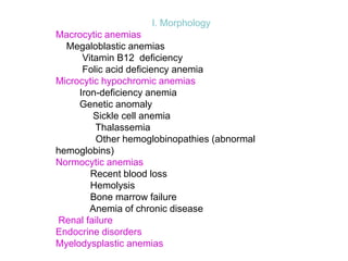
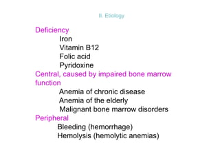
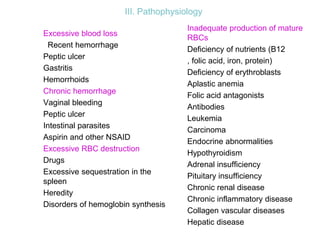
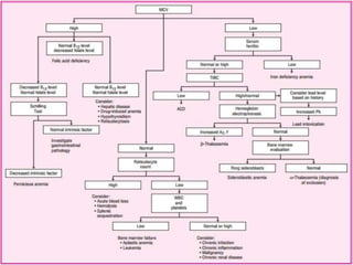
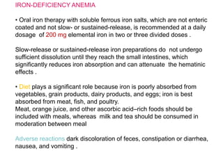
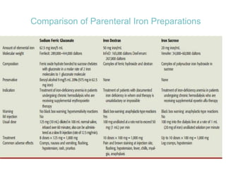
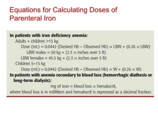
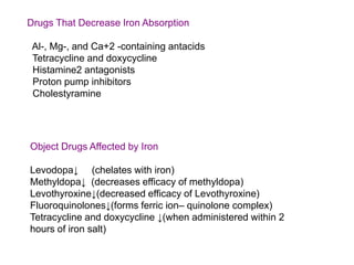
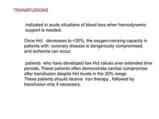
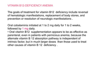
This document discusses anemia, including its definition, classification, symptoms, diagnosis, and treatment. Anemia is characterized by low hemoglobin and red blood cell counts, resulting in reduced oxygen-carrying capacity of blood. It is classified based on cell morphology, etiology, and pathophysiology. Common symptoms include fatigue, weakness, and shortness of breath. Diagnosis involves laboratory tests of hemoglobin, hematocrit, red blood cell indices, iron, vitamin B12, and folate levels. Treatment depends on the underlying cause, and may involve oral or intravenous iron supplementation for iron-deficiency anemia.
anaemia and its classification, blood transfusion, blood group, erythroblastosis foetalis, blood component , use of blood components in human diseases. blood group reaction
Iron deficiency anemia (IDA) is caused by not having enough iron available to make hemoglobin, limiting red blood cell and hemoglobin production and resulting in less oxygen delivery to tissues. IDA is common where meat intake is low and intestinal parasites are present. Symptoms include pallor, fatigue, and weakness. Studies in Saudi Arabia found IDA prevalence of 8.5-55.4% among children and 31.9-32% among pregnant women. Treatment involves iron supplementation and addressing underlying causes, while prevention focuses on iron-rich foods and supplements during pregnancy and breastfeeding.
This document discusses anemia, specifically iron deficiency anemia and megaloblastic anemia. It defines anemia and provides classifications. It then describes iron metabolism, daily iron requirements, sources of iron, and factors affecting iron absorption. It discusses the causes, clinical features, laboratory findings, and management of iron deficiency anemia and megaloblastic anemia. Key points covered include the role of vitamin B12 and folic acid in megaloblastic anemia, and morphological and etiological classifications of anemia.
Haemolytic anaemias are a group of anemias caused by the premature breakdown of red blood cells in the bloodstream or spleen. There are two main types - intrinsic defects that cause red blood cell damage from within, such as hereditary spherocytosis, and extrinsic defects that cause damage from outside factors like immune mediated hemolysis. Symptoms include anemia, jaundice, splenomegaly and gallstones. Laboratory tests show signs of increased red blood cell breakdown like elevated bilirubin and LDH, as well as signs of the bone marrow attempting to compensate with reticulocytosis and nucleated red blood cells. Intravascular hemolysis specifically causes hemoglobinemia,
Anemia Causes, Types, Symptoms, Diet, and Treatment Dr Medical
https://userupload.net/0gv9ijneu7hf
Anemia is a condition that develops when your blood lacks enough healthy red blood cells or hemoglobin. Hemoglobin is a main part of red blood cells and binds oxygen. If you have too few or abnormal red blood cells, or your hemoglobin is abnormal or low, the cells in your body will not get enough oxygen.
Anemia is a decrease in red blood cells (RBCs), hemoglobin (Hgb), or hematocrit (HCT) levels compared to normal levels for age and sex. Anemias can be classified based on RBC size and hemoglobin content as normocytic normochromic, microcytic hypochromic, or macrocytic normocytic. Common causes of anemia include iron deficiency, anemia of chronic disease, thalassemia, vitamin B12 or folate deficiency, blood loss, and aplastic anemia.
Anemia of chronic disease (ACD), also known as anemia of inflammation, is a common type of anemia associated with chronic infections, inflammatory disorders, and some cancers. It is characterized by inadequate red blood cell production, low serum iron levels, and low iron binding capacity. The anemia is usually mild to moderate in severity. Treatment involves addressing the underlying chronic condition causing the inflammation.
This document discusses megaloblastic anemia, its causes, symptoms, and treatments. It is characterized by abnormally large red blood cells due to a deficiency in vitamin B12 or folic acid, which is needed for DNA synthesis. The deficiencies can result from inadequate intake, malabsorption, or increased demand. Treatments include injections or supplements of vitamin B12, folic acid, or erythropoietin to stimulate red blood cell production. Adverse reactions are also discussed.
This presentation is about anemia of chronic disease, nowadays also called as anemia of Inflammation. I have dealt with anemia in CKD and malignancy in detail.
Megaloblastic anemia is a type of macrocytic anemia caused by a failure of DNA synthesis, resulting in asynchronous maturation of red blood cell nuclei and cytoplasm. The most common causes of megaloblastic anemia are vitamin B12 and folic acid deficiencies. Treatment involves treating the underlying deficiency, with vitamin B12 therapy for B12 deficiency and oral folic acid supplementation for folic acid deficiency. Response to treatment is monitored through improvement of hematological markers and symptoms over 1-8 weeks.
Aplastic anemia is a rare blood disorder where the bone marrow fails to produce sufficient new blood cells, leading to anemia, low white blood cell count, and low platelet count. It can be inherited or acquired due to exposure to toxins, medications, viruses, or autoimmune disorders. Symptoms include fatigue, bruising, and increased risk of infection. Treatment involves blood transfusions, immunosuppressive drugs, bone marrow transplant, or growth factors to stimulate blood cell production.
Hereditary spherocytosis is an inherited condition related to RBC destruction. its diagnosis is require to differentiate immune hemolytic anemia and G-6-P-D deficiency anemia
Hemolytic anemia is characterized by accelerated red blood cell destruction and vigorous blood regeneration. It can be classified as intrinsic or extrinsic, congenital or acquired. The site of red blood cell destruction can be intravascular or extravascular. Common causes of hemolytic anemia include hereditary spherocytosis, thalassemias, sickle cell anemia, glucose-6-phosphate dehydrogenase deficiency, paroxysmal nocturnal hemoglobinuria, and immune-mediated hemolytic anemia. Evaluation of hemolytic anemia involves determining whether the anemia is hemolytic, the site of red blood cell destruction, the etiology, and severity through blood smears, reticulocyte counts, LDH and
The document discusses anemia, including its definition, causes, types, symptoms, diagnosis, treatment, nursing care, and prevention. It provides details on iron deficiency anemia, anemia of chronic disease, thalassemia, sickle cell anemia, and the nursing process for patients with anemia. The document serves as an overview of anemia and aims to educate on this common blood disorder.
This document discusses aplastic anemia, a condition characterized by pancytopenia and bone marrow hypocellularity. It defines aplastic anemia, discusses its causes (acquired from things like radiation, drugs, viruses, immune diseases, or inherited genetic syndromes), epidemiology, pathophysiology, clinical features including symptoms and examination findings, diagnostic testing including blood tests and bone marrow biopsy, and treatment options including hematopoietic stem cell transplantation, immunosuppression, supportive care, and prognosis.
This document summarizes anemia, including its definition, classification, causes, signs and symptoms, diagnostic evaluation, and treatment. Anemia is defined as a deficiency in red blood cells, hemoglobin, or hematocrit. It is classified based on size, color, and cause of the red blood cells. Common causes include blood loss, decreased red blood cell production, and increased red blood cell breakdown. Signs and symptoms vary based on severity but can include fatigue, paleness, shortness of breath, and heart palpitations. Diagnostic testing includes complete blood count and iron studies. Treatment involves treating the underlying cause, oral or intravenous iron supplementation, blood transfusions, and medications.
The document discusses different types of anemias classified based on morphology, etiology, and pathophysiology. It describes macrocytic anemias like megaloblastic anemia caused by vitamin B12 or folate deficiency. Microcytic anemias discussed include iron deficiency anemia and sickle cell anemia. Normocytic anemias can result from blood loss, hemolysis, or bone marrow failure. Etiologies include deficiencies, impaired bone marrow function, or peripheral causes like bleeding or hemolysis. Diagnosis and treatment of various anemias like iron deficiency, vitamin B12 deficiency, and folate deficiency anemia are also summarized.
Iron deficiency anemia is a common type of microcytic anemia caused by low levels of iron available to produce hemoglobin. It affects people with high iron demands like pregnant women, young children, and menstruating women. Symptoms include fatigue, weakness, and shortness of breath. Treatment involves oral iron supplementation and dietary changes to restore iron stores. Prevention focuses on adequate iron intake through diet or supplements in at-risk groups.
Anaemia of Chronic Disease (ACD) is the most common type of anaemia seen in hospitalized patients. It develops mildly to moderately in patients with chronic infections, inflammatory diseases, cancers or kidney disease due to immune system activation. The main features of ACD include low serum iron levels, normal or elevated ferritin, and a blunted response to erythropoietin. Treatment focuses on managing the underlying condition causing chronic inflammation. For severe anaemia, erythropoiesis-stimulating agents may be used along with supplemental iron to maintain iron stores.
1. Aplastic anemia is a condition characterized by pancytopenia (low red blood cells, white blood cells, and platelets) due to bone marrow failure.
2. It can be caused by exposure to toxins, radiation, viruses, or immune system attacks on the bone marrow. The bone marrow is hypocellular with fatty replacement of hematopoietic tissue.
3. Symptoms include anemia, increased risk of infection, bruising/bleeding due to low blood cell counts. Diagnosis involves blood tests showing pancytopenia and a bone marrow biopsy revealing a hypocellular marrow. Treatment options include supportive care, immunosuppressants, bone marrow transplant, or androgens
This document discusses hemolytic anemia and focuses on sickle cell disease. It defines hemolytic anemia as increased destruction of red blood cells outside the bone marrow. Key diagnostic findings include increased reticulocyte count, hyperbilirubinemia, decreased haptoglobin, and increased lactate dehydrogenase. Hemolytic anemias are classified as hereditary defects within red blood cells or acquired external causes. Sickle cell disease results from a hereditary hemoglobinopathy and causes chronic hemolytic anemia. Complications include infections, acute chest syndrome, stroke, leg ulcers, splenic sequestration, and retinopathy. Diagnosis is made by finding sickle cells on peripheral smear and abnormal hem
This document discusses iron deficiency anemia, including its normal hemoglobin ranges, classification, etiological factors, signs and symptoms, diagnostic tests, and management. It is classified based on red blood cell morphology and etiology. Causes include low iron diet, growth spurts, GI problems, and blood loss. Diagnostic tests include hemoglobin measurement, complete blood count, peripheral smear, and iron studies. Management involves oral and parental iron therapy, blood transfusion, dietary changes including iron-rich foods, and preventive measures like exclusive breastfeeding and iron-fortified formula.
Megaloblastic anaemia is a red blood cell disorder due to the inhibition of DNA synthesis during erythropioesis.
Mitotically, the inhibition of the DNA synthesis impaires the progression of the cell cycle development from G2 to (M) stage.
Anemia - Types, Pathophysiology, Clinical Manifestations, Etiology, TreatmentMd Altamash Ahmad
Anaemia can be defined as a reduction from normal of the quantity of haemoglobin in the blood.
It is not one disease, but a condition that results from a number of different pathologies.
The World Health Organisation defines anaemia in adults as haemoglobin levels less than 13g/dL for males and less than 12g/dL for females.
The low haemoglobin level results in a corresponding decrease in the oxygen-carrying capacity of the blood.
Anaemia is possibly one of the most common conditions in the world and results in significant morbidity and mortality, particularly in the developing world.
This document provides an overview of hematopoiesis, erythropoiesis, and anemia. It discusses where blood cell formation occurs, the lifespan and production rate of red blood cells, and how hypoxia stimulates erythropoietin production. It defines anemia, lists global and country prevalence data, and compensatory mechanisms. It describes classifications of anemia including morphological and etiological, and covers causes such as blood loss, bone marrow disorders, nutritional deficiencies, and hemolytic anemias. Laboratory evaluation of anemia and peripheral blood smear findings are also summarized.
Iron deficiency anemia is the most common type of anemia worldwide. It occurs when iron levels in the body are low, preventing adequate hemoglobin production. Common causes include deficient diet, blood loss from menstruation or gastrointestinal issues, and increased needs during pregnancy or lactation. Symptoms include fatigue, palpitations, and pale skin. Diagnosis involves blood tests showing low ferritin, increased total iron-binding capacity, and transferrin saturation below 16%. Treatment focuses on oral iron supplementation, but parenteral iron may be used if oral iron is not tolerated or absorption is impaired.
This document defines anemia and describes its causes and symptoms. Anemia is a reduction in red blood cells or hemoglobin below normal levels. It can be classified based on red blood cell morphology or etiology. Common causes include blood loss, red blood cell destruction, nutritional deficiencies, and bone marrow disorders. Symptoms result from reduced oxygen carrying capacity and include fatigue, paleness, weakness, and increased heart rate. Severe anemia places greater workload on the heart and can lead to cardiac failure during exercise due to extreme tissue hypoxia.
Anemia is a condition where the hemoglobin level or red blood cell count is lower than normal. It can be caused by blood loss, decreased red blood cell production, or increased red blood cell destruction. Globally, anemia affects over 1.6 billion people. Common symptoms include fatigue, weakness, and pale skin. Diagnosis involves blood tests to measure hemoglobin, red blood cell count, and other indicators. Treatment depends on the underlying cause but may involve iron supplementation, vitamin B12/folate, blood transfusions, or treating the primary disease.
This document provides information on anemias, including their classification, red blood cell indices used in diagnosis, red blood cell morphology on peripheral smears, and descriptions of various types of anemias. It begins with definitions of anemia and classifications based on mean corpuscular volume. It then discusses various red cell indices and how they are used in differential diagnosis. The remainder of the document describes different types of anemias in more detail and abnormalities that may be seen on peripheral smear.
This document discusses anemia, including its definition, classification, and evaluation. It begins by defining anemia based on reductions in hemoglobin, hematocrit and red blood cell count. Anemia can be classified kinetically based on decreased red blood cell production, increased destruction, or blood loss. Morphologically, anemia is classified as macrocytic, microcytic, or normocytic based on mean corpuscular volume. The evaluation of a patient with anemia includes a complete blood count, reticulocyte count, peripheral smear, and further tests depending on suspected cause such as iron studies for iron deficiency anemia. Iron deficiency anemia is then discussed in detail, covering iron metabolism, stages of iron deficiency,
Iron deficiency anemia is the most common type of anemia globally. It is caused by low iron levels which prevents adequate hemoglobin production. Common symptoms include fatigue, weakness, and pale skin. Treatment involves oral or intravenous iron supplementation to replenish iron stores depending on the severity and underlying cause of the anemia. Oral iron is usually the first line treatment but intravenous iron may be needed if oral iron is not absorbed or anemia does not improve. Both forms can cause side effects but are generally safe and effective ways to treat iron deficiency anemia.
Hematologic markers such as hemoglobin (Hb), hematocrit (Ht), mean corpuscular volume (MCV), and mean corpuscular hemoglobin concentration (MCHC) provide information about red blood cell (RBC) counts, size, and hemoglobin levels. Iron deficiency anemia results in microcytic, hypochromic RBCs and low levels of serum ferritin, iron, and transferrin saturation due to insufficient iron intake or absorption to support normal hemoglobin synthesis. Clinical presentation includes pallor, fatigue, and cardiovascular symptoms. Laboratory evaluation reveals microcytic RBCs on peripheral smear along with low Hb, Ht, MCV, MCH, and iron stores.
This document discusses iron deficiency anemia. It begins by classifying anemias based on red blood cell morphology and etiology. Iron deficiency anemia is then explained in more detail. The symptoms, absorption of iron, and laboratory tests are summarized. The risks factors, treatment involving iron supplements, and prevention through an iron-rich diet are highlighted.
7 Anemia full lecture notes for preparing examRAFIULLAHRAFI14
This document defines anemia and describes methods for evaluating and classifying different types of anemia. Anemia is defined as a deficiency in the oxygen-carrying capacity of blood due to low red blood cell mass or hemoglobin. Anemia can be caused by blood loss, decreased red blood cell production, or increased red blood cell destruction. Lab tests like hemoglobin, hematocrit, red blood cell count, mean corpuscular volume, and red cell distribution width are used to classify anemia as microcytic, normocytic, or macrocytic. Specific deficiencies, diseases, and conditions are described as underlying causes of anemia. A systematic approach to evaluating a patient with anemia involves considering possible causes, reviewing lab results
Anemia is a common blood disorder in children characterized by a lack of hemoglobin in the blood, resulting in fewer red blood cells and diminished oxygen delivery to tissues. There are several types of anemia classified based on etiology including iron deficiency, megaloblastic, and sickle cell anemia. The document discusses the introduction, causes, signs and symptoms, diagnosis, and management of these different types of pediatric anemias.
The document discusses various types of anemia including their causes, symptoms, classification, and prevalence rates. It notes that according to WHO, approximately 25% of the global population, or 1.62 billion people, have anemia. In Pakistan, the prevalence of anemia is 62.3% among children aged 6-59 months, 52.1% among women aged 15-49 years, and 62.8% among pregnant women. Common types of anemia include iron deficiency anemia, anemia of chronic disease, thalassemia, and sideroblastic anemia. The document provides details on evaluating, diagnosing, and managing different anemias.
- Anemia is defined as a reduction in red blood cells (RBCs) or hemoglobin. It has many potential underlying causes and is a common complication in many disorders.
- A complete blood count provides important information about RBCs like hemoglobin levels, RBC count, MCV, and reticulocyte count. This helps classify anemias and determine if the bone marrow is responding appropriately.
- Taking a thorough history and physical exam can provide clues to the potential causes of anemia, like nutritional deficiencies, infections, hereditary disorders, medications, or other underlying illnesses. Certain physical findings may point to specific conditions like hepatosplenomegaly in hemolytic anem
This document provides an overview of anaemia, including its definition, causes, risk factors, pathophysiology, classification, clinical manifestations, diagnostic tests, medical management, and nursing care. Key points include:
- Anaemia is a reduction in red blood cells, haemoglobin, or hematocrit, causing tissue hypoxia. It can be caused by blood loss, decreased red blood cell production, or increased red blood cell destruction.
- Common causes include iron, vitamin B12, or folate deficiencies, blood loss, bone marrow disorders, and haemolytic disorders.
- Signs and symptoms vary depending on severity but can include pallor, fatigue, weakness, and shortness of breath.
- Diagnostic
This document provides an overview of anemia, including its definition, cut-off levels used to diagnose it, common causes, classification approaches, and key details about specific types like iron deficiency anemia, megaloblastic anemias, sickle cell disease, and thalassemias. It covers diagnostic testing and clinical manifestations, emphasizing the importance of considering a patient's red blood cell morphology, erythropoiesis, and underlying pathophysiology when evaluating the cause of an anemia.
1. Anaemia is caused by a reduction in red blood cells, haemoglobin, and hematocrit due to decreased red blood cell production, increased destruction, or blood loss.
2. Anaemia can be classified morphologically based on red blood cell size (MCV) and colour (MCHC) as normocytic normochromic, macrocytic normochromic, macrocytic hypochromic, or microcytic hypochromic.
3. Hemorrhagic anaemia results from blood loss and can be acute, from a sudden large loss, or chronic, from internal or external bleeding over a long period.
This document provides an overview of iron deficiency anemia, including its definition, pathophysiology, detection, and management. It begins by defining anemia and describing the various causes, including blood loss, inadequate red blood cell production, and excessive red blood cell destruction. Common signs and symptoms of iron deficiency anemia are then outlined. The document concludes by discussing the evaluation, diagnosis, and management of iron deficiency anemia through a case study, focusing on identifying risk factors, signs and symptoms, laboratory findings, and treating with iron supplementation.
Anemia is a decrease in red blood cells or hemoglobin in the blood. It impairs the body's ability to transport oxygen. There are several types of anemia based on cause, including iron deficiency, megaloblastic, pernicious, hemolytic, thalassemia, and sickle cell anemia. Anemia is diagnosed through a complete blood count. Treatment depends on the underlying cause but may involve iron supplements, vitamin B12/folate, managing the disease triggering an anemia, blood transfusions, or medication.
Vitamin B12 deficiency, also known as pernicious anemia, is an autoimmune disorder where antibodies block intrinsic factor and prevent vitamin B12 absorption from the stomach, leading to megaloblastic anemia. It presents with weakness, sore tongue, and neurological or gastrointestinal issues. Laboratory findings include macrocytic anemia, elevated MCV, hypersegmented neutrophils, and low serum B12 levels. The gold standard test is gastric biopsy showing parietal cell atrophy. Treatment is lifelong vitamin B12 supplementation by injection or oral administration.
Basavarajeeyam is a Sreshta Sangraha grantha (Compiled book ), written by Neelkanta kotturu Basavaraja Virachita. It contains 25 Prakaranas, First 24 Chapters related to Rogas& 25th to Rasadravyas.
Knee anatomy and clinical tests 2024.pdfvimalpl1234
This includes all relevant anatomy and clinical tests compiled from standard textbooks, Campbell,netter etc..It is comprehensive and best suited for orthopaedicians and orthopaedic residents.
Local Advanced Lung Cancer: Artificial Intelligence, Synergetics, Complex Sys...Oleg Kshivets
Overall life span (LS) was 1671.7±1721.6 days and cumulative 5YS reached 62.4%, 10 years – 50.4%, 20 years – 44.6%. 94 LCP lived more than 5 years without cancer (LS=2958.6±1723.6 days), 22 – more than 10 years (LS=5571±1841.8 days). 67 LCP died because of LC (LS=471.9±344 days). AT significantly improved 5YS (68% vs. 53.7%) (P=0.028 by log-rank test). Cox modeling displayed that 5YS of LCP significantly depended on: N0-N12, T3-4, blood cell circuit, cell ratio factors (ratio between cancer cells-CC and blood cells subpopulations), LC cell dynamics, recalcification time, heparin tolerance, prothrombin index, protein, AT, procedure type (P=0.000-0.031). Neural networks, genetic algorithm selection and bootstrap simulation revealed relationships between 5YS and N0-12 (rank=1), thrombocytes/CC (rank=2), segmented neutrophils/CC (3), eosinophils/CC (4), erythrocytes/CC (5), healthy cells/CC (6), lymphocytes/CC (7), stick neutrophils/CC (8), leucocytes/CC (9), monocytes/CC (10). Correct prediction of 5YS was 100% by neural networks computing (error=0.000; area under ROC curve=1.0).
Here is the updated list of Top Best Ayurvedic medicine for Gas and Indigestion and those are Gas-O-Go Syp for Dyspepsia | Lavizyme Syrup for Acidity | Yumzyme Hepatoprotective Capsules etc
ABDOMINAL TRAUMA in pediatrics part one.drhasanrajab
Abdominal trauma in pediatrics refers to injuries or damage to the abdominal organs in children. It can occur due to various causes such as falls, motor vehicle accidents, sports-related injuries, and physical abuse. Children are more vulnerable to abdominal trauma due to their unique anatomical and physiological characteristics. Signs and symptoms include abdominal pain, tenderness, distension, vomiting, and signs of shock. Diagnosis involves physical examination, imaging studies, and laboratory tests. Management depends on the severity and may involve conservative treatment or surgical intervention. Prevention is crucial in reducing the incidence of abdominal trauma in children.
share - Lions, tigers, AI and health misinformation, oh my!.pptxTina Purnat
• Pitfalls and pivots needed to use AI effectively in public health
• Evidence-based strategies to address health misinformation effectively
• Building trust with communities online and offline
• Equipping health professionals to address questions, concerns and health misinformation
• Assessing risk and mitigating harm from adverse health narratives in communities, health workforce and health system
Title: Sense of Smell
Presenter: Dr. Faiza, Assistant Professor of Physiology
Qualifications:
MBBS (Best Graduate, AIMC Lahore)
FCPS Physiology
ICMT, CHPE, DHPE (STMU)
MPH (GC University, Faisalabad)
MBA (Virtual University of Pakistan)
Learning Objectives:
Describe the primary categories of smells and the concept of odor blindness.
Explain the structure and location of the olfactory membrane and mucosa, including the types and roles of cells involved in olfaction.
Describe the pathway and mechanisms of olfactory signal transmission from the olfactory receptors to the brain.
Illustrate the biochemical cascade triggered by odorant binding to olfactory receptors, including the role of G-proteins and second messengers in generating an action potential.
Identify different types of olfactory disorders such as anosmia, hyposmia, hyperosmia, and dysosmia, including their potential causes.
Key Topics:
Olfactory Genes:
3% of the human genome accounts for olfactory genes.
400 genes for odorant receptors.
Olfactory Membrane:
Located in the superior part of the nasal cavity.
Medially: Folds downward along the superior septum.
Laterally: Folds over the superior turbinate and upper surface of the middle turbinate.
Total surface area: 5-10 square centimeters.
Olfactory Mucosa:
Olfactory Cells: Bipolar nerve cells derived from the CNS (100 million), with 4-25 olfactory cilia per cell.
Sustentacular Cells: Produce mucus and maintain ionic and molecular environment.
Basal Cells: Replace worn-out olfactory cells with an average lifespan of 1-2 months.
Bowman’s Gland: Secretes mucus.
Stimulation of Olfactory Cells:
Odorant dissolves in mucus and attaches to receptors on olfactory cilia.
Involves a cascade effect through G-proteins and second messengers, leading to depolarization and action potential generation in the olfactory nerve.
Quality of a Good Odorant:
Small (3-20 Carbon atoms), volatile, water-soluble, and lipid-soluble.
Facilitated by odorant-binding proteins in mucus.
Membrane Potential and Action Potential:
Resting membrane potential: -55mV.
Action potential frequency in the olfactory nerve increases with odorant strength.
Adaptation Towards the Sense of Smell:
Rapid adaptation within the first second, with further slow adaptation.
Psychological adaptation greater than receptor adaptation, involving feedback inhibition from the central nervous system.
Primary Sensations of Smell:
Camphoraceous, Musky, Floral, Pepperminty, Ethereal, Pungent, Putrid.
Odor Detection Threshold:
Examples: Hydrogen sulfide (0.0005 ppm), Methyl-mercaptan (0.002 ppm).
Some toxic substances are odorless at lethal concentrations.
Characteristics of Smell:
Odor blindness for single substances due to lack of appropriate receptor protein.
Behavioral and emotional influences of smell.
Transmission of Olfactory Signals:
From olfactory cells to glomeruli in the olfactory bulb, involving lateral inhibition.
Primitive, less old, and new olfactory systems with different path
NVBDCP.pptx Nation vector borne disease control programSapna Thakur
NVBDCP was launched in 2003-2004 . Vector-Borne Disease: Disease that results from an infection transmitted to humans and other animals by blood-feeding arthropods, such as mosquitoes, ticks, and fleas. Examples of vector-borne diseases include Dengue fever, West Nile Virus, Lyme disease, and malaria.
Ozempic: Preoperative Management of Patients on GLP-1 Receptor Agonists Saeid Safari
Preoperative Management of Patients on GLP-1 Receptor Agonists like Ozempic and Semiglutide
ASA GUIDELINE
NYSORA Guideline
2 Case Reports of Gastric Ultrasound
3. Anemias are a group of diseases characterized by a
decrease in hemoglobin(Hb) or the volume of red blood
cells (RBCs) , resulting in decreased oxygen-carrying
capacity of blood.
4. Anemias are generally a sign of underlying pathology;
therefore, determining the cause of the anemia is
important
Possible consequences of chronic anemia include
reduced quality-of-life, decreased survival, and increased
risk of cardiac complications, neurologic dysfunction, and
surgical complications.
Awareness of anemia, its detection, investigation, and
management must be raised.
5. Clinical presentation of anemia
■Patients may be asymptomatic or have vague
complaints.
■Patients with vitamin B12 deficiency may develop
neurologic consequences.
■In anemia of chronic disease, signs and symptoms
of the underlying disorder often overshadow those of
the anemia .
8. I. Morphology
Macrocytic anemias
Megaloblastic anemias
Vitamin B12 deficiency
Folic acid deficiency anemia
Microcytic hypochromic anemias
Iron-deficiency anemia
Genetic anomaly
Sickle cell anemia
Thalassemia
Other hemoglobinopathies (abnormal
hemoglobins)
Normocytic anemias
Recent blood loss
Hemolysis
Bone marrow failure
Anemia of chronic disease
Renal failure
Endocrine disorders
Myelodysplastic anemias
9. II. Etiology
Deficiency
Iron
Vitamin B12
Folic acid
Pyridoxine
Central, caused by impaired bone marrow
function
Anemia of chronic disease
Anemia of the elderly
Malignant bone marrow disorders
Peripheral
Bleeding (hemorrhage)
Hemolysis (hemolytic anemias)
10. III. Pathophysiology
Excessive blood loss
Recent hemorrhage
Peptic ulcer
Gastritis
Hemorrhoids
Chronic hemorrhage
Vaginal bleeding
Peptic ulcer
Intestinal parasites
Aspirin and other NSAID
Excessive RBC destruction
Drugs
Excessive sequestration in the
spleen
Heredity
Disorders of hemoglobin synthesis
Inadequate production of mature
RBCs
Deficiency of nutrients (B12
, folic acid, iron, protein)
Deficiency of erythroblasts
Aplastic anemia
Folic acid antagonists
Antibodies
Leukemia
Carcinoma
Endocrine abnormalities
Hypothyroidism
Adrenal insufficiency
Pituitary insufficiency
Chronic renal disease
Chronic inflammatory disease
Collagen vascular diseases
Hepatic disease
12. LABORATORY EVALUATION
1. RBC production failure (hypoproliferative)
2. Cell maturation ineffectiveness
3. Increase in RBC destruction
13.
14. Hemoglobin
Hb represent the amount of Hb per volume of whole
blood.
The higher values seen in males are due to stimulation
of RBC production by androgenic steroids, whereas the
lower values in females are due to decrease in Hb as a
result of blood loss during menstruation.
The Hb estimate of the oxygen-carrying capacity of
blood.
Hb levels may be diminished because of a decreased
quantity of Hb per RBC or because of a decrease in the
actual number of RBCs.
In pregnancy Hb may not reflect red cell mass
changes.
15. Hematocrit
Expressed as a percentage.
hematocrit (Hct) is the actual volume of RBCs in a
unit volume of whole blood .
abnormal cell size or shape and indicates the
pathology.
A low Hct indicates a reduction in either the number
or the size of RBCs or an increase in plasma volume.
The RBC count is an indirect
estimate of the Hb content of the
blood; it is an actual count of
RBCs per unit of blood.
16. Red Blood Cell Indices
indices describe the size and Hb content of the RBCs
and are calculated from the Hb, Hct, and RBC count.
RBC indices, such as mean corpuscular volume (MCV)
and mean corpuscular Hb (MCH), are single mean
values that do not express the variation that can occur
in cells.
.
17. Mean Corpuscular Volume (Hct/RBC Count)MCV
represents the average volume of RBCs.
It may reflect changes in MCH, but it can be confounded.
Cells are considered macrocytic if they are larger than normal,
microcytic if they are smaller than normal, and normocytic if their
size falls within normal limits.
Folic acid and vitamin B12 deficiency anemias yield macrocytic
morphology, whereas iron deficiency and thalassemia are
examples of microcytic anemias.
MCV is falsely elevated in the presence of cold agglutinins and
hyperglycemia.
When IDA (decreased MCV) is accompanied by folate deficiency
(increased MCV), failure to understand that the MCV represents an
average RBC size creates the potential for overlooking the real
cause of the anemia.
18. Mean Corpuscular Hemoglobin (Hb/RBC Count)MCH
Is defined as the volume of Hb in an RBC.
It reflects the adequacy of iron supply to developing
erythron.
Two morphologic changes, microcytosis and
hypochromia, can reduce MCH .
19. Mean Corpuscular Hemoglobin Concentration
(Hb/Hct)
The weight of Hb per volume of cells is the mean
corpuscular Hb concentration (MCHC).
Because MCHC is independent of cell size, it is more
useful than MCH in distinguishing between
microcytosis and hypochromia.
A low MCHC always indicates hypochromia; a
microcyte with a normal Hb concentration will have a
low MCH but a normal MCHC.
A decreased MCHC is seen most often in
association with IDA.
20. Total Reticulocyte Count
Although an indirect assessment, the total reticulocyte
count is an indication of new RBC production.
It measures how quickly immature RBCs
(reticulocytes) are produced by bone marrow and
released into the blood.
the reticulocyte count usually are low.
Reticulocytes circulate in the blood approximately 2
days before maturing into RBCs
21. Red Blood Cell Distribution Width
The higher the red blood cell distribution width (RDW),
the more variable the size of the RBCs.
The RDW increases in early IDA because of the release
of large, immature, nucleated RBCs to compensate for
the anemia, but this change is not specific for IDA.
The RDW also can be helpful in the diagnosis of a mixed
anemia. A patient can have a normal MCV yet have a
wide RDW.
This finding indicates the presence of microcytes and
macrocytes, which would yield a “normal” average RBC
size.
22. Serum Iron
The level of serum iron is the concentration of iron bound to
transferrin. Normally, transferrin is approximately one-third bound
(saturated) to iron.
Total Iron-Binding Capacity
An indirect measurement of the iron-binding capacity of serum
transferrin, TIBC evaluation is performed by adding an excess
of iron to plasma to saturate all transferrin with iron .
Folic Acid
The results of folic acid measurements vary depending on the
assay method used.
Vitamin B12
Low levels of vitamin B12(cyanocobalamin) indicate vitamin B12
deficiency.
23. Coombs Test
Antiglobulin tests, also called Coombs tests ,
indicate hemolytic anemia caused by an immune
response.
Schilling Test
The purpose of the rarely used Schilling urinary
excretion test is to diagnose vitamin B12 deficiency
anemia caused by a B12 absorption defect resulting
from a lack of intrinsic factor (pernicious anemia) .
25. Homocysteine
Vitamin B12 and folate both are required for conversion of
homocysteine to methionine. Increased serum homocysteine
may suggest vitamin B12 or folate deficiency.
Homocysteine levels also can be elevated in patients with
vitamin B6 deficiency, renal failure and hypothyroidism .
Methylmalonic Acid
A vitamin B12 coenzyme is needed to convert methylmalonyl
coenzyme A to succinyl coenzyme A. Patients with vitamin B12
Deficiency may have increased concentrations of serum
methylmalonic acid (MMA), which is a more specific marker for
vitamin B12 deficiency .
26. ■Hb, hematocrit (Hct), and RBC indices may remain normal early in
the disease and then decrease as the anemia progresses .
■Serum iron is low in IDA and ACD .
■Ferritin levels are low in IDA and normal to increased in ACD
■TIBC is high in IDA and is low or normal in ACD .
■Mean corpuscular volume is elevated in vitamin B12 deficiency and
folate deficiency .
■Vitamin B12 and folate levels are low in their respective types of
anemia .
■Homocysteine is elevated in vitamin B12 deficiency and folate
deficiency .
■Methylmalonic acid is elevated in vitamin B12 deficiency .
29. IRON-DEFICIENCY ANEMIA
• Oral iron therapy with soluble ferrous iron salts, which are not enteric
coated and not slow- or sustained-release, is recommended at a daily
dosage of 200 mg elemental iron in two or three divided doses .
Slow-release or sustained-release iron preparations do not undergo
sufficient dissolution until they reach the small intestines, which
significantly reduces iron absorption and can attenuate the hematinic
effects .
• Diet plays a significant role because iron is poorly absorbed from
vegetables, grain products, dairy products, and eggs; iron is best
absorbed from meat, fish, and poultry.
Meat, orange juice, and other ascorbic acid–rich foods should be
included with meals, whereas milk and tea should be consumed in
moderation between meal
Adverse reactions dark discoloration of feces, constipation or diarrhea,
nausea, and vomiting .
30. • Parenteral iron may be required for patients with iron
malabsorption, intolerance of oral iron therapy, or
noncompliance.
The replacement dose depends on etiology of anemia
and Hb concentration
• Available parenteral iron preparations have similar
efficacy but different pharmacologic, pharmacokinetic,
and safety profile.
The newer products, sodium ferric gluconateand iron
sucrose, appear to be better tolerated than iron dextran.
33. Drugs That Decrease Iron Absorption
Al-, Mg-, and Ca+2 -containing antacids
Tetracycline and doxycycline
Histamine2 antagonists
Proton pump inhibitors
Cholestyramine
Object Drugs Affected by Iron
Levodopa↓ (chelates with iron)
Methyldopa↓ (decreases efficacy of methyldopa)
Levothyroxine↓(decreased efficacy of Levothyroxine)
Fluoroquinolones↓(forms ferric ion– quinolone complex)
Tetracycline and doxycycline ↓(when administered within 2
hours of iron salt)
34. TRANSFUSIONS
indicated in acute situations of blood loss when hemodynamic
support is needed.
Once Hct decreases to <30%, the oxygen-carrying capacity in
patients with coronary disease is dangerously compromised,
and ischemia can occur.
patients who have developed low Hct values over extended time
periods, These patients often demonstrate cardiac compromise
after transfusion despite Hct levels in the 20% range.
These patients should receive iron therapy , followed by
transfusion only if necessary.
35. VITAMIN B12-DEFICIENCY ANEMIA
The goals of treatment for vitamin B12 deficiency include reversal
of hematologic manifestations, replacement of body stores, and
prevention or resolution of neurologic manifestations .
Oral cobalaminis initiated at 1 to 2 mg daily for 1 to 2 weeks,
followed by 1 mg daily.
• Oral vitamin B12 supplementation appears to be as effective as
parenteral, even in patients with pernicious anemia, because the
alternate vitamin B 12 absorption pathway is independent of
intrinsic factor, but in much larger doses than those used to treat
other causes of vitamin B 12 deficiency .
36. • Parenteral therapy is more rapid acting than oral therapy
and should be used if neurologic symptoms are present.
A popular regimen is cyanocobalamin 1,000 mcg daily for 1
week, then weekly for 1 month, and then monthly.
When symptoms resolve, daily oral administration can be
initiated.
• Adverse events are rare with vitamin B 12 therapy,
Uncommon side effects include hyperuricemia and
hypokalemia due to marked increase in potassium utilization
during production of new hematopoietic cells.
37. FOLATE-DEFICIENCY ANEMIA
folic acid deficiency include inadequate intake, decreased
absorption, hyperutilization, and inadequate utilization .
Hyperutilization of folic acid may occur when the rate of cellular
division is increased, as seen in pregnant women; patients with
hemolytic anemia, myelofibrosis, malignancy, chronic
inflammatory disorders such as Crohn disease, rheumatoid
arthritis, or psoriasis; patients undergoing long-term dialysis;
burn patients; and
in adolescents and infants during their growth spurts .
38. Dose: Oral folate1 mg daily for 4 months is usually sufficient for
treatment of folate-deficiency anemia, unless the etiology cannot
be corrected.
If malabsorption is present, the daily dose should be increased to
5 mg.
Several drugs have been reported to cause a folic acid deficiency
megaloblastic anemia. Some drugs (e.g., azathioprine, 6-
mercaptopurine, 5-fluorouracil, hydroxyurea, and zidovudine) directly
inhibit DNA synthesis.
Other drugs are folate antagonists; the most toxic is methotrexate
(other examples include trimethoprim, and triamterene).
A number of drugs (e.g., phenytoin and phenobarbital ) antagonize
folate via poorly understood mechanisms but are thought to reduce
vitamin absorption by the intestine
39. humans are unable to synthesize sufficient folate to meet total
daily requirements, they depend on dietary sources.
Major dietary sources of folate include fresh, green, leafy
vegetables, citrus fruits, yeast, mushrooms, dairy products, and
animal organs such as liver and kidney
Because the body stores approximately 5 to 10 mg of
folate, primarily in the liver, cessation of dietary folate
intake can result in megaloblastosis within 4 to 5 months.
Periconceptional folic acid supplementation is
recommended to decrease the occurrence and recurrence of
neural tube defects, specifically anencephaly and spina bifida.
41. Hemolytic anemia results in decreased survival time of
RBCs secondary to destruction in the spleen or
circulation.
Hemolytic anemias usually are normocytic and
normochromic, with increased levels of reticulocytes,
lactate dehydrogenase, and indirect bilirubin.
Treatment is directed toward correcting or controlling the
underlying pathology.
43. • Treatment of hemolytic anemia should focus on
correcting the underlying cause.
There is no specific therapy for G6PD deficiency, so
treatment consists of avoiding oxidant medications
and chemicals.
Steroids, other immunosuppressants, and even
splenectomy can be indicated to reduce RBC
destruction.
44.
45. Patients with sickle cell disease( SCD) require lifelong
multidisciplinary care.
All patients should receive regularly scheduled comprehensive
medical evaluations.
The goal of comprehensive care is to reduce hospitalizations,
complications, and mortality.
Because of the complexity of the disease, a multidisciplinary
team is needed to provide medical care, education, counseling,
and psychosocial support.
Appropriate comprehensive care can have a positive impact on
both longevity and general quality of life. This care includes the
use of traditional prophylactic and general symptomatic
supportive care and the use of newer, more specific therapies
aimed at altering hematologic capacity and function.
46. Treatment for patients with SCD involves the use of
general measures to meet the unique demands for
increased erythropoiesis.
Additional interventions can be aimed at preventing or
treating complications of the disease.
When crises occur, the type and severity of the crisis
determine the appropriate therapeutic plan .
47. Hemolysis and Vaso-occlusion
Vaso-occlusion:
Occurs when the rigid
sickle shaped cells fail to
move through the small
blood vessels, blocking
local blood flow to a
microscopic region of
tissue. Amplified many
times, these episodes
produce tissue hypoxia.
The result is pain, and
often damage to organs.
Hemolysis:
The anemia in SCD is
caused by red cell
destruction, or hemolysis,
and the degree of anemia
varies widely between
patients. The production of
red cells by the bone
marrow increases
dramatically, but is unable
to keep pace with the
destruction.
48.
49. Hemolysis and Vaso-occlusion
Acute Manifestations:
Bacterial Sepsis or
meningitis*
Recurrent vaso-occlusive
pain (dactylitis, muscoskeletal
or abdominal pain)
Splenic Sequestration*
Aplastic Crisis*
Acute Chest Syndrome*
Stroke*
Priapism
Hematuria, including papillary
necrosis
Chronic Manifestations:
Anemia
Jaundice
Splenomegaly
Cardiomegaly and functional
murmurs
Proteinemia
Cholelithiasis
Delayed growth and sexual
maturation
Restrictive lung disease*
Pulmonary Hypertension*
Avascular necrosis
Proliferative retinopathy
Leg ulcers
Transfusional hemosiderosis*
*Potential cause of mortality
50. TREATMENT
GENERAL PRINCIPLES
• Patients with SCD require lifelong multidisciplinary care.
Interventions include general measures, preventive strategies,
and treatment of complications and acute crises.
• Patients with SCD should receive routine immunizations plus
influenza, meningococcal, and pneumococcal vaccinations.
• Prophylactic penicillinis recommended for children with SCD
until they are 5 years old. Beginning at age 2 months or earlier,
the dosage is penicillin V potassium, 125 mg orally twice daily
until 3 years of age and then 250 mg twice daily until age 5
years, or benzathine penicillin, 600,000 units intramuscularly
every 4 weeks from age 6 months to 6 years.
• Folic acid, 1 mg daily, is recommended in adult patients.
51. FETAL HEMOGLOBIN (HbF) INDUCERS
Increases in HbF correlate with decreased RBC sickling
and adhesion. Patients with low HbF levels have more
frequent crises and higher mortality.
• Hydroxyurea, a chemotherapeutic agent, has many
effects on blood cells, including the stimulation of HbF
production.
It is indicated for patients with frequent painful episodes,
severe symptomatic anemia, acute chest syndrome, or
other severe vasoocclusive complications.
The dosage should be individualized based on response
and toxicity.
52. TREATMENT OF COMPLICATIONS
• Patients should be educated to recognize conditions that
require urgent evaluation.
To avoid exacerbation during acute illness, patients should
maintain balanced fluid status and oxygen saturation of at
least 92%.
• RBC transfusions are indicated for acute exacerbation of
baseline anemia (e.g., aplastic crisis, hepatic or splenic
sequestration, severe hemolysis), severe vasoocclusive
episodes, and procedures requiring general anesthesia or
ionic contrast .
53. Fever of 38.5°C (101.3°F) or higher should be evaluated
promptly. A low threshold for empiric antibiotic therapy with
coverage against encapsulated organisms is recommended
(e.g., ceftriaxonefor outpatients and cefotaximefor inpatients).
• Patients with acute chest syndrome should receive incentive
spirometry; appropriate fluid therapy; broad-spectrum
antibiotics including a macrolideor quinolone; and, for hypoxia
or acute distress, oxygen therapy.
Steroids and nitric oxide are being evaluated.
• Priapism has been treated with analgesics, antianxiety
agents, and vasoconstrictors to force blood out of the corpus
cavernosum (e.g., epinephrine), and vasodilators to relax
smooth muscle (e.g., hydralazine).
54. TREATMENT OF SICKLE CELL CRISIS
• Treatment is primarily supportive. Blood transfusions may be indicated for
severe or symptomatic anemia. Antibiotic therapy is not warranted because the
most common etiology is viral, not bacterial, infection.
• Treatment options for splenic sequestrationinclude observation alone,
especially for adults because they tend to have milder episodes; chronic
transfusion to delay splenectomy; and splenectomy after a life-threatening
crisis, after repetitive episodes, or for chronic hypersplenism.
• Hydration and analgesics are the mainstays of treatment for vasoocclusive
(painful) crisis. Fluid replacement should be 1.5 times the maintenance
requirement, can be administered IV or orally, and should be monitored to avoid
volume overload. An infectious etiology should be considered; if appropriate,
empiric therapy should be initiated.
55. • Mild to moderate pain should be treated with nonsteroidal
antiinflammatory drugs or acetaminophen.
• Severe pain should be treated aggressively with an opioid, such as
morphine, hydromorphone, fentanyl,and methadone. Moderate pain
should be treated with a weak opioid, such as codeineor
hydrocodone.
• Severe pain should be treated with an IV opioid titrated to pain
relief and then administered on a scheduled basis with as-needed
dosing for breakthrough pain. Patient-controlled analgesia is
commonly utilized.
• Suspicion of addiction commonly leads to suboptimal pain control.
Factors that minimize dependence include aggressive pain control,
frequent monitoring, and tapering medication according to response.
57. A hereditary condition, drug-induced oxidative hemolytic anemia,
most often accompanies a glucose-6-phosphate dehydrogenase
(G6PD) enzyme deficiency, but it can occur because of other
enzyme defects .
A G6PD deficiency is a disorder of the hexose monophosphate
shunt, which is responsible for producing NADPH in RBCs,
which in turn keeps glutathione in a reduced state.
Reduced glutathione is a substrate for glutathione peroxidase,
an enzyme that removes peroxide from RBCs, thus protecting
them from oxidative stress.
Without reduced glutathione, oxidative drugs can oxidize
the sulfhydryl groups of hemoglobin, removing them prematurely
from the circulation (i.e., causing hemolysis).
58. Although severe hemolysis is rare, any drug that places
oxidative stress on RBCs can cause drug-induced
oxidative hemolytic anemia.
One case of drug-induced oxidative hemolytic anemia
has been reported in a child when dapsone (an oxidizing
agent) was transferred through the breast milk of the
mother, who was taking the drug.
59.
60. ANEMIA OF CHRONIC DISEASE
Anemia of chronic disease is a diagnosis of exclusion, It results
from chronic inflammation, infection, or malignancy and can occur as
early as 1 to 2 months after the onset of these processes.
The serum iron level usually is decreased, but in contrast to IDA , the
serum ferritin concentration is normal or increased and TIBC is normal
or decreased.
• Treatment of anemia of chronic disease is less specific than that of
other anemias and should focus on correcting reversible causes
. Iron therapy is not effective when inflammation is present.
RBC transfusions are effective but should be limited to episodes
of inadequate oxygen transport and Hb of 8 to 10 g/dL .
61. • Epoetin alfa (recompinant human erythropoitin)can be considered,
especially if cardiovascular status is compromised, but the
response can be impaired in patients with anemia of chronic
disease (off-label use).
The initial dosage is 50 to 100 units/kg three times weekly.
If Hb does not increase after 6 to 8 weeks, the dosage can be
increased to 150 units/kg three times weekly.
• Epoetin alfa is usually well tolerated. The hypertension seen in
patients with end-stage kidney disease is less common in patients
with acquired immune deficiency syndrome.
62. Diseases Causing Anemia of Chronic
Disease
Less common causes
Alcoholic liver disease
Congestive heart failure
Thrombophlebitis
Chronic obstructive pulmonary
disease
Ischemic heart disease
Common causes
Chronic infections
Tuberculosis
HIV
Subacute bacterial endocarditis
Osteomyelitis
Chronic UTI
Chronic inflammation
Rheumatoid arthritis
SLE
Inflammatory bowel disease
Inflammatory osteoarthritis
Gout
Chronic inflammatory liver diseases
Malignancies
63. OTHER TYPES OF ANEMIAS
• Patients with other types of anemias require appropriate
supplementation depending on the etiology of anemia.
• In patients with anemia of critical illness, parenteral iron is often utilized
but is associated with a theoretical risk of infection.
Routine use of epoetin alfa or RBC transfusions is not supported by
clinical studies.
• Anemia of prematurity is usually treated with RBC transfusions. The
use of epoetin is controversial.
• In the pediatric population, the daily dose of elemental iron,
administered as iron sulfate, is 3 mg/kg for infants and 6 mg/kg for older
children for 4 weeks.
If response is seen, iron should be continued for 2 to 3 months. The
dose and schedule of vitamin B 12 should be titrated according to
clinical and laboratory response.
The daily dose of folate is 1 to 3 mg.
64. EVALUATION OF THERAPEUTIC OUTCOMES
In iron-deficiency anemia, iron therapy should cause reticulocytosis
in 5 to 7 days and raise Hb by 2 to 4 g/dL every 3 weeks.
The patient should be reevaluated if reticulocytosis does not occur
or if Hb does not increase by 2 g/dL within 3 weeks.
Iron therapy is continued until iron stores are replenished, which
usually requires at least 3 to 6 months.
bleeding may require iron replacement therapy
for only 1 month after correction of the underlying lesion.
65. In megaloblastic anemia, signs and symptoms usually improve
within a few days after starting vitamin B12 or folate therapy.
Neurologic symptoms can take longer to improve or can be
irreversible, but they should not progress during therapy.
Reticulocytosis should occur within 2 to 5 days.
A week after starting vitamin B12 therapy, Hb should rise and
leukocyte and platelet counts should normalize .
A CBC count and serum cobalamin level usually are drawn 1 to 2
months after initiation of therapy and 3 to 6 months thereafter for
surveillance monitoring .
66. Slow response to therapy or failure to observe
normalization of laboratory results may suggest the
presence of an additional abnormality such as iron
deficiency, thalassemia trait, infection, malignancy, or
misdiagnosis .
67. Folic acid
Symptomatic improvement, as evidenced by increased alertness,
appetite, and cooperation, often occurs early during the course of
treatment.
Reticulocytosis occurs within 2 to 3 days and peaks
within 5 to 8 days after beginning therapy. Hct begins to rise within 2
weeks and should reach normal levels within 2 months.
MCV initially increases because of an increase in reticulocytes but
gradually decreases to normal .
68. In anemia of chronic disease, reticulocytosis should occur a few
days after starting epoetin alfa therapy.
Iron, TIBC, transferrin saturation, or ferritin levels should be
monitored periodically because iron depletion is a major reason for
treatment failure.
If clinical response does not occur by 8 weeks, epoetin should be
discontinued.