Dr .Touqeer Ahmed Siddiqui

 Epidemiology,
 Clinical manifestations,
 Diagnosis and staging,
 Treatment and prognosis.
Lay out

INTRODUCTION

INTRODUCTION
Ampullary carcinomas are
defined as those that arise
within the ampullary
complex, distal to the
confluence of the distal
common bile duct and the
pancreatic duct .

 Difficult to distinguish a primary ampullary carcinoma
from other periampullary tumors preoperatively.
 Prognosis better for ampullary carcinomas.
INTRODUCTION

 Neoplastic transformation more commonly near the
ampulla than at any other site in the small intestine.
 Both benign and malignant ampullary tumors can
occur sporadically or in the setting of a genetic
syndrome
 average age at diagnosis of sporadic ampullary
carcinomas is 60 to 70 years old
EPIDEMIOLOGY AND BIOLOGIC BEHAVIOR

 The histology of primary ampullary neoplasms more often
resembles that of adenomas and adenocarcinomas of
intestinal origin rather than pancreaticobiliary origin.
 In one study of 170 ampullary carcinomas, the most
common histologic subtype was
 intestinal (47 percent), followed by pancreatobiliary (24
percent),
 poorly differentiated adenocarcinomas (13 percent),
intestinal-mucinous (8 percent), and
 invasive papillary (5 percent)
EPIDEMIOLOGY AND BIOLOGIC BEHAVIOR

 K-ras mutations are an early event in ampullary
carcinogenesis, with an incidence (37 percent) that is
similar to that in colon cancer (up to 50 percent) .
 Expression profiling of cyclooxygenase-2 (COX-2) by
ampullary carcinomas is more consistent with a neoplasm
of intestinal origin than pancreaticobiliary origin.
 High COX-2 expression has been detected in 78 percent
of ampullary carcinomas
EPIDEMIOLOGY AND BIOLOGIC BEHAVIOR

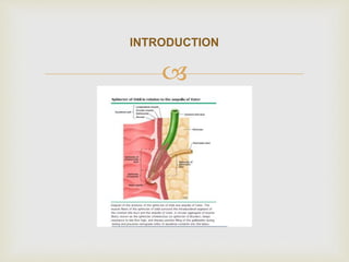
 Subdividing adenocarcinomas of the ampulla of Vater according to
histologic subtype and immunohistochemical staining pattern into
distinct subsets with differing biologic behavior has prognostic
importance.
 In a retrospective study of 208 patients treated for ampullary
adenocarcinoma in Sydney, Australia,
 those with a histomolecular pancreaticobiliary phenotype (CDX-
negative, MUC1 positive) had a significantly worse outcome
 than did those with an intestinal phenotype (CDX-positive, MUC1-
negative), with median survival of 16 versus 116 months [22].
EPIDEMIOLOGY AND BIOLOGIC BEHAVIOR
 When histomolecular phenotype was combined with the lymph node status, three
subsets of ampullary adenocarcinomas emerged with significantly different survival
outcomes:
 Patients with a node-negative, non-pancreaticobiliary histomolecular phenotype
tumor had an excellent prognosis (five-year survival 88 percent).
 Patients with a node-positive pancreaticobiliary phenotype had a poor prognosis
(five-year survival 20 percent).
 The remaining patients (node-positive non-pancreaticobiliary phenotype, node-
negative pancreaticobiliary phenotype) had an intermediate prognosis (five-year
survival 47 percent).
 The results were comparable in two additional independent cohorts of 90 patients
from Glasgow, Scotland, and 46 from Verona, Italy
EPIDEMIOLOGY AND BIOLOGIC BEHAVIOR
 Most common presenting symptom of ampullary carcinoma is obstructive jaundice
(80 percent)
 Additional symptoms may include diarrhea due to fat malabsorption (steatorrhea),
mild weight loss, and fatigue.
 Up to one-third of patients have chronic, frequently occult gastrointestinal blood loss
with an associated microcytic anemia or heme-positive stools.
 occasionally present with frank bleeding due to sloughing of the tumor, a condition
exacerbated by the use of antiplatelet agents such as aspirin and clopidogrel.
 In one report, nonspecific symptoms include abdominal pain (45 percent), fever (45
percent), mild nausea, and dyspepsia [25].
 Large lesions may produce gastric outlet obstruction associated with severe nausea
and vomiting
CLINICAL MANIFESTATIONS

 The most commonly used staging system is the tumor-
node-metastasis (TNM) system of the combined AJCC
(American Joint Committee
on Cancer)/UICC (International Union Against Cancer)
 In the absence of metastases, the prognosis of an
ampullary carcinoma depends primarily upon two factors:
 the degree of local tumor invasion, as reflected by the T
stage, and the presence of lymphatic spread, as reflected
by the N stage
DIAGNOSIS AND STAGING


 The diagnostic evaluation of a jaundiced patient with
a suspected malignant bile duct obstruction is
designed to
 eliminate benign tumors or gallstones from the
differential,
 to establish the extent of tumor invasion and spread.
DIAGNOSIS AND STAGING

 A transabdominal ultrasound (US) is a reasonable first test in patients
presenting with obstructive jaundice, but it will generally not show the
tumor.
 Helical computed tomography (CT) scanning should be obtained to
visualize the pancreas and surrounding structures.
 Although its spatial resolution is inadequate to determine the degree of
local tumor invasion, it is the most useful test to exclude the presence
of distant metastases.

 Endoscopic retrograde cholangiopancreatography (ERCP) is the single
most useful endoscopic study since it permits identification of the
tumor, biopsy, and decompression, if needed.
 While endoscopic ultrasonography (EUS) is as sensitive as ERCP and
superior to CT and transabdominal US for detecting small ampullary
tumors, it is typically not required for diagnosis.
 It may have a role in preoperative staging but may result in over-
staging.
 As a result, we do not routinely employ EUS for the diagnosis and
staging of ampullary carcinoma.

 Liver biochemical tests —
 Blood chemistries cannot establish the diagnosis of ampullary
carcinoma, but may reflect the presence of cholestasis when an
ampullary neoplasm results in partial or complete biliary
obstruction.
 Patients generally have a cholestatic pattern of liver
biochemical test abnormalities, although aminotransferases
may also be elevated .
 The prothrombin time may be elevated due to impaired
absorption of fat-soluble vitamins including vitamin K .

 Serum tumor markers —
 not specific for ampullary carcinomas and have
limited diagnostic application.
 Nevertheless, some ampullary cancers are
associated with increased serum levels of
carbohydrate antigen CA 19-
9 and/or carcinoembryonic antigen (CEA) , and serial
assay of these tumor markers may be useful for
posttreatment follow-up.

 The outcome of resected ampullary cancer depends
upon
 1. the extent of local invasion,
 2. status of the surgical margins,
 3. the presence or absence of nodal metastases
PROGNOSIS —

 Histology
 Nodal metastases
 Obstructive jaundice
 Intraoperative blood transfusion
 Tumor marker elevation
Prognostic factors

 It can be difficult to distinguish a primary ampullary carcinoma
from other periampullary tumors (mainly pancreatic carcinoma or
distal cholangiocarcinoma) preoperatively.
 However, true ampullary cancers have a better prognosis than
pancreatic head cancers or distal cholangiocarcinomas.
 Resectability rates are higher,
 five-year survival rates are 30 to 50 percent in selected patients
with limited lymph node involvement.
 Thus, an aggressive approach to diagnosis and treatment of
periampullary tumors is needed to ensure that patients with these
comparatively favorable cancers are treated optimally.
Treatment

 TREATMENT FOR LOCALIZED DISEASE — The
only potentially curative treatment for ampullary
carcinoma is surgical resection. Complete tumor
resection with negative margins (R0 resection) is a
prerequisite for cure.
 Pancreaticoduodenectomy —
Pancreaticoduodenectomy (Whipple operation) is
considered the standard approach for ampullary
cancer

 Local resection — Many patients with ampullary
cancer are elderly and have significant comorbidities.
This has generated interest in less aggressive
surgical options, such as local resection or
ampullectomy for selected patients
 In the aggregate, the available data indicate that
local resection is associated with lower morbidity
than pancreaticoduodenectomy, but at the expense
of higher recurrence rates and inferior survival, at
least in the setting of invasive disease

 Early, low-grade tumors — In contrast to
pancreaticoduodenectomy, ampullectomy does not
accomplish removal of the regional lymph nodes.
 Because of the low rate of nodal metastases (less
than 4 percent in most series), some have suggested
that local resection is a reasonable approach for
well-differentiated small (<6 mm) tumors that do not
penetrate through the ampullary musculature (ie, Tis,
T1, .

 Minimally-invasive nonsurgical therapies —
Minimally invasive nonsurgical therapies for
ampullary carcinoma include endoscopic snare
resection, Nd:YAG laser ablation, and photodynamic
therapy. The literature on these techniques in the
setting of ampullary carcinoma is limited to single-
case reports and small series.

 Endoscopic snare resection (papillectomy) is an effective means of treating
ampullary adenomas.
 Endoscopic papillectomy has been attempted in early stage (Tis, T1) well
differentiated ampullary cancers without angiolymphatic invasion
 Endoscopic debulking has been used mainly preoperatively to permit stent
insertion and decompression of the biliary tree.
 Laser ablation offers the potential for control of local tumor growth in
patients who are unfit for more aggressive therapy. In one such series of
12 patients with ampullary cancer, duodenal obstruction was relieved in
one, and the longest survival was 36 months (median 21 months) [45].
 ●Compared with laser ablation, photodynamic therapy (PDT) eradicates
local tumor with less surrounding tissue destruction. PDT uses a
photosensitizing drug (a hematoporphyrin derivative, Photofrin®), which is
disproportionately retained by malignant tissue after intravenous
administration.
 Pancreaticoduodenectomy rather than local resection for most patients with invasive
ampullary carcinomas (Grade 1B).
 Local ampullary excision rather than pancreaticoduodenectomy for patients with
noninvasive ampullary tumors.
Ampullectomy is also a reasonable approach for poor surgical candidates who have
a well-differentiated T1 tumor that is less than 6 mm in size (based upon endoscopic
ultrasound [EUS]).
 However, a more aggressive surgical approach is preferred for patients who are
candidates for pancreaticoduodenectomy because of better outcomes.
 Nonsurgical treatment modalities (ie, endoscopic snare resection, laser ablation,
photodynamic therapy) provide palliative rather than curative benefit for patients with
ampullary carcinoma.
 These methods should be restricted to patients who are not operative candidates
and those who refuse surgery.
Primary treatment

 There is no consensus regarding the optimal management of
patients after resection of an ampullary adenocarcinoma.
 There are scant data to guide adjuvant treatment decisions, and
the true benefit of such therapy remains uncertain.
 Nevertheless, many clinicians, treat these patients in a similar
manner as those with resected pancreatic head
adenocarcinomas.
 Offer adjuvant therapy to all patients with resected ampullary
cancer stage IB or higher
Adjuvant therapy —

European clinicians
 ESPAC-1 trial, showed that 5-FU-containing chemotherapy (but not
chemoradiotherapy) prolongs survival in resected pancreatic cancer.
 German CONKO trial showing a survival benefit from
adjuvant gemcitabine in the same patient population,
 preliminary report of the ESPAC-3 trial, suggesting a potentially clinically
meaningful but statistically insignificant improvement in overall survival with
adjuvant gemcitabine or leucovorin-modulated 5-FU in ampullary cancer,
 most use chemotherapy alone after resection of an ampullary neoplasm.
The approach differs in Europe and in the United
States:

 The American approach more often includes
chemoradiotherapy as well as adjuvant
chemotherapy.
The approach differs in Europe and in the United
States:

 Recommended approach
 •Eligible patients should be encouraged to enroll in clinical trials evaluating
the potential benefits of chemotherapy and/or chemoradiotherapy as well
as new therapies.
 •Off-protocol, combination of concurrent chemoradiotherapy and
chemotherapy for patients with resected ampullary cancers stage IB or
higher ,rather than observation alone (Grade 2C).
 During the concurrent chemoradiotherapy portion, prefer infusional 5-FU,
and use gemcitabine alone for the chemotherapy portion.
 The optimal way to sequence 5-FU-based chemoradiotherapy
and gemcitabine chemotherapy is unclear.
 An acceptable regimen is that used in the RTOG 9704 trial , which consists of three
weekly doses of gemcitabine alone (1000 mg/m2 per week), followed by
chemoradiotherapy using concurrent infusional 5-FU.
 Although the RTOG trial used 250 mg/m2 daily continuously, many clinicians,
consider this too toxic and use 225 mg/m2 daily, five days per week.
 Following chemoradiotherapy and starting three to five weeks later, three months of
single agent gemcitabine are given.
 An alternative approach that is often used for pancreatic cancer is to administer all
six months of chemotherapy upfront and to pursue concurrent fluoropyrimidine-based
chemoradiotherapy only for patients who remain free of metastatic disease.

 Distinction as to whether periampullary tumors are of intestinal,
biliary, or pancreatic origin is particularly important in patients
with metastatic disease as the approach differs.
 The optimal regimen for systemic therapy of true ampullary
tumors has not been established.
 Based upon the results of the ABC trial, the combination
of gemcitabine plus cisplatin is a reasonable approach for
patients who are able to tolerate it.
 Others use a single agent gemcitabine initially ,followed by an
oxaliplatin-based regimen, or vice versa, in a manner similar to
treatment of pancreatic cancer.
Metastatic disease

 The results of clinical trials are not definitive, and
there is no consensus regarding the optimal
management of patients after resection of an
ampullary cancer.
 Recommendations for management of ampullary
carcinoma are not included in published guidelines
from either the NCCN or ESMO .

Layout

Ampullary carcinoma

  • 1.
  • 2.
      Epidemiology,  Clinicalmanifestations,  Diagnosis and staging,  Treatment and prognosis. Lay out
  • 3.
  • 4.
     INTRODUCTION Ampullary carcinomas are definedas those that arise within the ampullary complex, distal to the confluence of the distal common bile duct and the pancreatic duct .
  • 5.
      Difficult todistinguish a primary ampullary carcinoma from other periampullary tumors preoperatively.  Prognosis better for ampullary carcinomas. INTRODUCTION
  • 6.
      Neoplastic transformationmore commonly near the ampulla than at any other site in the small intestine.  Both benign and malignant ampullary tumors can occur sporadically or in the setting of a genetic syndrome  average age at diagnosis of sporadic ampullary carcinomas is 60 to 70 years old EPIDEMIOLOGY AND BIOLOGIC BEHAVIOR
  • 7.
      The histologyof primary ampullary neoplasms more often resembles that of adenomas and adenocarcinomas of intestinal origin rather than pancreaticobiliary origin.  In one study of 170 ampullary carcinomas, the most common histologic subtype was  intestinal (47 percent), followed by pancreatobiliary (24 percent),  poorly differentiated adenocarcinomas (13 percent), intestinal-mucinous (8 percent), and  invasive papillary (5 percent) EPIDEMIOLOGY AND BIOLOGIC BEHAVIOR
  • 8.
      K-ras mutationsare an early event in ampullary carcinogenesis, with an incidence (37 percent) that is similar to that in colon cancer (up to 50 percent) .  Expression profiling of cyclooxygenase-2 (COX-2) by ampullary carcinomas is more consistent with a neoplasm of intestinal origin than pancreaticobiliary origin.  High COX-2 expression has been detected in 78 percent of ampullary carcinomas EPIDEMIOLOGY AND BIOLOGIC BEHAVIOR
  • 9.
      Subdividing adenocarcinomasof the ampulla of Vater according to histologic subtype and immunohistochemical staining pattern into distinct subsets with differing biologic behavior has prognostic importance.  In a retrospective study of 208 patients treated for ampullary adenocarcinoma in Sydney, Australia,  those with a histomolecular pancreaticobiliary phenotype (CDX- negative, MUC1 positive) had a significantly worse outcome  than did those with an intestinal phenotype (CDX-positive, MUC1- negative), with median survival of 16 versus 116 months [22]. EPIDEMIOLOGY AND BIOLOGIC BEHAVIOR
  • 10.
     When histomolecularphenotype was combined with the lymph node status, three subsets of ampullary adenocarcinomas emerged with significantly different survival outcomes:  Patients with a node-negative, non-pancreaticobiliary histomolecular phenotype tumor had an excellent prognosis (five-year survival 88 percent).  Patients with a node-positive pancreaticobiliary phenotype had a poor prognosis (five-year survival 20 percent).  The remaining patients (node-positive non-pancreaticobiliary phenotype, node- negative pancreaticobiliary phenotype) had an intermediate prognosis (five-year survival 47 percent).  The results were comparable in two additional independent cohorts of 90 patients from Glasgow, Scotland, and 46 from Verona, Italy EPIDEMIOLOGY AND BIOLOGIC BEHAVIOR
  • 11.
     Most commonpresenting symptom of ampullary carcinoma is obstructive jaundice (80 percent)  Additional symptoms may include diarrhea due to fat malabsorption (steatorrhea), mild weight loss, and fatigue.  Up to one-third of patients have chronic, frequently occult gastrointestinal blood loss with an associated microcytic anemia or heme-positive stools.  occasionally present with frank bleeding due to sloughing of the tumor, a condition exacerbated by the use of antiplatelet agents such as aspirin and clopidogrel.  In one report, nonspecific symptoms include abdominal pain (45 percent), fever (45 percent), mild nausea, and dyspepsia [25].  Large lesions may produce gastric outlet obstruction associated with severe nausea and vomiting CLINICAL MANIFESTATIONS
  • 12.
      The mostcommonly used staging system is the tumor- node-metastasis (TNM) system of the combined AJCC (American Joint Committee on Cancer)/UICC (International Union Against Cancer)  In the absence of metastases, the prognosis of an ampullary carcinoma depends primarily upon two factors:  the degree of local tumor invasion, as reflected by the T stage, and the presence of lymphatic spread, as reflected by the N stage DIAGNOSIS AND STAGING
  • 13.
  • 14.
      The diagnosticevaluation of a jaundiced patient with a suspected malignant bile duct obstruction is designed to  eliminate benign tumors or gallstones from the differential,  to establish the extent of tumor invasion and spread. DIAGNOSIS AND STAGING
  • 15.
      A transabdominalultrasound (US) is a reasonable first test in patients presenting with obstructive jaundice, but it will generally not show the tumor.  Helical computed tomography (CT) scanning should be obtained to visualize the pancreas and surrounding structures.  Although its spatial resolution is inadequate to determine the degree of local tumor invasion, it is the most useful test to exclude the presence of distant metastases.
  • 16.
      Endoscopic retrogradecholangiopancreatography (ERCP) is the single most useful endoscopic study since it permits identification of the tumor, biopsy, and decompression, if needed.  While endoscopic ultrasonography (EUS) is as sensitive as ERCP and superior to CT and transabdominal US for detecting small ampullary tumors, it is typically not required for diagnosis.  It may have a role in preoperative staging but may result in over- staging.  As a result, we do not routinely employ EUS for the diagnosis and staging of ampullary carcinoma.
  • 17.
      Liver biochemicaltests —  Blood chemistries cannot establish the diagnosis of ampullary carcinoma, but may reflect the presence of cholestasis when an ampullary neoplasm results in partial or complete biliary obstruction.  Patients generally have a cholestatic pattern of liver biochemical test abnormalities, although aminotransferases may also be elevated .  The prothrombin time may be elevated due to impaired absorption of fat-soluble vitamins including vitamin K .
  • 18.
      Serum tumormarkers —  not specific for ampullary carcinomas and have limited diagnostic application.  Nevertheless, some ampullary cancers are associated with increased serum levels of carbohydrate antigen CA 19- 9 and/or carcinoembryonic antigen (CEA) , and serial assay of these tumor markers may be useful for posttreatment follow-up.
  • 19.
      The outcomeof resected ampullary cancer depends upon  1. the extent of local invasion,  2. status of the surgical margins,  3. the presence or absence of nodal metastases PROGNOSIS —
  • 20.
      Histology  Nodalmetastases  Obstructive jaundice  Intraoperative blood transfusion  Tumor marker elevation Prognostic factors
  • 21.
      It canbe difficult to distinguish a primary ampullary carcinoma from other periampullary tumors (mainly pancreatic carcinoma or distal cholangiocarcinoma) preoperatively.  However, true ampullary cancers have a better prognosis than pancreatic head cancers or distal cholangiocarcinomas.  Resectability rates are higher,  five-year survival rates are 30 to 50 percent in selected patients with limited lymph node involvement.  Thus, an aggressive approach to diagnosis and treatment of periampullary tumors is needed to ensure that patients with these comparatively favorable cancers are treated optimally. Treatment
  • 22.
      TREATMENT FORLOCALIZED DISEASE — The only potentially curative treatment for ampullary carcinoma is surgical resection. Complete tumor resection with negative margins (R0 resection) is a prerequisite for cure.  Pancreaticoduodenectomy — Pancreaticoduodenectomy (Whipple operation) is considered the standard approach for ampullary cancer
  • 23.
      Local resection— Many patients with ampullary cancer are elderly and have significant comorbidities. This has generated interest in less aggressive surgical options, such as local resection or ampullectomy for selected patients  In the aggregate, the available data indicate that local resection is associated with lower morbidity than pancreaticoduodenectomy, but at the expense of higher recurrence rates and inferior survival, at least in the setting of invasive disease
  • 24.
      Early, low-gradetumors — In contrast to pancreaticoduodenectomy, ampullectomy does not accomplish removal of the regional lymph nodes.  Because of the low rate of nodal metastases (less than 4 percent in most series), some have suggested that local resection is a reasonable approach for well-differentiated small (<6 mm) tumors that do not penetrate through the ampullary musculature (ie, Tis, T1, .
  • 25.
      Minimally-invasive nonsurgicaltherapies — Minimally invasive nonsurgical therapies for ampullary carcinoma include endoscopic snare resection, Nd:YAG laser ablation, and photodynamic therapy. The literature on these techniques in the setting of ampullary carcinoma is limited to single- case reports and small series.
  • 26.
      Endoscopic snareresection (papillectomy) is an effective means of treating ampullary adenomas.  Endoscopic papillectomy has been attempted in early stage (Tis, T1) well differentiated ampullary cancers without angiolymphatic invasion  Endoscopic debulking has been used mainly preoperatively to permit stent insertion and decompression of the biliary tree.  Laser ablation offers the potential for control of local tumor growth in patients who are unfit for more aggressive therapy. In one such series of 12 patients with ampullary cancer, duodenal obstruction was relieved in one, and the longest survival was 36 months (median 21 months) [45].  ●Compared with laser ablation, photodynamic therapy (PDT) eradicates local tumor with less surrounding tissue destruction. PDT uses a photosensitizing drug (a hematoporphyrin derivative, Photofrin®), which is disproportionately retained by malignant tissue after intravenous administration.
  • 27.
     Pancreaticoduodenectomy ratherthan local resection for most patients with invasive ampullary carcinomas (Grade 1B).  Local ampullary excision rather than pancreaticoduodenectomy for patients with noninvasive ampullary tumors. Ampullectomy is also a reasonable approach for poor surgical candidates who have a well-differentiated T1 tumor that is less than 6 mm in size (based upon endoscopic ultrasound [EUS]).  However, a more aggressive surgical approach is preferred for patients who are candidates for pancreaticoduodenectomy because of better outcomes.  Nonsurgical treatment modalities (ie, endoscopic snare resection, laser ablation, photodynamic therapy) provide palliative rather than curative benefit for patients with ampullary carcinoma.  These methods should be restricted to patients who are not operative candidates and those who refuse surgery. Primary treatment
  • 28.
      There isno consensus regarding the optimal management of patients after resection of an ampullary adenocarcinoma.  There are scant data to guide adjuvant treatment decisions, and the true benefit of such therapy remains uncertain.  Nevertheless, many clinicians, treat these patients in a similar manner as those with resected pancreatic head adenocarcinomas.  Offer adjuvant therapy to all patients with resected ampullary cancer stage IB or higher Adjuvant therapy —
  • 29.
     European clinicians  ESPAC-1trial, showed that 5-FU-containing chemotherapy (but not chemoradiotherapy) prolongs survival in resected pancreatic cancer.  German CONKO trial showing a survival benefit from adjuvant gemcitabine in the same patient population,  preliminary report of the ESPAC-3 trial, suggesting a potentially clinically meaningful but statistically insignificant improvement in overall survival with adjuvant gemcitabine or leucovorin-modulated 5-FU in ampullary cancer,  most use chemotherapy alone after resection of an ampullary neoplasm. The approach differs in Europe and in the United States:
  • 30.
      The Americanapproach more often includes chemoradiotherapy as well as adjuvant chemotherapy. The approach differs in Europe and in the United States:
  • 31.
      Recommended approach •Eligible patients should be encouraged to enroll in clinical trials evaluating the potential benefits of chemotherapy and/or chemoradiotherapy as well as new therapies.  •Off-protocol, combination of concurrent chemoradiotherapy and chemotherapy for patients with resected ampullary cancers stage IB or higher ,rather than observation alone (Grade 2C).  During the concurrent chemoradiotherapy portion, prefer infusional 5-FU, and use gemcitabine alone for the chemotherapy portion.
  • 32.
     The optimalway to sequence 5-FU-based chemoradiotherapy and gemcitabine chemotherapy is unclear.  An acceptable regimen is that used in the RTOG 9704 trial , which consists of three weekly doses of gemcitabine alone (1000 mg/m2 per week), followed by chemoradiotherapy using concurrent infusional 5-FU.  Although the RTOG trial used 250 mg/m2 daily continuously, many clinicians, consider this too toxic and use 225 mg/m2 daily, five days per week.  Following chemoradiotherapy and starting three to five weeks later, three months of single agent gemcitabine are given.  An alternative approach that is often used for pancreatic cancer is to administer all six months of chemotherapy upfront and to pursue concurrent fluoropyrimidine-based chemoradiotherapy only for patients who remain free of metastatic disease.
  • 33.
      Distinction asto whether periampullary tumors are of intestinal, biliary, or pancreatic origin is particularly important in patients with metastatic disease as the approach differs.  The optimal regimen for systemic therapy of true ampullary tumors has not been established.  Based upon the results of the ABC trial, the combination of gemcitabine plus cisplatin is a reasonable approach for patients who are able to tolerate it.  Others use a single agent gemcitabine initially ,followed by an oxaliplatin-based regimen, or vice versa, in a manner similar to treatment of pancreatic cancer. Metastatic disease
  • 34.
      The resultsof clinical trials are not definitive, and there is no consensus regarding the optimal management of patients after resection of an ampullary cancer.  Recommendations for management of ampullary carcinoma are not included in published guidelines from either the NCCN or ESMO .
  • 35.