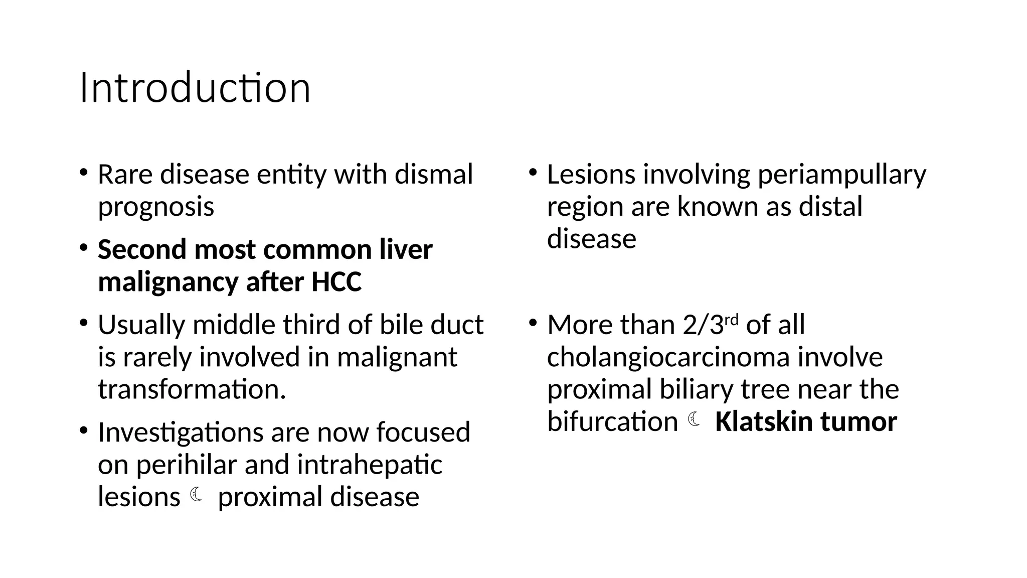
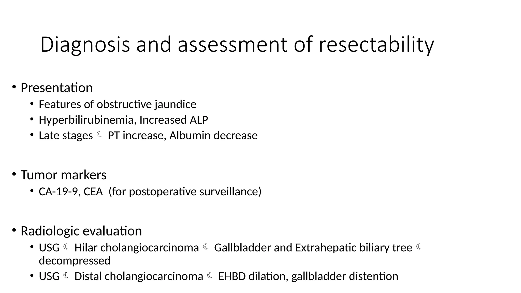
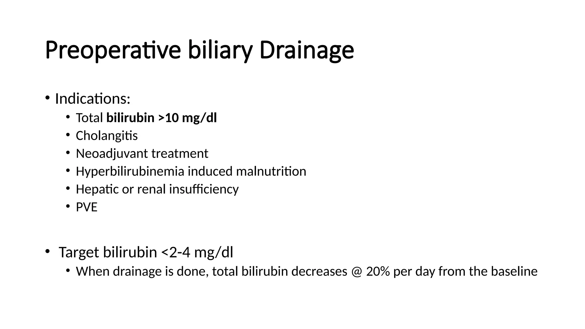
Malignant biliary disease
Nabin Paudyal
MRCP findings during stricture
Gallbladder cancer
• Aggressive disease
• Poor prognosis
• Late stage presentation
• Earlier stage patients More surgical approach is warranted
Incidence
• 6th
and 7th
decade
• 2-3 times Women>> Men
• Highest in SEAR
• USA GB cancer is most common cancer of the biliary tract and the
fifth most common gastrointestinal cancer.
Cause
• Prevailing theory of gall bladder cancer focuses on chronic inflammation as a prime
cause for development of neoplasia.
• Presence of gallstones is therefore a primary risk factor and stones > 3 cm carry
increased risk of cancer development.
• > 80% of patients with gallbladder cancer have CHOLELITHIASIS.
• Gallstone patients are at 7 times more risk of having gallbladder cancer
• Other risk factors APBJ, choledochal cyst, Primary sclerosing cholangitis, Porcelain
gallbladder.
Adenoma-carcinoma sequence in gallbladder
carcinoma
• Severe dysplasia and Carcinoma in situ form gallbladder carcinoma
• Polyposis do not carry increased risk of malignancy
• Single gallbladder polyp > 10 mm carries an increased risk for
malignancy and hence is indicated for cholecystectomy.
Pathology and staging
• GB cancer spreads via lymphatics, hematogenously and peritoneal cavity OR
along biopsy or surgical wound tracts
• Morphologically
• Infiltrative (most common) spread in subserosal plane and invade entire GB and even
porta hepatis
• Nodular more circumscribed growth and invade liver
• Papillary limited to GB wall
• Combined
• Lymph nodes
• Drain into cystic and peri choledochal LN Pass into retro portal and pancreaticoduodenal
LN
• From here, the lymph drains into Celiac, superior mesenteric and aortocaval LN.
• Kocherization at the time of surgery is essential to properly stage GB cancer.
Gall bladder has rapid infiltration because it doesn’t have submucosa and serosal covering (at liver)
Clinical features
• Most patients are asymptomatic as more than 90% of gallbladder cancers are
originating in the fundus or body of gallbladder.
• Most gallbladder have symptoms at the time of presentation with 35% nodal
disease and distant metastasis in 40%.
• By chance if any patient has symptoms of acute cholecystitis with obstruction
of neck of the gallbladder the prognosis may be better as the disease is
caught in earlier stage [भागयमनी को भूतै कमारो]
• Symptoms are similar to hilar cholangiocarcinoma
• At late stages Weight loss, jaundice, abdominal mass are present
• Other symptoms Chronic epigastric pain, early satiety, sense of fullness.
Diagnosis
• Not helpful in early stages
• Helps identify advanced disease Anemia, hypoalbuminemia, leukocytosis
and elevated ALP or bilirubin levels are present
• Other increased markers CEA, Carbohydrate 19-9
• Radiological investigations
• USG irregularly shaped lesion in the subhepatic space/ heterogeneous mass in the
gallbladder lumen, asymmetrically thickened gallbladder wall.
• Polyp of more than 10 mm should raise the suspicion of gallbladder cancer
• CT/MRI  Provides information on local extent of cancer and distant metastasis
[peritoneal, hepatic parenchymal, lymphadenopathy, adjacent vascular involvement]
Diagnosis
• PET search for metastatic disease when CT or MRI provides limited
information about the primary tumor.
• NEVER BIOPSY
• If diagnosis is suspected Surgeon and patient must be prepared for
definitive operation.
• If unresectable (vascular encasement, extensive hepatic involvement) /
incurable (hepatic OR peritoneal metastasis) biopsy can be taken
Mass may be found as
a. Incidental finding on pathological review
b. Mass on imaging
Presentation of patients may be as follows
• Patients with gallbladder polyp
• Patients with an incidental finding of gallbladder cancer at the time of
or after cholecystectomy
• Patients suspected of having gallbladder cancer preoperatively
• Patients with advanced disease at presentation.
a. Gallbladder polyp
• Adenomatous polyp are the only polyp with malignant potential
• Predictors of malignancy
• Single polyp
• Size greater than 1 cm
• Age older than 50 years
• Polyp > 1cm Perform laparoscopically
• Polyp < 1cm  undergo surveillance to demonstrate stability, except PSC
where the threshold for cholecystectomy should be lower.
b. Gallbladder cancer after cholecystectomy
• Treatment depends upon the depth of penetration of the gallbladder
wall and surgical margins
• T1a Cholecystectomy will suffice.
• T1b and above As perineural, lymphatic or vascular invasion will
increase likelihood of nodal disease extended cholecystectomy is
done in such patients cholecystectomy and en-bloc resection
adjacent liver parenchyma in segments Ivb and V and portal
lymphadenopathy
• Extended cholecystectomy directed at obtaining R0 resection disease
including LN basins. Henceforth, removal of hepatoduodenal,
gastrohepatic and retro duodenal LN should be done.
• Cystic duct margin
• 2 cm of apparently normal hepatic parenchyma from gb fossa
• Port site excision [recommended by some surgeons]
• For T2 and more Radical cholecystectomy is done.
• Radical cholecystectomy is cholecystectomy with removal of LN.
• Includes segments IV b and V, lymphadenectomy and extended hepatic or biliary
resection to obtain negative margin
c. Patients suspected of having gallbladder
cancer preoperatively
• Offer an attempt at resection
• Patients tend to present at an advanced stage of locoregional disease
• Often require extended liver resection
• Radical resection should be considered for adequate operative candidates
• Begin with diagnostic laparoscopy to identify hepatic OR small-volume
peritoneal metastasis conclude inoperable (if present)
• Metastatic disease Non-operative strategies and palliative care is
considered
• For stage T3 and T4 lesions resect at least IV B and V but more often
requires central hepatectomy [segment IV B, V and VIII].
Trisegmentectomy may be required to achieve R0 status.
• Debulking with complete resection of disease is MAINSTAY of
management of gallbladder cancer.
d. Patients with advanced disease at
presentation
• Palliation is the goal
• Jaundice, pain and intestinal obstruction are the main presenting
complaints
• Jaundice ERCP, Self expanding Endobiliary metal stents
• Pain Oral narcotics, percutaneous neurolysis of celiac ganglion
• Intestinal obstruction occurs due to gastric outlet obstruction from
local extension of tumor and managed by endoscopic duodenal wall
stent.
Adjuvant Therapy
• Majority of gallbladder carcinoma recurrences include distant sites as
a part of recurrence pattern.
• Gemcitabine-based therapy often combined with platinum agent are
typically used for treatment of gallbladder cancer
• T4 lesions, positive LN status, R1 resection  indications for adjuvant
therapy
Survival
• Survival depends on stage of presentation, degree of surgical resection
performed
• T status, N status, histologic differentiation, CBD involvement, R0 resection 
independent factors affecting survival
• Overall survival of gallbladder cancer < 15%
Stage of the cancer Prognosis and survival
T1a -1b Excellent prognosis
T2 Depends on extent of nodal status and
radical resection. Survival (5 yr) 20-60%
T3 5 yr survival < 20%
T4 Months
Cholangiocarcinoma
Introduction
• Rare disease entity with dismal
prognosis
• Second most common liver
malignancy after HCC
• Usually middle third of bile duct
is rarely involved in malignant
transformation.
• Investigations are now focused
on perihilar and intrahepatic
lesions proximal disease
• Lesions involving periampullary
region are known as distal
disease
• More than 2/3rd
of all
cholangiocarcinoma involve
proximal biliary tree near the
bifurcation Klatskin tumor
Risk factors
• Congenital lesions
• Choledochal cysts
• Exposure of biliary epithelium to
toxic pancreatic secretions
• 10-20% of cysts turn into malignant if
cyst is not resected by age 20
• Infections/inflammation
• Clonorchis sinensis
• Opisthorchis viverrine
• Cause chronic biliary inflammation,
obstruction and stricture
• Primary sclerosing cholangitis
• Annual risk of 1.5% per year
• Medications and chemical
carcinogens
• Thorotrast
• Oral contraceptive pills
• Asbestos
• Cigarette smoke
• Cirrhosis of liver
Staging and classification
• Pathologic subtypes
• Sclerosing
• Nodular
• Papillary (@PNS)
• Sclerosing variety are mostly located in proximal
region
• Papillary and nodular are located in distal region
and are manifested as intraluminal growth
• Nodular A firm mass based in the duct wall
can be seen growing into the duct lumen
• Papillary Polypoidal lesion that is soft with less
periductal fibrosis
• TNM classification
• 3 staging subdivisions
• Intrahepatic
• Perihilar
• Distal bile duct
• Tumors within bile duct and those
extending outside the duct but not
invading any nearby structure usually have
good prognosis.
• Factors influencing prognosis
• R0 resection
• LN invasion
Clinical features
•Depends on site of obstruction
• Painless jaundice
• If unilobar obstruction
• Ipsilateral lobar atrophy, with contralateral hypertrophy
• Liver compensates until late stages
• If obstruction at or below hepatic bifurcation
• Earlier manifestations
• Features of obstructive jaundice
• Submucosal spread of tumor may lead to perineural invasion
Diagnosis and assessment of resectability
• Presentation
• Features of obstructive jaundice
• Hyperbilirubinemia, Increased ALP
• Late stages PT increase, Albumin decrease
• Tumor markers
• CA-19-9, CEA (for postoperative surveillance)
• Radiologic evaluation
• USG Hilar cholangiocarcinoma Gallbladder and Extrahepatic biliary tree
decompressed
• USG Distal cholangiocarcinoma EHBD dilation, gallbladder distention
• CT scan in cholangiocarcinoma
• Triphasic CT
• Assessment of metastasis, Evaluation of resectability
• Location of tumor
• Relation with vascular structures
• Identification of aberrant anatomy and determination of segmental or lobar
involvement
• Cholangiography
• ERCP/ PTC / MRCP
• Helps determine the proximal extent of resection
Imaging
Ultrasound :
• Used as a screening examination in patients with RUQ pain, a palpable
mass, or unexplained jaundice.
• Nonspecific appearance as a hypoechoic hepatic mass.
• Satellite lesions and capsular retraction may be seen.
• Hypovascular with minimal Doppler evidence of internal blood flow.
• Biliary dilatation, portal venous invasion, hepatic venous invasion and
portal lymphadenopathy.
Imaging
• Multiphasic CT with IV contrast of the abdomen and pelvis.
• Hypodense with irregular, infiltrative margins.
• Variable delayed enhancement in the portal venous phase.
• Biliary dilatation, portal or hepatic venous involvement , lobar atrophy.
• Identification of aberrant anatomy and segmental or lobar involvement
• Helpful for preoperative planning.
• Chest CT (with or without contrast) should be performed.
Imaging: MRI
• Hypointense on T1-weighted, hyperintense on T2-weighted images
• Pooling of contrast within the lesions on delayed images (6 to 8
minutes)
• Venous and arterial involvement by tumor
• Cholangiopancreatography is a noninvasive method of obtaining
cholangiograms.
• Tissue diagnosis before resection of the tumor in operative patients is
unnecessary.
• Brush cytology Reports unreliable Negative cytology thus does not
alter subsequent management
Preoperative biliary Drainage
• Distal bile duct cancer- single catheter, whereas in perihilar- multiple
• When intrahepatic bile ducts are isolated , PTC and multiple direct
segmental transhepatic drainages may be necessary.
• Future liver remnant should be assessed prior to drainage.
Preoperative biliary Drainage
• Indications:
• Total bilirubin >10 mg/dl
• Cholangitis
• Neoadjuvant treatment
• Hyperbilirubinemia induced malnutrition
• Hepatic or renal insufficiency
• PVE
• Target bilirubin <2-4 mg/dl
• When drainage is done, total bilirubin decreases @ 20% per day from the baseline
Management
Pre-operative Portal venous embolization
• Important in the presurgical management.
• Patients who undergo extensive liver resection.
• Increase the functional liver by inducing compensatory hypertrophy.
• Sizable compensatory hypertrophy of the non embolized segments
• In right PVE, the volume of the left lobe increases by 130 cm3
on
average within 2 weeks after embolization.
• Estimated resection volume decreases by approximately 10%.
Operative management
• Even with clinical suspicion of the disease Adequate surgical
candidates' exploration should proceed even in the absence of
confirmed tissue diagnosis
• 7-15% of patients with suspected malignant disease will have benign
disease
• Staging laparoscopy is important initial step at the time of resection
to reduce the incidence of nontherapeutic laparotomy
Management of distal CC
• Operative management includes Pancreaticoduodenectomy
• Intra-operative frozen section to determine R0 resection status is
preferred.
• 5 year survival – up to 50% when R0 is achieved.
Management of Proximal CC
• Regional nodal tissue and en bloc resection of the CBD with resection
of hepatic parenchyma as necessary to achieve negative margins
• Resection depends upon Bismuth-Corlette classification
Type of cholangiocarcinoma Operation procedure
a. Type I and II lesions Common duct resection
Cholecystectomy
5-10 mm of resection margin
[Type II resection also may require partial hepatic resection (commonly including caudate lobe). Resection of bile
duct and nodal tissue requires complete skeletonization of hepatic artery and portal vein]
b. Type III and IV lesions • Complex resection and reconstruction
of portal vein, hepatic artery or both.
• If secondary biliary radicals are also
resected Trans anastomotic stenting
• Hepatic resection in selected cases to
achieve negative margins
Negative margin status is the most important variable associated with outcome of patients with cholangiocarcinoma
Prognosis and survival rate
• As high as 59% have been reported
• With vascular resection and reconstruction techniques
• Role of lymph node resection is debated.
• There has been no demonstrable benefit of routine lymph node dissection
however LN are one of the most important prognostic factors in
cholangiocarcinoma.
Palliation
• In unresectable cases or incurable diseases all attempts to palliate their
symptoms non-operatively should be used
• Goals
• Relief of jaundice
• Alleviation of pain Oral/ IV narcotics
• Relief of duodenal obstruction Endoscopic duodenal stenting
• Palliation may be
• Endoscopic ERCP for distal CC
• Percutaneous PTC for proximal CC
• In either cases, placement of self expanding metallic stent is used
Medical therapy
• No improved survival by chemotherapy
• Radiotherapy has also no benefit in survival
• Studies report < 10% of clinical response rate Radiotherapy
• Neither radiotherapy nor chemotherapy is used routinely in patients
in adjuvant/ neoadjuvant setting
Outcomes
• Depend on stage of presentation and completeness of surgical
resection to negative margin
• With use of CBD resection with partial hepatectomy negative
margin rates is as high as 75%
• 5 year survival rate 20-45% in most series with available expertise.
• Defined by LN metastasis.
• Unresected cases Median survival is 5-8 months.
Should we go for liver replacement?
• Controversial
• Limited to research protocols
Intrahepatic cholangiocarcinoma
Introduction
• 40-60% of the bile duct malignancies are IHC where biliary confluence is
the most commonly involved area (Klatskin tumor)
• Second most common primary hepatic neoplasm
• MC risk factors include
• PSC
• Choledochal cyst
• Hepatolithiasis
• Recurrent pyogenic cholangitis
• Hepatitis B/ Hepatitis C
• NASH
• Diabetes
Clinical Features
• Similar to HCC
• Asymptomatic Early stages
• Symptoms include
• RUQ pain
• Weight loss
• Jaundice (relatively uncommon)
• Incidental mass
Laboratory investigations
• Normal ALP
• CEA and CA 19-9 may be elevated
• May be normal as well
• IHC is a diagnosis of exclusion
• Search for primary tumor with UGI / LGI
endoscopy, CT abdomen/pelvis, chest
• CT/ MRI Focal hepatic mass that
may be associated with biliary dilation
• Contrast study shows
• peripheral/ central enhancement
• persistent enhancement on delayed
phase due to fibrotic nature of
cholangiocarcinoma
Management
• Complete resection
• Focused on R0 resection margin
• Generally up to 60% resectable
• Completely resected cases have 3 year survival rates from 16-65%, 5 year
survival rates from 24-44%
• Factors associated with poor outcome
• Multifocal tumors
• LN metastasis
• Vascular invasion
• Positive margin
• @ V-L-M-P
• Because of rarity of the tumor little is known about the effectiveness of
radiotherapy and chemotherapy
• Application of radiotherapy and chemotherapy is not done in routine use
• BILCAP study Role of adjuvant capecitabine for IHC
25% reduced death risk was noted in the study
What's new in intrahepatic cholangiocarcinoma?
Malignant biliary disease -introduction and management.pptx

Malignant biliary disease -introduction and management.pptx

  • 1.
  • 2.
  • 3.
    Gallbladder cancer • Aggressivedisease • Poor prognosis • Late stage presentation • Earlier stage patients More surgical approach is warranted
  • 4.
    Incidence • 6th and 7th decade •2-3 times Women>> Men • Highest in SEAR • USA GB cancer is most common cancer of the biliary tract and the fifth most common gastrointestinal cancer.
  • 5.
    Cause • Prevailing theoryof gall bladder cancer focuses on chronic inflammation as a prime cause for development of neoplasia. • Presence of gallstones is therefore a primary risk factor and stones > 3 cm carry increased risk of cancer development. • > 80% of patients with gallbladder cancer have CHOLELITHIASIS. • Gallstone patients are at 7 times more risk of having gallbladder cancer • Other risk factors APBJ, choledochal cyst, Primary sclerosing cholangitis, Porcelain gallbladder.
  • 6.
    Adenoma-carcinoma sequence ingallbladder carcinoma • Severe dysplasia and Carcinoma in situ form gallbladder carcinoma • Polyposis do not carry increased risk of malignancy • Single gallbladder polyp > 10 mm carries an increased risk for malignancy and hence is indicated for cholecystectomy.
  • 7.
    Pathology and staging •GB cancer spreads via lymphatics, hematogenously and peritoneal cavity OR along biopsy or surgical wound tracts • Morphologically • Infiltrative (most common) spread in subserosal plane and invade entire GB and even porta hepatis • Nodular more circumscribed growth and invade liver • Papillary limited to GB wall • Combined • Lymph nodes • Drain into cystic and peri choledochal LN Pass into retro portal and pancreaticoduodenal LN • From here, the lymph drains into Celiac, superior mesenteric and aortocaval LN. • Kocherization at the time of surgery is essential to properly stage GB cancer.
  • 8.
    Gall bladder hasrapid infiltration because it doesn’t have submucosa and serosal covering (at liver)
  • 9.
    Clinical features • Mostpatients are asymptomatic as more than 90% of gallbladder cancers are originating in the fundus or body of gallbladder. • Most gallbladder have symptoms at the time of presentation with 35% nodal disease and distant metastasis in 40%. • By chance if any patient has symptoms of acute cholecystitis with obstruction of neck of the gallbladder the prognosis may be better as the disease is caught in earlier stage [भागयमनी को भूतै कमारो] • Symptoms are similar to hilar cholangiocarcinoma • At late stages Weight loss, jaundice, abdominal mass are present • Other symptoms Chronic epigastric pain, early satiety, sense of fullness.
  • 10.
    Diagnosis • Not helpfulin early stages • Helps identify advanced disease Anemia, hypoalbuminemia, leukocytosis and elevated ALP or bilirubin levels are present • Other increased markers CEA, Carbohydrate 19-9 • Radiological investigations • USG irregularly shaped lesion in the subhepatic space/ heterogeneous mass in the gallbladder lumen, asymmetrically thickened gallbladder wall. • Polyp of more than 10 mm should raise the suspicion of gallbladder cancer • CT/MRI  Provides information on local extent of cancer and distant metastasis [peritoneal, hepatic parenchymal, lymphadenopathy, adjacent vascular involvement]
  • 11.
    Diagnosis • PET searchfor metastatic disease when CT or MRI provides limited information about the primary tumor. • NEVER BIOPSY • If diagnosis is suspected Surgeon and patient must be prepared for definitive operation. • If unresectable (vascular encasement, extensive hepatic involvement) / incurable (hepatic OR peritoneal metastasis) biopsy can be taken
  • 12.
    Mass may befound as a. Incidental finding on pathological review b. Mass on imaging
  • 15.
    Presentation of patientsmay be as follows • Patients with gallbladder polyp • Patients with an incidental finding of gallbladder cancer at the time of or after cholecystectomy • Patients suspected of having gallbladder cancer preoperatively • Patients with advanced disease at presentation.
  • 16.
    a. Gallbladder polyp •Adenomatous polyp are the only polyp with malignant potential • Predictors of malignancy • Single polyp • Size greater than 1 cm • Age older than 50 years • Polyp > 1cm Perform laparoscopically • Polyp < 1cm  undergo surveillance to demonstrate stability, except PSC where the threshold for cholecystectomy should be lower.
  • 17.
    b. Gallbladder cancerafter cholecystectomy • Treatment depends upon the depth of penetration of the gallbladder wall and surgical margins • T1a Cholecystectomy will suffice. • T1b and above As perineural, lymphatic or vascular invasion will increase likelihood of nodal disease extended cholecystectomy is done in such patients cholecystectomy and en-bloc resection adjacent liver parenchyma in segments Ivb and V and portal lymphadenopathy
  • 18.
    • Extended cholecystectomydirected at obtaining R0 resection disease including LN basins. Henceforth, removal of hepatoduodenal, gastrohepatic and retro duodenal LN should be done. • Cystic duct margin • 2 cm of apparently normal hepatic parenchyma from gb fossa • Port site excision [recommended by some surgeons] • For T2 and more Radical cholecystectomy is done. • Radical cholecystectomy is cholecystectomy with removal of LN. • Includes segments IV b and V, lymphadenectomy and extended hepatic or biliary resection to obtain negative margin
  • 20.
    c. Patients suspectedof having gallbladder cancer preoperatively • Offer an attempt at resection • Patients tend to present at an advanced stage of locoregional disease • Often require extended liver resection • Radical resection should be considered for adequate operative candidates • Begin with diagnostic laparoscopy to identify hepatic OR small-volume peritoneal metastasis conclude inoperable (if present) • Metastatic disease Non-operative strategies and palliative care is considered
  • 21.
    • For stageT3 and T4 lesions resect at least IV B and V but more often requires central hepatectomy [segment IV B, V and VIII]. Trisegmentectomy may be required to achieve R0 status. • Debulking with complete resection of disease is MAINSTAY of management of gallbladder cancer.
  • 22.
    d. Patients withadvanced disease at presentation • Palliation is the goal • Jaundice, pain and intestinal obstruction are the main presenting complaints • Jaundice ERCP, Self expanding Endobiliary metal stents • Pain Oral narcotics, percutaneous neurolysis of celiac ganglion • Intestinal obstruction occurs due to gastric outlet obstruction from local extension of tumor and managed by endoscopic duodenal wall stent.
  • 23.
    Adjuvant Therapy • Majorityof gallbladder carcinoma recurrences include distant sites as a part of recurrence pattern. • Gemcitabine-based therapy often combined with platinum agent are typically used for treatment of gallbladder cancer • T4 lesions, positive LN status, R1 resection  indications for adjuvant therapy
  • 25.
    Survival • Survival dependson stage of presentation, degree of surgical resection performed • T status, N status, histologic differentiation, CBD involvement, R0 resection  independent factors affecting survival • Overall survival of gallbladder cancer < 15% Stage of the cancer Prognosis and survival T1a -1b Excellent prognosis T2 Depends on extent of nodal status and radical resection. Survival (5 yr) 20-60% T3 5 yr survival < 20% T4 Months
  • 26.
  • 27.
    Introduction • Rare diseaseentity with dismal prognosis • Second most common liver malignancy after HCC • Usually middle third of bile duct is rarely involved in malignant transformation. • Investigations are now focused on perihilar and intrahepatic lesions proximal disease • Lesions involving periampullary region are known as distal disease • More than 2/3rd of all cholangiocarcinoma involve proximal biliary tree near the bifurcation Klatskin tumor
  • 28.
    Risk factors • Congenitallesions • Choledochal cysts • Exposure of biliary epithelium to toxic pancreatic secretions • 10-20% of cysts turn into malignant if cyst is not resected by age 20 • Infections/inflammation • Clonorchis sinensis • Opisthorchis viverrine • Cause chronic biliary inflammation, obstruction and stricture • Primary sclerosing cholangitis • Annual risk of 1.5% per year • Medications and chemical carcinogens • Thorotrast • Oral contraceptive pills • Asbestos • Cigarette smoke • Cirrhosis of liver
  • 30.
    Staging and classification •Pathologic subtypes • Sclerosing • Nodular • Papillary (@PNS) • Sclerosing variety are mostly located in proximal region • Papillary and nodular are located in distal region and are manifested as intraluminal growth • Nodular A firm mass based in the duct wall can be seen growing into the duct lumen • Papillary Polypoidal lesion that is soft with less periductal fibrosis • TNM classification • 3 staging subdivisions • Intrahepatic • Perihilar • Distal bile duct • Tumors within bile duct and those extending outside the duct but not invading any nearby structure usually have good prognosis. • Factors influencing prognosis • R0 resection • LN invasion
  • 34.
    Clinical features •Depends onsite of obstruction • Painless jaundice • If unilobar obstruction • Ipsilateral lobar atrophy, with contralateral hypertrophy • Liver compensates until late stages • If obstruction at or below hepatic bifurcation • Earlier manifestations • Features of obstructive jaundice • Submucosal spread of tumor may lead to perineural invasion
  • 35.
    Diagnosis and assessmentof resectability • Presentation • Features of obstructive jaundice • Hyperbilirubinemia, Increased ALP • Late stages PT increase, Albumin decrease • Tumor markers • CA-19-9, CEA (for postoperative surveillance) • Radiologic evaluation • USG Hilar cholangiocarcinoma Gallbladder and Extrahepatic biliary tree decompressed • USG Distal cholangiocarcinoma EHBD dilation, gallbladder distention
  • 36.
    • CT scanin cholangiocarcinoma • Triphasic CT • Assessment of metastasis, Evaluation of resectability • Location of tumor • Relation with vascular structures • Identification of aberrant anatomy and determination of segmental or lobar involvement • Cholangiography • ERCP/ PTC / MRCP • Helps determine the proximal extent of resection
  • 37.
    Imaging Ultrasound : • Usedas a screening examination in patients with RUQ pain, a palpable mass, or unexplained jaundice. • Nonspecific appearance as a hypoechoic hepatic mass. • Satellite lesions and capsular retraction may be seen. • Hypovascular with minimal Doppler evidence of internal blood flow. • Biliary dilatation, portal venous invasion, hepatic venous invasion and portal lymphadenopathy.
  • 38.
    Imaging • Multiphasic CTwith IV contrast of the abdomen and pelvis. • Hypodense with irregular, infiltrative margins. • Variable delayed enhancement in the portal venous phase. • Biliary dilatation, portal or hepatic venous involvement , lobar atrophy. • Identification of aberrant anatomy and segmental or lobar involvement • Helpful for preoperative planning. • Chest CT (with or without contrast) should be performed.
  • 41.
    Imaging: MRI • Hypointenseon T1-weighted, hyperintense on T2-weighted images • Pooling of contrast within the lesions on delayed images (6 to 8 minutes) • Venous and arterial involvement by tumor • Cholangiopancreatography is a noninvasive method of obtaining cholangiograms.
  • 44.
    • Tissue diagnosisbefore resection of the tumor in operative patients is unnecessary. • Brush cytology Reports unreliable Negative cytology thus does not alter subsequent management
  • 45.
    Preoperative biliary Drainage •Distal bile duct cancer- single catheter, whereas in perihilar- multiple • When intrahepatic bile ducts are isolated , PTC and multiple direct segmental transhepatic drainages may be necessary. • Future liver remnant should be assessed prior to drainage.
  • 46.
    Preoperative biliary Drainage •Indications: • Total bilirubin >10 mg/dl • Cholangitis • Neoadjuvant treatment • Hyperbilirubinemia induced malnutrition • Hepatic or renal insufficiency • PVE • Target bilirubin <2-4 mg/dl • When drainage is done, total bilirubin decreases @ 20% per day from the baseline
  • 47.
  • 48.
    Pre-operative Portal venousembolization • Important in the presurgical management. • Patients who undergo extensive liver resection. • Increase the functional liver by inducing compensatory hypertrophy. • Sizable compensatory hypertrophy of the non embolized segments • In right PVE, the volume of the left lobe increases by 130 cm3 on average within 2 weeks after embolization. • Estimated resection volume decreases by approximately 10%.
  • 49.
    Operative management • Evenwith clinical suspicion of the disease Adequate surgical candidates' exploration should proceed even in the absence of confirmed tissue diagnosis • 7-15% of patients with suspected malignant disease will have benign disease • Staging laparoscopy is important initial step at the time of resection to reduce the incidence of nontherapeutic laparotomy
  • 50.
    Management of distalCC • Operative management includes Pancreaticoduodenectomy • Intra-operative frozen section to determine R0 resection status is preferred. • 5 year survival – up to 50% when R0 is achieved.
  • 51.
    Management of ProximalCC • Regional nodal tissue and en bloc resection of the CBD with resection of hepatic parenchyma as necessary to achieve negative margins • Resection depends upon Bismuth-Corlette classification
  • 53.
    Type of cholangiocarcinomaOperation procedure a. Type I and II lesions Common duct resection Cholecystectomy 5-10 mm of resection margin [Type II resection also may require partial hepatic resection (commonly including caudate lobe). Resection of bile duct and nodal tissue requires complete skeletonization of hepatic artery and portal vein] b. Type III and IV lesions • Complex resection and reconstruction of portal vein, hepatic artery or both. • If secondary biliary radicals are also resected Trans anastomotic stenting • Hepatic resection in selected cases to achieve negative margins Negative margin status is the most important variable associated with outcome of patients with cholangiocarcinoma
  • 55.
    Prognosis and survivalrate • As high as 59% have been reported • With vascular resection and reconstruction techniques • Role of lymph node resection is debated. • There has been no demonstrable benefit of routine lymph node dissection however LN are one of the most important prognostic factors in cholangiocarcinoma.
  • 56.
    Palliation • In unresectablecases or incurable diseases all attempts to palliate their symptoms non-operatively should be used • Goals • Relief of jaundice • Alleviation of pain Oral/ IV narcotics • Relief of duodenal obstruction Endoscopic duodenal stenting • Palliation may be • Endoscopic ERCP for distal CC • Percutaneous PTC for proximal CC • In either cases, placement of self expanding metallic stent is used
  • 57.
    Medical therapy • Noimproved survival by chemotherapy • Radiotherapy has also no benefit in survival • Studies report < 10% of clinical response rate Radiotherapy • Neither radiotherapy nor chemotherapy is used routinely in patients in adjuvant/ neoadjuvant setting
  • 58.
    Outcomes • Depend onstage of presentation and completeness of surgical resection to negative margin • With use of CBD resection with partial hepatectomy negative margin rates is as high as 75% • 5 year survival rate 20-45% in most series with available expertise. • Defined by LN metastasis. • Unresected cases Median survival is 5-8 months.
  • 59.
    Should we gofor liver replacement? • Controversial • Limited to research protocols
  • 60.
  • 61.
    Introduction • 40-60% ofthe bile duct malignancies are IHC where biliary confluence is the most commonly involved area (Klatskin tumor) • Second most common primary hepatic neoplasm • MC risk factors include • PSC • Choledochal cyst • Hepatolithiasis • Recurrent pyogenic cholangitis • Hepatitis B/ Hepatitis C • NASH • Diabetes
  • 62.
    Clinical Features • Similarto HCC • Asymptomatic Early stages • Symptoms include • RUQ pain • Weight loss • Jaundice (relatively uncommon) • Incidental mass Laboratory investigations • Normal ALP • CEA and CA 19-9 may be elevated • May be normal as well • IHC is a diagnosis of exclusion • Search for primary tumor with UGI / LGI endoscopy, CT abdomen/pelvis, chest • CT/ MRI Focal hepatic mass that may be associated with biliary dilation • Contrast study shows • peripheral/ central enhancement • persistent enhancement on delayed phase due to fibrotic nature of cholangiocarcinoma
  • 63.
    Management • Complete resection •Focused on R0 resection margin • Generally up to 60% resectable • Completely resected cases have 3 year survival rates from 16-65%, 5 year survival rates from 24-44% • Factors associated with poor outcome • Multifocal tumors • LN metastasis • Vascular invasion • Positive margin • @ V-L-M-P
  • 64.
    • Because ofrarity of the tumor little is known about the effectiveness of radiotherapy and chemotherapy • Application of radiotherapy and chemotherapy is not done in routine use • BILCAP study Role of adjuvant capecitabine for IHC
  • 65.
    25% reduced deathrisk was noted in the study
  • 66.
    What's new inintrahepatic cholangiocarcinoma?