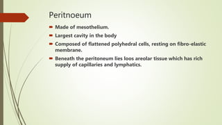
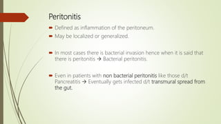
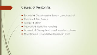
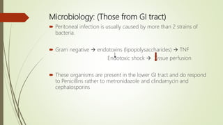
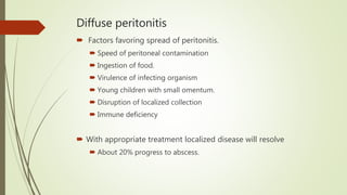
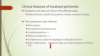
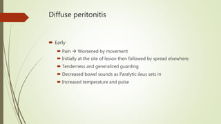
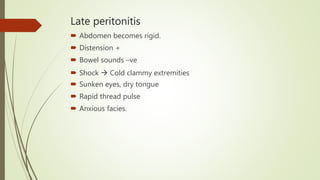
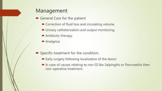
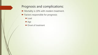
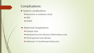
This document discusses acute peritonitis, including:
- Peritonitis is defined as inflammation of the peritoneum and can be localized or generalized. It is usually caused by bacterial infection entering through the gastrointestinal tract or other sources.
- Symptoms depend on whether it is localized or diffuse. Localized peritonitis causes pain specific to the affected organ, while diffuse peritonitis causes generalized abdominal pain and tenderness.
- Treatment involves antibiotics, fluid resuscitation, and sometimes surgery to address the underlying cause and drain any abscesses. Outcomes depend on several factors but mortality is around 10% with prompt treatment. Complications can include shock, bowel obstruction, and residual infections.
Intestinal fistulas pose the greatest challenge to the General Surgeon. The presentation provides abrief guideline for management of this complex problem.
Approach to Management of Upper Gastrointestinal (GI) BleedingArun Vasireddy
Upper gastrointestinal bleeding is gastrointestinal bleeding in the upper gastrointestinal tract, commonly defined as bleeding arising from the esophagus, stomach, or duodenum. Blood may be observed in vomit (hematemesis) or in altered form in the stool (melena). Depending on the severity of the blood loss, there may be symptoms of insufficient circulating blood volume and shock. As a result, upper gastrointestinal bleeding is considered a medical emergency and typically requires hospital care for urgent diagnosis and treatment. Upper gastrointestinal bleeding can be caused by peptic ulcers, gastric erosions, esophageal varices, and some rarer causes such as gastric cancer.
The initial assessment includes measurement of the blood pressure and heart rate, as well as blood tests to determine hemoglobin concentration. In significant bleeding, fluid replacement is often required, as well as blood transfusion, before the source of bleeding can be determined by endoscopy of the upper digestive tract with an esophagogastroduodenoscopy. Depending on the source, endoscopic therapy can be applied to reduce rebleeding risk. Specific medical treatments (such as proton pump inhibitors for peptic ulcer disease) or procedures (such as TIPS for variceal hemorrhage) may be used. Recurrent or refractory bleeding may lead to need for surgery, although this has become uncommon as a result of improved endoscopic and medical treatment.
MBBS STUDENTS UNDER GRADUATES ..WITH USES PROPERTIE IMAGES......FOLEYS CATHETER MALECOTS CATHETER ARTERY FORCEPS SMALL MEDIUM LARGE RETRACTOR KELLYS SURGERY SURGICAL INSTRUMENTS MBBS STUDENTS UNDERGRADUATES SUTURE MATERIALS MAYOS SCISSORS MBBS STUDENTS UNDER GRADUATES .......LISTERS SINUS FORCEPS....RIGHT ANGLED FORCEPS....KOCHERS CLAMP...suture material suture removal Jolls thyroid retractor .....WITH USES PROPERTIE IMAGES......FOLEYS CATHETER MALECOTS CATHETER ARTERY FORCEPS SMALL MEDIUM LARGE RETRACTOR KELLYS SURGERY SURGICAL INSTRUMENTS MBBS STUDENTS UNDERGRADUATES SUTURE MATERIALS MAYOS SCISSORS ..FOLEYS CATHETER ....3 WAY 2WAY RYLES TUBE.........HILTONS METHOD ..ALLIS .... MOSQUITO . LANES FORCEPS ....Lanes twin anastomosis clamp......MALECOTS CATHETER.. ARTERY FORCEPS..... SMALL MEDIUM LARGE ......RETRACTOR ....KELLYS ....SURGERY SURGICAL INSTRUMENTS MBBS STUDENTS UNDERGRADUATES..... SUTURE MATERIALS.... MAYOS SCISSORS
...........................MBBS STUDENTS UNDER GRADUATES ..COMPARISON WITH IMAGES NOTES FROM LECTURE CLASSES.............................................................................................................................................................................................................................................................................................................................................................................................................................................................................................................................................................................................................................................................................................................................................................................................
this presentation includes anatomy physiology function of peritoneum ,also includes cause of peritonitis its severity ,various scoring system investigation and treatment.It includes the recent advancement and latest articles from latest books of surgery.
These lecture slides, by Dr Sidra Arshad, offer a quick overview of physiological basis of a normal electrocardiogram.
Learning objectives:
1. Define an electrocardiogram (ECG) and electrocardiography
2. Describe how dipoles generated by the heart produce the waveforms of the ECG
3. Describe the components of a normal electrocardiogram of a typical bipolar leads (limb II)
4. Differentiate between intervals and segments
5. Enlist some common indications for obtaining an ECG
Study Resources:
1. Chapter 11, Guyton and Hall Textbook of Medical Physiology, 14th edition
2. Chapter 9, Human Physiology - From Cells to Systems, Lauralee Sherwood, 9th edition
3. Chapter 29, Ganong’s Review of Medical Physiology, 26th edition
4. Electrocardiogram, StatPearls - https://www.ncbi.nlm.nih.gov/books/NBK549803/
5. ECG in Medical Practice by ABM Abdullah, 4th edition
6. ECG Basics, http://www.nataliescasebook.com/tag/e-c-g-basics
HOT NEW PRODUCT! BIG SALES FAST SHIPPING NOW FROM CHINA!! EU KU DB BK substit...GL Anaacs
Contact us if you are interested:
Email / Skype : kefaya1771@gmail.com
Threema: PXHY5PDH
New BATCH Ku !!! MUCH IN DEMAND FAST SALE EVERY BATCH HAPPY GOOD EFFECT BIG BATCH !
Contact me on Threema or skype to start big business!!
Hot-sale products:
NEW HOT EUTYLONE WHITE CRYSTAL!!
5cl-adba precursor (semi finished )
5cl-adba raw materials
ADBB precursor (semi finished )
ADBB raw materials
APVP powder
5fadb/4f-adb
Jwh018 / Jwh210
Eutylone crystal
Protonitazene (hydrochloride) CAS: 119276-01-6
Flubrotizolam CAS: 57801-95-3
Metonitazene CAS: 14680-51-4
Payment terms: Western Union,MoneyGram,Bitcoin or USDT.
Deliver Time: Usually 7-15days
Shipping method: FedEx, TNT, DHL,UPS etc.Our deliveries are 100% safe, fast, reliable and discreet.
Samples will be sent for your evaluation!If you are interested in, please contact me, let's talk details.
We specializes in exporting high quality Research chemical, medical intermediate, Pharmaceutical chemicals and so on. Products are exported to USA, Canada, France, Korea, Japan,Russia, Southeast Asia and other countries.
micro teaching on communication m.sc nursing.pdfAnurag Sharma
Microteaching is a unique model of practice teaching. It is a viable instrument for the. desired change in the teaching behavior or the behavior potential which, in specified types of real. classroom situations, tends to facilitate the achievement of specified types of objectives.
MANAGEMENT OF ATRIOVENTRICULAR CONDUCTION BLOCK.pdfJim Jacob Roy
Cardiac conduction defects can occur due to various causes.
Atrioventricular conduction blocks ( AV blocks ) are classified into 3 types.
This document describes the acute management of AV block.
Report Back from SGO 2024: What’s the Latest in Cervical Cancer?bkling
Are you curious about what’s new in cervical cancer research or unsure what the findings mean? Join Dr. Emily Ko, a gynecologic oncologist at Penn Medicine, to learn about the latest updates from the Society of Gynecologic Oncology (SGO) 2024 Annual Meeting on Women’s Cancer. Dr. Ko will discuss what the research presented at the conference means for you and answer your questions about the new developments.
Tom Selleck Health: A Comprehensive Look at the Iconic Actor’s Wellness Journeygreendigital
Tom Selleck, an enduring figure in Hollywood. has captivated audiences for decades with his rugged charm, iconic moustache. and memorable roles in television and film. From his breakout role as Thomas Magnum in Magnum P.I. to his current portrayal of Frank Reagan in Blue Bloods. Selleck's career has spanned over 50 years. But beyond his professional achievements. fans have often been curious about Tom Selleck Health. especially as he has aged in the public eye.
Follow us on: Pinterest
Introduction
Many have been interested in Tom Selleck health. not only because of his enduring presence on screen but also because of the challenges. and lifestyle choices he has faced and made over the years. This article delves into the various aspects of Tom Selleck health. exploring his fitness regimen, diet, mental health. and the challenges he has encountered as he ages. We'll look at how he maintains his well-being. the health issues he has faced, and his approach to ageing .
Early Life and Career
Childhood and Athletic Beginnings
Tom Selleck was born on January 29, 1945, in Detroit, Michigan, and grew up in Sherman Oaks, California. From an early age, he was involved in sports, particularly basketball. which played a significant role in his physical development. His athletic pursuits continued into college. where he attended the University of Southern California (USC) on a basketball scholarship. This early involvement in sports laid a strong foundation for his physical health and disciplined lifestyle.
Transition to Acting
Selleck's transition from an athlete to an actor came with its physical demands. His first significant role in "Magnum P.I." required him to perform various stunts and maintain a fit appearance. This role, which he played from 1980 to 1988. necessitated a rigorous fitness routine to meet the show's demands. setting the stage for his long-term commitment to health and wellness.
Fitness Regimen
Workout Routine
Tom Selleck health and fitness regimen has evolved. adapting to his changing roles and age. During his "Magnum, P.I." days. Selleck's workouts were intense and focused on building and maintaining muscle mass. His routine included weightlifting, cardiovascular exercises. and specific training for the stunts he performed on the show.
Selleck adjusted his fitness routine as he aged to suit his body's needs. Today, his workouts focus on maintaining flexibility, strength, and cardiovascular health. He incorporates low-impact exercises such as swimming, walking, and light weightlifting. This balanced approach helps him stay fit without putting undue strain on his joints and muscles.
Importance of Flexibility and Mobility
In recent years, Selleck has emphasized the importance of flexibility and mobility in his fitness regimen. Understanding the natural decline in muscle mass and joint flexibility with age. he includes stretching and yoga in his routine. These practices help prevent injuries, improve posture, and maintain mobilit
Lung Cancer: Artificial Intelligence, Synergetics, Complex System Analysis, S...Oleg Kshivets
RESULTS: Overall life span (LS) was 2252.1±1742.5 days and cumulative 5-year survival (5YS) reached 73.2%, 10 years – 64.8%, 20 years – 42.5%. 513 LCP lived more than 5 years (LS=3124.6±1525.6 days), 148 LCP – more than 10 years (LS=5054.4±1504.1 days).199 LCP died because of LC (LS=562.7±374.5 days). 5YS of LCP after bi/lobectomies was significantly superior in comparison with LCP after pneumonectomies (78.1% vs.63.7%, P=0.00001 by log-rank test). AT significantly improved 5YS (66.3% vs. 34.8%) (P=0.00000 by log-rank test) only for LCP with N1-2. Cox modeling displayed that 5YS of LCP significantly depended on: phase transition (PT) early-invasive LC in terms of synergetics, PT N0—N12, cell ratio factors (ratio between cancer cells- CC and blood cells subpopulations), G1-3, histology, glucose, AT, blood cell circuit, prothrombin index, heparin tolerance, recalcification time (P=0.000-0.038). Neural networks, genetic algorithm selection and bootstrap simulation revealed relationships between 5YS and PT early-invasive LC (rank=1), PT N0—N12 (rank=2), thrombocytes/CC (3), erythrocytes/CC (4), eosinophils/CC (5), healthy cells/CC (6), lymphocytes/CC (7), segmented neutrophils/CC (8), stick neutrophils/CC (9), monocytes/CC (10); leucocytes/CC (11). Correct prediction of 5YS was 100% by neural networks computing (area under ROC curve=1.0; error=0.0).
CONCLUSIONS: 5YS of LCP after radical procedures significantly depended on: 1) PT early-invasive cancer; 2) PT N0--N12; 3) cell ratio factors; 4) blood cell circuit; 5) biochemical factors; 6) hemostasis system; 7) AT; 8) LC characteristics; 9) LC cell dynamics; 10) surgery type: lobectomy/pneumonectomy; 11) anthropometric data. Optimal diagnosis and treatment strategies for LC are: 1) screening and early detection of LC; 2) availability of experienced thoracic surgeons because of complexity of radical procedures; 3) aggressive en block surgery and adequate lymph node dissection for completeness; 4) precise prediction; 5) adjuvant chemoimmunoradiotherapy for LCP with unfavorable prognosis.
These simplified slides by Dr. Sidra Arshad present an overview of the non-respiratory functions of the respiratory tract.
Learning objectives:
1. Enlist the non-respiratory functions of the respiratory tract
2. Briefly explain how these functions are carried out
3. Discuss the significance of dead space
4. Differentiate between minute ventilation and alveolar ventilation
5. Describe the cough and sneeze reflexes
Study Resources:
1. Chapter 39, Guyton and Hall Textbook of Medical Physiology, 14th edition
2. Chapter 34, Ganong’s Review of Medical Physiology, 26th edition
3. Chapter 17, Human Physiology by Lauralee Sherwood, 9th edition
4. Non-respiratory functions of the lungs https://academic.oup.com/bjaed/article/13/3/98/278874
- Video recording of this lecture in English language: https://youtu.be/lK81BzxMqdo
- Video recording of this lecture in Arabic language: https://youtu.be/Ve4P0COk9OI
- Link to download the book free: https://nephrotube.blogspot.com/p/nephrotube-nephrology-books.html
- Link to NephroTube website: www.NephroTube.com
- Link to NephroTube social media accounts: https://nephrotube.blogspot.com/p/join-nephrotube-on-social-media.html
Title: Sense of Smell
Presenter: Dr. Faiza, Assistant Professor of Physiology
Qualifications:
MBBS (Best Graduate, AIMC Lahore)
FCPS Physiology
ICMT, CHPE, DHPE (STMU)
MPH (GC University, Faisalabad)
MBA (Virtual University of Pakistan)
Learning Objectives:
Describe the primary categories of smells and the concept of odor blindness.
Explain the structure and location of the olfactory membrane and mucosa, including the types and roles of cells involved in olfaction.
Describe the pathway and mechanisms of olfactory signal transmission from the olfactory receptors to the brain.
Illustrate the biochemical cascade triggered by odorant binding to olfactory receptors, including the role of G-proteins and second messengers in generating an action potential.
Identify different types of olfactory disorders such as anosmia, hyposmia, hyperosmia, and dysosmia, including their potential causes.
Key Topics:
Olfactory Genes:
3% of the human genome accounts for olfactory genes.
400 genes for odorant receptors.
Olfactory Membrane:
Located in the superior part of the nasal cavity.
Medially: Folds downward along the superior septum.
Laterally: Folds over the superior turbinate and upper surface of the middle turbinate.
Total surface area: 5-10 square centimeters.
Olfactory Mucosa:
Olfactory Cells: Bipolar nerve cells derived from the CNS (100 million), with 4-25 olfactory cilia per cell.
Sustentacular Cells: Produce mucus and maintain ionic and molecular environment.
Basal Cells: Replace worn-out olfactory cells with an average lifespan of 1-2 months.
Bowman’s Gland: Secretes mucus.
Stimulation of Olfactory Cells:
Odorant dissolves in mucus and attaches to receptors on olfactory cilia.
Involves a cascade effect through G-proteins and second messengers, leading to depolarization and action potential generation in the olfactory nerve.
Quality of a Good Odorant:
Small (3-20 Carbon atoms), volatile, water-soluble, and lipid-soluble.
Facilitated by odorant-binding proteins in mucus.
Membrane Potential and Action Potential:
Resting membrane potential: -55mV.
Action potential frequency in the olfactory nerve increases with odorant strength.
Adaptation Towards the Sense of Smell:
Rapid adaptation within the first second, with further slow adaptation.
Psychological adaptation greater than receptor adaptation, involving feedback inhibition from the central nervous system.
Primary Sensations of Smell:
Camphoraceous, Musky, Floral, Pepperminty, Ethereal, Pungent, Putrid.
Odor Detection Threshold:
Examples: Hydrogen sulfide (0.0005 ppm), Methyl-mercaptan (0.002 ppm).
Some toxic substances are odorless at lethal concentrations.
Characteristics of Smell:
Odor blindness for single substances due to lack of appropriate receptor protein.
Behavioral and emotional influences of smell.
Transmission of Olfactory Signals:
From olfactory cells to glomeruli in the olfactory bulb, involving lateral inhibition.
Primitive, less old, and new olfactory systems with different path
2. Peritnoeum
Made of mesothelium.
Largest cavity in the body
Composed of flattened polyhedral cells, resting on fibro-elastic
membrane.
Beneath the peritoneum lies loos areolar tissue which has rich
supply of capillaries and lymphatics.
3. Visceral Peritoneum: Poorly supplied by blood vessels hence
cannot localize pain properly.
Parietal Peritoneum: Richly supplied by blood vessels can
localize pain better
4. Peritonitis
Defined as inflammation of the peritoneum.
May be localized or generalized.
In most cases there is bacterial invasion hence when it is said that
there is peritonitis Bacterial peritonitis.
Even in patients with non bacterial peritonitis like those d/t
Pancreatitis Eventually gets infected d/t transmural spread from
the gut.
7. Microbiology: (Those from GI tract)
Peritoneal infection is usually caused by more than 2 strains of
bacteria.
Gram negative endotoxins (lipopolysaccharides) TNF
Endotoxic shock Tissue perfusion
These organisms are present in the lower GI tract and do respond
to Penicillins rather to metronidazole and clindamycin and
cephalosporins
8. Non gastrointestinal causes of Peritonitis
Pelvic infection via fallopian tubes are one of the major causes of
Non GI cause of peritonitis.
The most common organisms being Chlamydia or gonococcus.
Chlamydia Fitz Hugh Curtis Syndrome (perihepatitis)
Fungal Peritonitis In severely ill patients or
Immunocompramised patients.
10. Localized Peritonitis
Anatomical and pathological factors help confining infection to
localized areas.
Greater sac is divided into
Subphrenic space
The pelvis
Peritoneal cavity proper.
Supracolic and infracolic (division by transverse colon and transverse
mesocolon)
When supracolic compartment overflows, it does so over to
infracolic region/paracolic gutters/pelvis.
11. Pathological
Peritoneum
• Inflammed peritoneum loses sheen
Fibrin
• Flakes of fibrin appear loops of intestine become adherent to each other
Leukocytes
• Outpouring of serous fluid rich in leukocytes which later becomes frank pus
Ileus Prevents spread of infection Greater omentum seals the area.
12. Diffuse peritonitis
Factors favoring spread of peritonitis.
Speed of peritoneal contamination
Ingestion of food.
Virulence of infecting organism
Young children with small omentum.
Disruption of localized collection
Immune deficiency
With appropriate treatment localized disease will resolve
About 20% progress to abscess.
13. Clinical features of localized peritonitis
Symptoms and signs are those of the affected organ.
Abdominal pain, specific GI symptoms, malaise, anorexia & nausea.
Then peritoneum gets inflamed
Pain worsens,
Increased temp and pulse rate.
Localized guarding ++
Rebound tenderness ++
If inflammation under the diaphragm Shoulder tip Pain+
Pelvic inflammation: Abdominal signs but severe tenderness of P/R or
P/V
14. Diffuse peritonitis
Early
Pain Worsened by movement
Initially at the site of lesion then followed by spread elsewhere.
Tenderness and generalized guarding
Decreased bowel sounds as Paralytic ileus sets in
Increased temperature and pulse
17. Imaging
Erect X-ray abdomen – Air under the diaphragm
Supine X-ray – Distended bowel loops
CECT – To localize the condition.
USG abdomen – To localize the condition.
18. Management
General Care for the patient
Correction of fluid loss and circulating volume.
Urinary catheterization and output monitoring.
Antibiotic therapy.
Analgesia
Specific treatment for the condition.
Early surgery following localization of the lesion
In case of causes relating to non GI like Salpingitis or Pancreatitis then
non-operative treatment.
22. Bile peritonitis:
Usually occurs following Lap. Cholecystectomy on damaging the
biliary tract or a duodenal stump blow out.
Extravasated bile gets collected and causes local chemical
peritonitis laparotomy and evaluation
Source of bile leak should be identified and treated.
Laparotomy wound is not closed unless the leak is dealt with.
Usually dealt with placement of drain and ERCP and stenting of the
CBD.
23. Primary peritonitis or Spontaneous bacterial
peritonitis:
D/t Pneumococci occurs in Cirrhosis or Nephrotic syndrome.
Rarely in Female children (3-9 yrs)
Sudden onset with pain over lower abdomen
Raised temp
Vomiting but after 24-48 hrs Profuse diarrhea
Peritonism + but less than perforation peritonitis.
Investigations:
Leukocytes >30k with > 90 % polymorphs
If peritoneal fluid is odourless and sticky then almost certain diagnosis
Peritoneal fluid can be sent for evaluation