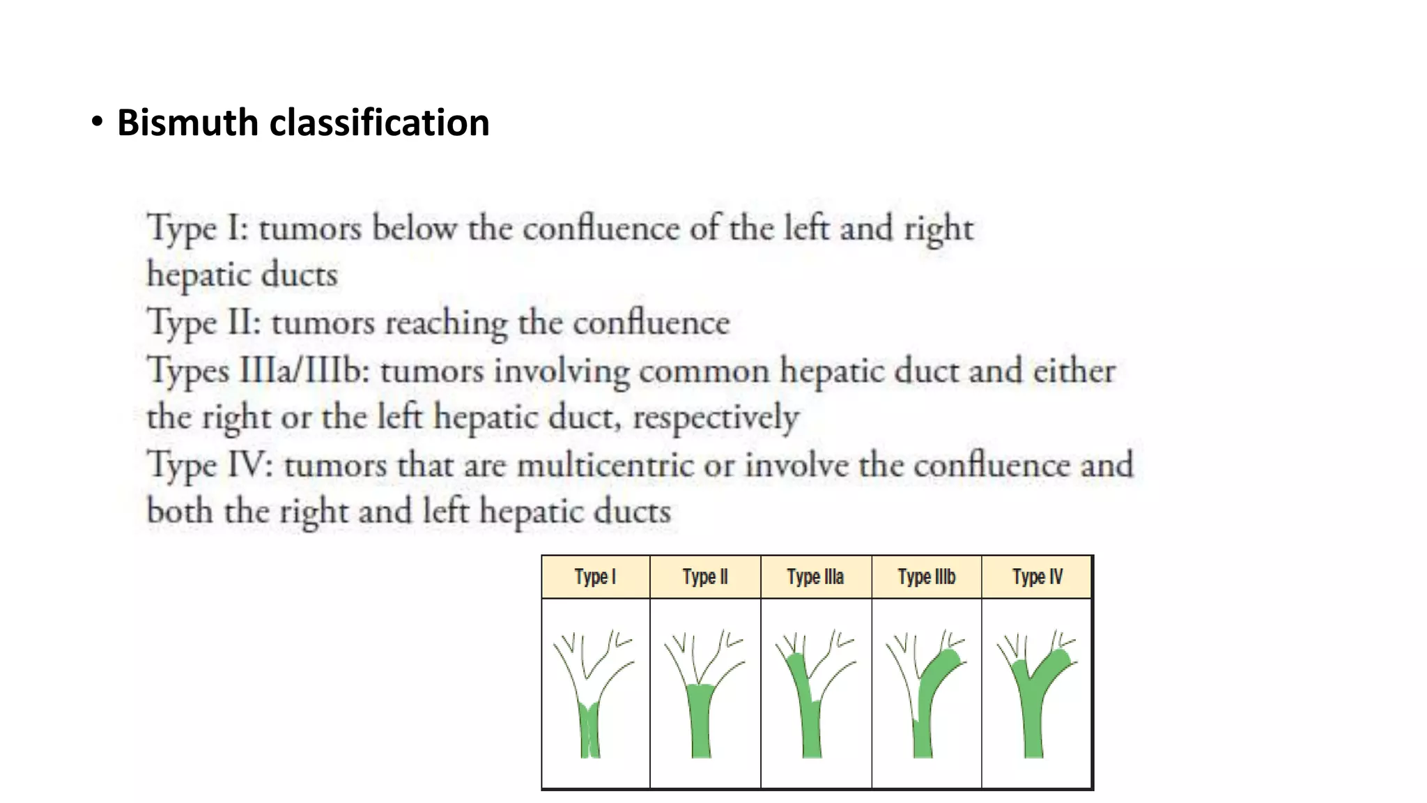
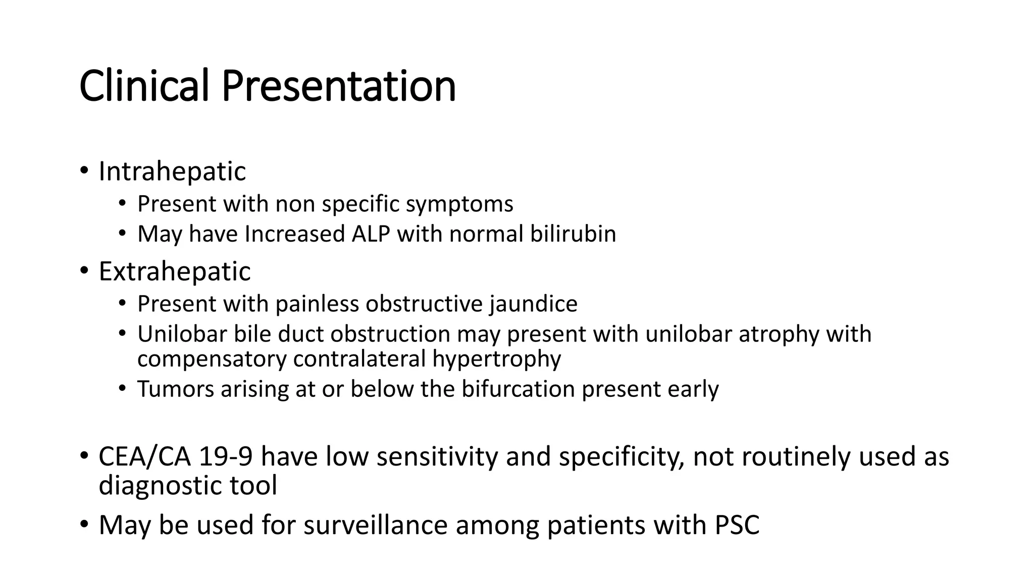
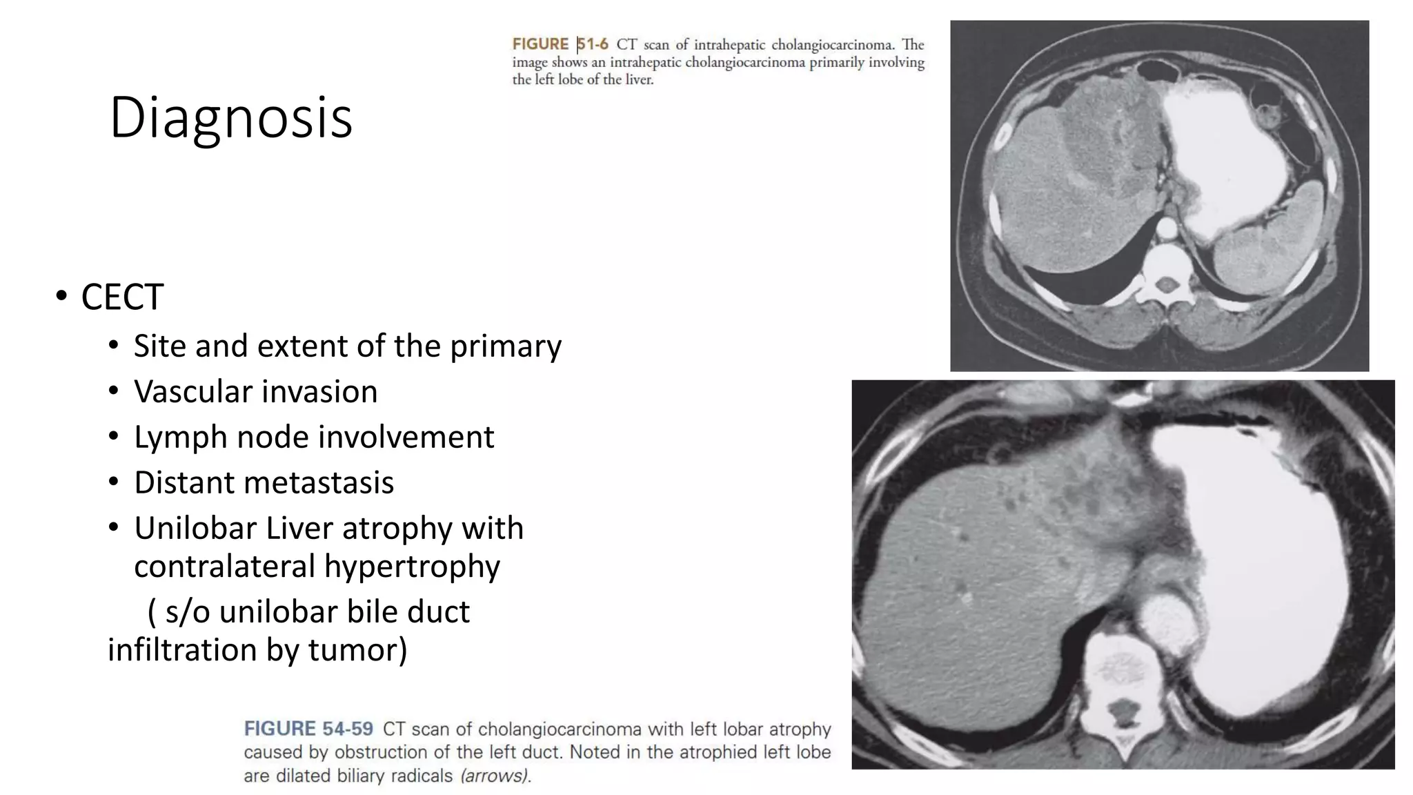
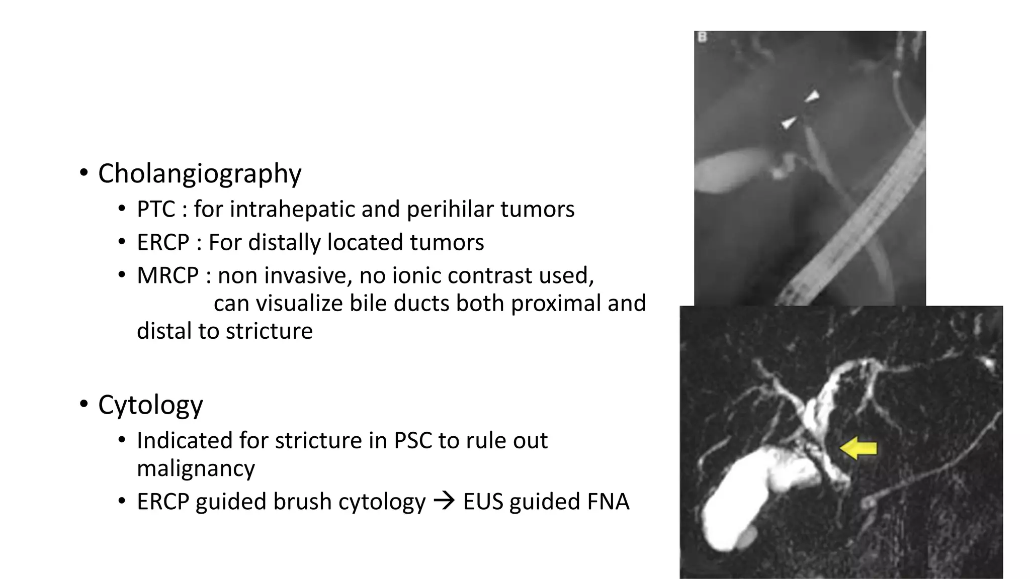
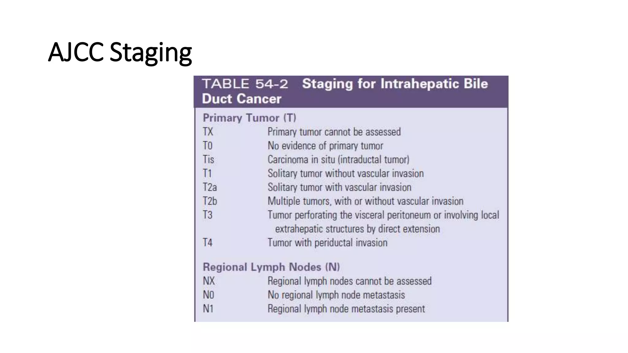
Biliary Tract Cancer
Presented By : Dr Ankit Lalchandani
Moderated By : Dr MP Singh
GB Cancer
• High prevalence in north India
• Incidence increases with age
• Women > men
• Risk Factor : chronic inflammation due to gall stones
• Gall stone present in 70-90% patients
• But only 0.5-3 % patients with gall stones develop GB cancer
• Others :
• Porcelain GB
• Adenomatous polyps
• S. Typhi infection
• Radon exposure
• APBDJ
• Abnormal Pancreaticobiliary Duct Junction
• Long common channel and increased tone of Sphincter of Oddi
• Reflux of pancreatic secretions into CBD chronic inflammation
• Carcinoma occur at a younger age
• Not associated with cholelithiasis
Pathogenesis
p53
CarcinomaCISDysplasia
Chronic
inflammation
APBDJ
Gall
Stone
K ras
• Histology
• Adenocarcinoma ( 80%)
• Small cell
• Squamous cell CA
• Lymphoma
• Morphology
• Infiltrative : diffuse growth, difficult to recognize on imaging, metastasize
early
• Papillary : Project into the lumen, less likely to metastasize , best prognosis
Clinical Presentation and diagnosis
• Symptoms
• Early : asymptomatic / mild abdominal pain, anorexia, nausea
• Advanced : weight loss, hepatomegaly, ascites
• Blood investigations
• Suggest obstructive jaundice
• Tumor markers CEA/ CA 19-9 may be elevated ( low sensitivity and specificity)
Diagnostic Imaging
• USG abdomen
• Asymmetrical wall thickening
• GB mass
• Loss of normal GB- liver interface
• CECT Abdomen
• To assess local invasion
• Vascular invasion
• Lymph node involvement
• Distant Metastasis
• MRI/MRCP
• Delineates invasion into porta hepatis
• ERCP/PTC :
• used primarily for palliation or preoperative management of obstructive
jaundice
• FNAC/ Biopsy :
• Contraindicated if imaging features suggestive of resectable disease
AJCC Staging
Surgical Management
• Macroscopically complete surgical resection with negative margin
(R0) remains the only curative treatment
T1a
• Tumors confined to lamina propria
• Incidental finding post cholecystectomy
• Simple cholecystectomy alone is definitive
( 5yr survival 97-99%, Recurrence 0.6-3.4 %)
T1b
• Tumors invade muscularis propria
• Rates of residual disease in GB fossa after simple cholecystectomy ~10%
• Rate of lymph node positivity ~15%
• Extended cholecystectomy with en bloc resection of adjacent liver
parenchyma to include segment IVb and V + Regional LN dissection
• Bile duct resection only if cystic duct margin is positive
T2
• Tumors extend through perimuscular connective
tissue
• Nodal positivity rate 39-46%
• Recommended Tx : same as T1b
• Simple cholecystectomy is done in subserosal plane,
increased risk of residual disease
• If diagnosed postcholecystectomy : Re exploration
and radical resection and re excision of all port sites
T3
• Tumors invade GB serosa and/or invade
liver or an adjacent organ
• Major hepatic resections may provide
survival advantage if disease is limited
to periportal lymph nodes
• 5 yr survival 16-39%
• As GB fossa bridges IVb and V ,
extended right hepatectomy may be
required
T4
• Tumors invade 2 or more adjacent organs or invade main portal vein/hepatic
artery
• Unresectable
Lymph Node dissection :
• Portal LN dissection recommended for T1b –T4
(porta hepatis, gastrohepatic ligament, retroduodenal)
• AHPBA recommends at least 6 LN to be dissected
• N2 disease ( celiac, retropancreatic, inter aortocaval) is unresectable
Staging Laparoscopy:
• Identifies those with unresectable disease when imaging studies are
equivocal
• High yield in T3 disease (30-50%)
Adjuvant Therapy :
• EBRT +/- 5 FU is associated with low rates of local recurrence
• Not standard recommendation
Palliation
• Goals :
• Relieve pain : Opioid analgesics
• Biliary obstruction : ERCP and stenting/ PTBD
• Bowel obstruction : Endoscopic Duodenal stenting
• Chemotherapy : Gemcitabine + cisplatin ( ABC-02 trial)
Summary
Cholecystectomy
Extended
Chole.
Major hepatic
resection
Palliation
Cholangiocarcinoma
• Involves intrahepatic and extrahepatic biliary channels
• Incidence men > women
• Risk Factors
• Primary Sclerosing cholangitis
• Liver flukes (Opisthorchis and chlonorchis)
• Choledochal cyst
• Carolis disease
• Hepatolithiasis
• Thorotrast
• Hepatitis C
• 90% are adenocarcinomas
• Morphology :
• Sclerosing : intense desmoplastic reaction, highly invasive, low resectability
• Nodular : constriction annular lesions, low resectability
• Papillary : rare, bulky masses projecting into the lumen, cause jaundice early,
high resectability
• Location :
• Intrahepatic (10%)
• Perihilar (65%)
• Distal ( 25%)
• Bismuth classification
Clinical Presentation
• Intrahepatic
• Present with non specific symptoms
• May have Increased ALP with normal bilirubin
• Extrahepatic
• Present with painless obstructive jaundice
• Unilobar bile duct obstruction may present with unilobar atrophy with
compensatory contralateral hypertrophy
• Tumors arising at or below the bifurcation present early
• CEA/CA 19-9 have low sensitivity and specificity, not routinely used as
diagnostic tool
• May be used for surveillance among patients with PSC
Diagnosis
• CECT
• Site and extent of the primary
• Vascular invasion
• Lymph node involvement
• Distant metastasis
• Unilobar Liver atrophy with
contralateral hypertrophy
( s/o unilobar bile duct
infiltration by tumor)
• Cholangiography
• PTC : for intrahepatic and perihilar tumors
• ERCP : For distally located tumors
• MRCP : non invasive, no ionic contrast used,
can visualize bile ducts both proximal and
distal to stricture
• Cytology
• Indicated for stricture in PSC to rule out
malignancy
• ERCP guided brush cytology  EUS guided FNA
AJCC Staging
Surgical Management
Intrahepatic Cholangiocarcinoma
• Major hepatic resection with negative margins is curative
( +/- EHBD, vascular resction)
• 5 yr survival in R0(39%) vs R1(4.7%)
• Surgery recommended only if R0 possible
• Contraindications:
• Involvement of inflow and outflow bilaterally
• Multiple intrahepatic tumors
• Metastatic disease
• Lymph node dissection
• No therapeutic value
• May help in staging and prognosis
Perihilar Cholangiocarcinoma
• R0 resection may require partial hepatectomy along with EHBD
resection
• Include resection of caudate lobe for tumors involving confluence
• Frozen section should be performed to ensure negative margins
• Secondary R0 resection vs primary R0
• Survival is equivalent
• Increased incidence of biliary fistula after additional resection
• After R0, 5 yr survival (20-40%, median 36 months) with high
recurrence rates (68% within 24 months)
• Contraindications
• Hepatic duct involvement with tumor extension bilaterally to second order radilcles
• Encasement of main portal vein
• Lobar atrophy with tumor involvement of contralateral second order biliary radicles
• Lobar atrophy with tumor involvement of contralateral portal vein branches
• Distant metastasis
• Lymph Node dissection
• Include nodes along hepatoduodenal ligament
• Inclusion of lymph nodes along common hepatic or coeliac axis is not recommended
• For accurate staging, at least seven LN recommended
Distal Cholangiocarcinoma
• Most commonly along the pancreatic portion
• High rates of lymph node (63%) and pancreatic invasion (87%)
• Resection involves pancreaticoduodenectomy and lymphadenectomy
• After R0, 5 yr survival ( 27-44%), median survival 18 months
Palliation
• Goal : Relieve biliary obstruction
• Biliary stenting
• Percutaneous : proximal tumors
• Endoscopic : Distal tumors
• Bismuth Type 1 require single stent while others may require two or
more
• Plastic stents patency (3-6 months) vs metal stents ( 8-12 mo)
• Chemotherapy : Gemicitabine + cisplatin
THANK YOU

Biliary tract cancer

  • 1.
    Biliary Tract Cancer PresentedBy : Dr Ankit Lalchandani Moderated By : Dr MP Singh
  • 2.
    GB Cancer • Highprevalence in north India • Incidence increases with age • Women > men • Risk Factor : chronic inflammation due to gall stones • Gall stone present in 70-90% patients • But only 0.5-3 % patients with gall stones develop GB cancer
  • 3.
    • Others : •Porcelain GB • Adenomatous polyps • S. Typhi infection • Radon exposure • APBDJ
  • 4.
    • Abnormal PancreaticobiliaryDuct Junction • Long common channel and increased tone of Sphincter of Oddi • Reflux of pancreatic secretions into CBD chronic inflammation • Carcinoma occur at a younger age • Not associated with cholelithiasis
  • 5.
  • 6.
    • Histology • Adenocarcinoma( 80%) • Small cell • Squamous cell CA • Lymphoma • Morphology • Infiltrative : diffuse growth, difficult to recognize on imaging, metastasize early • Papillary : Project into the lumen, less likely to metastasize , best prognosis
  • 7.
    Clinical Presentation anddiagnosis • Symptoms • Early : asymptomatic / mild abdominal pain, anorexia, nausea • Advanced : weight loss, hepatomegaly, ascites • Blood investigations • Suggest obstructive jaundice • Tumor markers CEA/ CA 19-9 may be elevated ( low sensitivity and specificity)
  • 8.
    Diagnostic Imaging • USGabdomen • Asymmetrical wall thickening • GB mass • Loss of normal GB- liver interface
  • 9.
    • CECT Abdomen •To assess local invasion • Vascular invasion • Lymph node involvement • Distant Metastasis • MRI/MRCP • Delineates invasion into porta hepatis
  • 10.
    • ERCP/PTC : •used primarily for palliation or preoperative management of obstructive jaundice • FNAC/ Biopsy : • Contraindicated if imaging features suggestive of resectable disease
  • 11.
  • 13.
    Surgical Management • Macroscopicallycomplete surgical resection with negative margin (R0) remains the only curative treatment T1a • Tumors confined to lamina propria • Incidental finding post cholecystectomy • Simple cholecystectomy alone is definitive ( 5yr survival 97-99%, Recurrence 0.6-3.4 %)
  • 14.
    T1b • Tumors invademuscularis propria • Rates of residual disease in GB fossa after simple cholecystectomy ~10% • Rate of lymph node positivity ~15% • Extended cholecystectomy with en bloc resection of adjacent liver parenchyma to include segment IVb and V + Regional LN dissection • Bile duct resection only if cystic duct margin is positive
  • 15.
    T2 • Tumors extendthrough perimuscular connective tissue • Nodal positivity rate 39-46% • Recommended Tx : same as T1b • Simple cholecystectomy is done in subserosal plane, increased risk of residual disease • If diagnosed postcholecystectomy : Re exploration and radical resection and re excision of all port sites
  • 16.
    T3 • Tumors invadeGB serosa and/or invade liver or an adjacent organ • Major hepatic resections may provide survival advantage if disease is limited to periportal lymph nodes • 5 yr survival 16-39% • As GB fossa bridges IVb and V , extended right hepatectomy may be required
  • 17.
    T4 • Tumors invade2 or more adjacent organs or invade main portal vein/hepatic artery • Unresectable Lymph Node dissection : • Portal LN dissection recommended for T1b –T4 (porta hepatis, gastrohepatic ligament, retroduodenal) • AHPBA recommends at least 6 LN to be dissected • N2 disease ( celiac, retropancreatic, inter aortocaval) is unresectable
  • 18.
    Staging Laparoscopy: • Identifiesthose with unresectable disease when imaging studies are equivocal • High yield in T3 disease (30-50%) Adjuvant Therapy : • EBRT +/- 5 FU is associated with low rates of local recurrence • Not standard recommendation
  • 19.
    Palliation • Goals : •Relieve pain : Opioid analgesics • Biliary obstruction : ERCP and stenting/ PTBD • Bowel obstruction : Endoscopic Duodenal stenting • Chemotherapy : Gemcitabine + cisplatin ( ABC-02 trial)
  • 20.
  • 21.
    Cholangiocarcinoma • Involves intrahepaticand extrahepatic biliary channels • Incidence men > women • Risk Factors • Primary Sclerosing cholangitis • Liver flukes (Opisthorchis and chlonorchis) • Choledochal cyst • Carolis disease • Hepatolithiasis • Thorotrast • Hepatitis C
  • 22.
    • 90% areadenocarcinomas • Morphology : • Sclerosing : intense desmoplastic reaction, highly invasive, low resectability • Nodular : constriction annular lesions, low resectability • Papillary : rare, bulky masses projecting into the lumen, cause jaundice early, high resectability • Location : • Intrahepatic (10%) • Perihilar (65%) • Distal ( 25%)
  • 23.
  • 24.
    Clinical Presentation • Intrahepatic •Present with non specific symptoms • May have Increased ALP with normal bilirubin • Extrahepatic • Present with painless obstructive jaundice • Unilobar bile duct obstruction may present with unilobar atrophy with compensatory contralateral hypertrophy • Tumors arising at or below the bifurcation present early • CEA/CA 19-9 have low sensitivity and specificity, not routinely used as diagnostic tool • May be used for surveillance among patients with PSC
  • 25.
    Diagnosis • CECT • Siteand extent of the primary • Vascular invasion • Lymph node involvement • Distant metastasis • Unilobar Liver atrophy with contralateral hypertrophy ( s/o unilobar bile duct infiltration by tumor)
  • 26.
    • Cholangiography • PTC: for intrahepatic and perihilar tumors • ERCP : For distally located tumors • MRCP : non invasive, no ionic contrast used, can visualize bile ducts both proximal and distal to stricture • Cytology • Indicated for stricture in PSC to rule out malignancy • ERCP guided brush cytology  EUS guided FNA
  • 27.
  • 30.
    Surgical Management Intrahepatic Cholangiocarcinoma •Major hepatic resection with negative margins is curative ( +/- EHBD, vascular resction) • 5 yr survival in R0(39%) vs R1(4.7%) • Surgery recommended only if R0 possible
  • 31.
    • Contraindications: • Involvementof inflow and outflow bilaterally • Multiple intrahepatic tumors • Metastatic disease • Lymph node dissection • No therapeutic value • May help in staging and prognosis
  • 32.
    Perihilar Cholangiocarcinoma • R0resection may require partial hepatectomy along with EHBD resection • Include resection of caudate lobe for tumors involving confluence • Frozen section should be performed to ensure negative margins • Secondary R0 resection vs primary R0 • Survival is equivalent • Increased incidence of biliary fistula after additional resection • After R0, 5 yr survival (20-40%, median 36 months) with high recurrence rates (68% within 24 months)
  • 33.
    • Contraindications • Hepaticduct involvement with tumor extension bilaterally to second order radilcles • Encasement of main portal vein • Lobar atrophy with tumor involvement of contralateral second order biliary radicles • Lobar atrophy with tumor involvement of contralateral portal vein branches • Distant metastasis • Lymph Node dissection • Include nodes along hepatoduodenal ligament • Inclusion of lymph nodes along common hepatic or coeliac axis is not recommended • For accurate staging, at least seven LN recommended
  • 34.
    Distal Cholangiocarcinoma • Mostcommonly along the pancreatic portion • High rates of lymph node (63%) and pancreatic invasion (87%) • Resection involves pancreaticoduodenectomy and lymphadenectomy • After R0, 5 yr survival ( 27-44%), median survival 18 months
  • 35.
    Palliation • Goal :Relieve biliary obstruction • Biliary stenting • Percutaneous : proximal tumors • Endoscopic : Distal tumors • Bismuth Type 1 require single stent while others may require two or more • Plastic stents patency (3-6 months) vs metal stents ( 8-12 mo) • Chemotherapy : Gemicitabine + cisplatin
  • 36.