
Recommended
More Related Content
What's hot
What's hot (20)
Similar to Airway assessment in anaesthesia
Similar to Airway assessment in anaesthesia (20)
Recently uploaded
Recently uploaded (20)
Airway assessment in anaesthesia
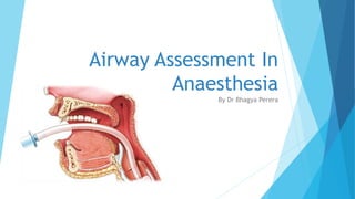
- 1. Airway Assessment In Anaesthesia By Dr Bhagya Perera
- 2. What is the Airway? ‘Airway’ refers to the normal passageway for air entry and exit in a human being for efficient gas exchange at the lungs. The airway includes the nose, mouth, pharynx, larynx and trachea. Maintenance of Airway patency and continuous gas exchange is of paramount importance in general anesthesia.
- 3. What is a difficult airway? A difficult airway is defined as the clinical situation in which a conventionally trained anesthesiologist experiences difficulty with facemask ventilation of the upper airway, difficulty with tracheal intubation, or both. The difficult airway represents a complex interaction between patient factors, the clinical setting, and the skills of the practitioner. Difficult tracheal intubation accounts for 17% of all respiratory injuries and results in significant morbidity and mortality due to respiratory injuries and hypoxia.
- 4. Statistics of Difficult Ventilation and Airway Intubation: Difficult Ventilation: The event where a trained Anesthetist is unable to maintain SpO2 at >90% on 100% inspired O2 via Facemask, provided Pre- induction SpO2 levels were normal. Occurs in 0.1- 5% cases. Difficult Intubation: More than 3 attempts, Longer than 10 mins, Optimal best attempt has failed. Difficult LMA – 0.2- 1% cases Difficult Intubation Normal Surgical Population: 1-2% cases Patients with compromised neck extension (RA, CS) : 40- 50% cases
- 5. Goals of Airway Assessment To identify potentially difficult airway intubations prior to surgery and anesthesia To identify possible risks associated with certain airway intubations in individual patients To minimize the risk of respiratory events both intra and post-operatively.
- 7. History Age Dentures/Loose teeth Snoring/ Sleep Apnoea Cervical Spine Abnormalities: RA, Cervical Spondylosis, Ankylosing Spondylosis Previous Surgeries in the neck : Tracheostomy/ Thyroidectomy Mandibular/ TMJ abnormalities: Prev Hemi-mandibulectomy, TMJ dysfunction Previous airway problems/ difficult intubations/ stenosis/ reaction to anaesthetics Examination Face: facial deformities : Receding chin, small submandibular space Teeth: Malocclusion overbite of anterior teeth Limited protrusion of the Mandible/ Limited mouth opening Loose teeth or dentures Large tongue Neck: Obesity, Short neck, Dorsocervical fat pad, Limited neck movement
- 8. Airway Assessment TESTS: Always better to use a combination of tests Oropharynx Atlanto-Occipital Joint Submandibular Space
- 9. Oropharynx : Mallampati Test Aims: To assess mouth opening and tongue and pharyngeal size Relative assessment of view of pharynx Patient seated in neutral position looking directly ahead Ask Patient to open mouth wide and protrude tongue as far as possible without phonation. Must be examined with eyes kept in line with the patient’s mouth. Findings are then compared against preset classification.
- 10. Mallampati Test: Classification Back of the Pharynx
- 11. Atlanto-Occipital Joint Movement Head and Neck Extension Head Extension at Atlanto-occipital joint and flexion at Atlanto-axial joint brings the laryngeal and pharyngeal planes almost to a straight line This must be examined in the seated position to avoid error due to bowing of the cervical spine. A neck extension of 35o degrees is considered normal.
- 12. Sterno-mental Distance The patient is asked to extend the neck and look at the ceiling and the distance from the supra-sternal notch to the tip of the chin (mandible) is measured. The critical distance is 12.5cm
- 13. Submandibular Space - Thyro-mental distance With the patient’s head extended, the sub-mandibular space is measured by the distance between the tip of the chin and the thyroid notch in cm or finger breaths. Normal finding would be >6.5cm or >3finger breadths. <6cm is considered difficult intubation. Smaller sub-mandibular space may mean obstruction of line of vision of pharyngeal space. A Miller-blade may be preferred in laryngoscopy.
- 15. In the field: 3,3,2
- 17. Cormack-Lehane Direct Laryngoscopy classification Epiglottis Vocal Cords Arytenoids
- 19. LEMON RULE The LEMON rule allows us to remember to look externally and to look at those parameters that will make the intubation simple or difficult. L.E.M.O.N stands for: L – Look externally – Is the patient obese, do they have a high arched palate, a short neck, facial or neck trauma? Extensive beard? E – Evaluate the 3:3:2 rule – 3cm/finger breaths mouth opening, 3cm thyro-mental distance, 2cm between hyoid bone and thyroid notch. M – Mallampati Score –a Mallampati Class 4 is associated with a >10% chance of difficult airway O – Obstruction – Is there a tumour, epiglottitis, recent neck surgery? N – Neck mobility – Is the patient in a cervical collar, are they elderly?
- 20. Wilson scoring Predicts difficult airway intubations using five factors Each factor is scored between 0- 2 A total score is calculated between 0-10 A higher score indicating a higher incidence of difficulty intubating the patient. Factors Weight Head and Neck Movement Jaw Movement Receding Mandible Buck Teeth
- 21. What to do when suspecting a Difficult Airway Intubation
- 22. What to do when suspecting a Difficult Airway Intubation Get help. Confer with a senior colleague or Consultant Anesthetist. Get advice on how to proceed with the intubation. Be ready for every case scenario Check and be prepared with the difficult intubation trolley Confer with Surgical team. Consider alternative methods or anesthesia such as regional anesthesia (Ex Epidural, Spinal Anesthesia, Local anesthesia) Consider Supraglottic Airway (LMAs)
- 23. Difficult Intubation Strategy A: • Airway Management B: • Breathing: Oxygenation and Ventilation C: • Continue Surgery/ Awaken and Postpone D: • Danger! CVCI Plan: Rescue Techniques E: • Extubation Strategy: Awake Extubation F: • Follow up
- 25. Difficult Intubation Trolley Oro and nasopharyngeal airways of all sizes Laryngoscopes 2 – Batteries/ Light source checked Blades- 4 sizes, Mackintosh, Miller, Mc Coy blades, straight blades Endotracheal tubes many sizes Stylet and Bougie LMAs all sizes, ILMA Combi tube (blind insertion) Wee’s Oesophageal detector Video Laryngoscope Tracheostomy tube + Set Cricothyroidotomy Set
- 27. Take Home Messages Analyse all prospective GA patients for risk of difficult intubation. Identifying the risk early gives Anesthetist time to anticipate and prepare for the problem. If suspicious of possible difficult airway: Secure the airway while awake. Not maintaining airway? Oxygenate and restore Spontaneous breathing. Attempt to secure airway with more skilled personnel. Cannot ventilate Cannot intubate?: Move to Rescue measures. Follow up to ensure future procedures undergo proper risk assessment of patient.
- 28. References British Journal Of Anesthesia – Oxford University Press Online Article Archive Handbook of Anesthesia- College of Anesthesiologists, Sri Lanka Practice Guidelines for Difficult Airway Management- American Society of Anesthesiologists (Special Issue 2013) NCBI- NIH- US National Library of Medicine Elsevier Open Access Journals Smith and Atkinhead’s Textbook of Anesthesia East Midlands Emergency Medicine edu media. Resus Austrailia