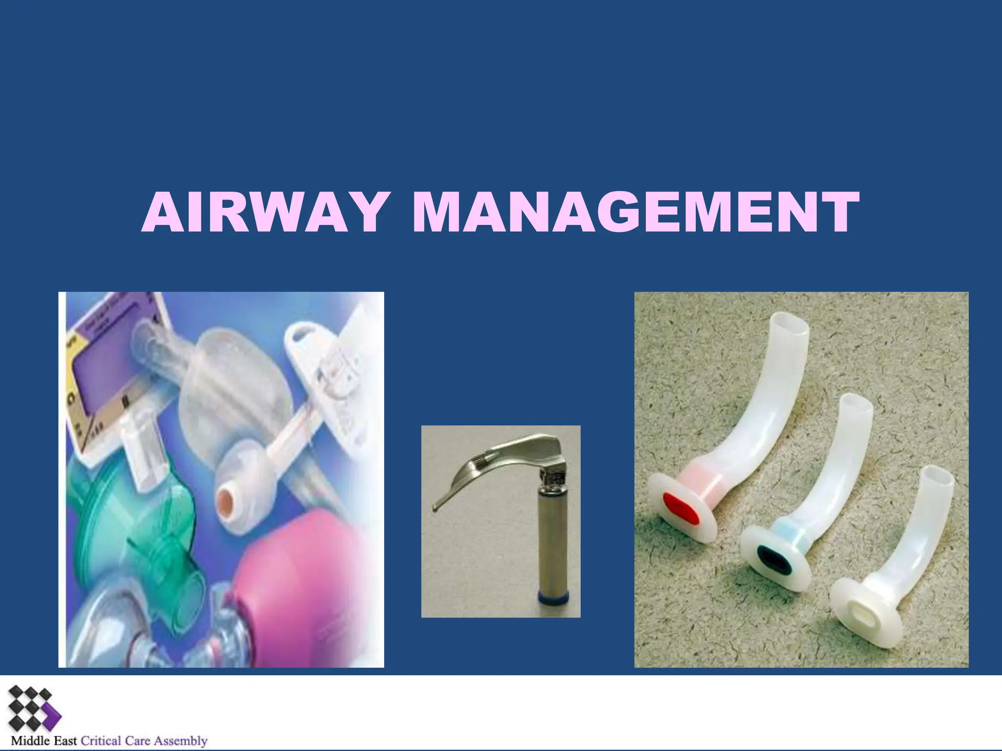
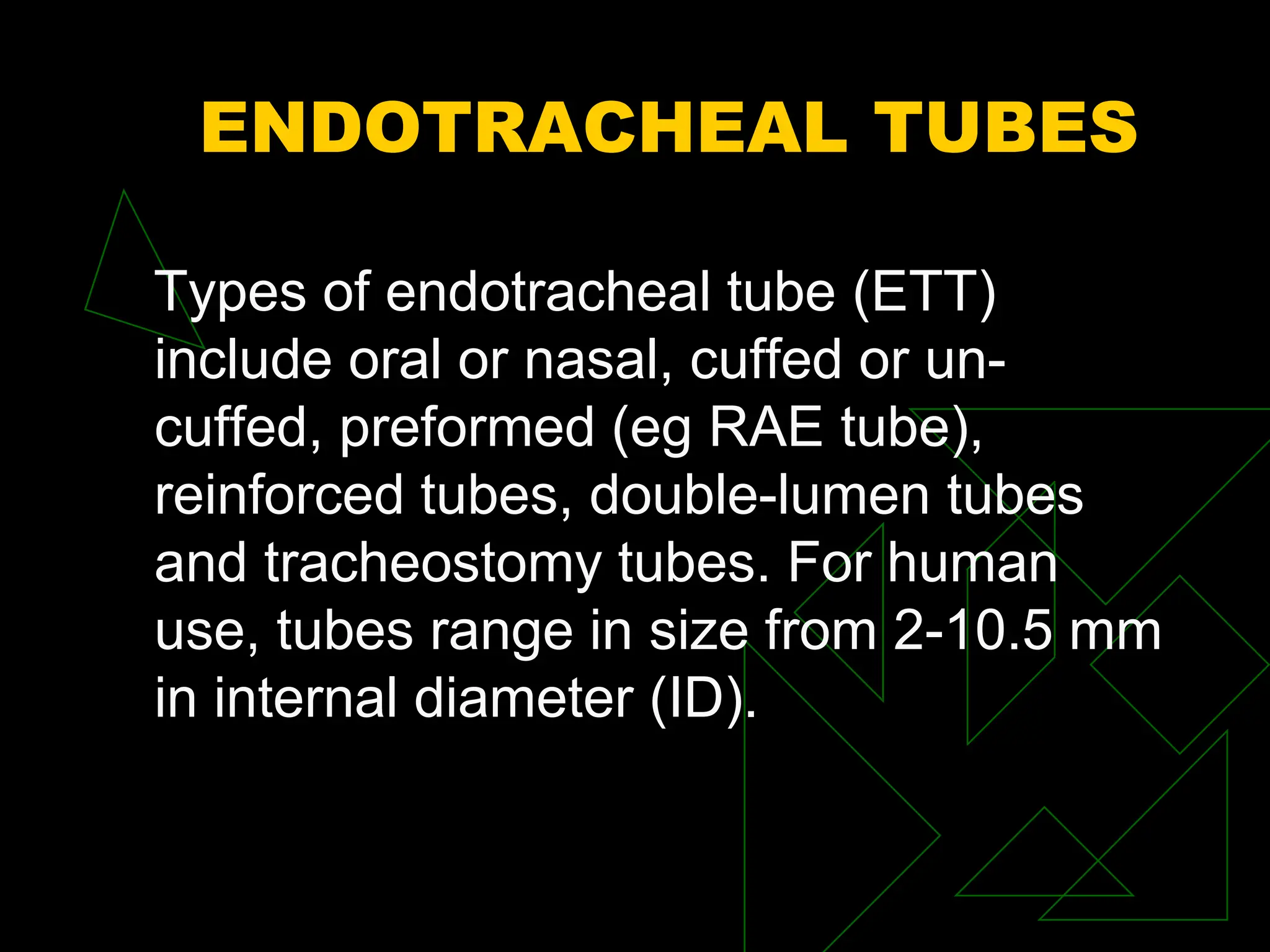
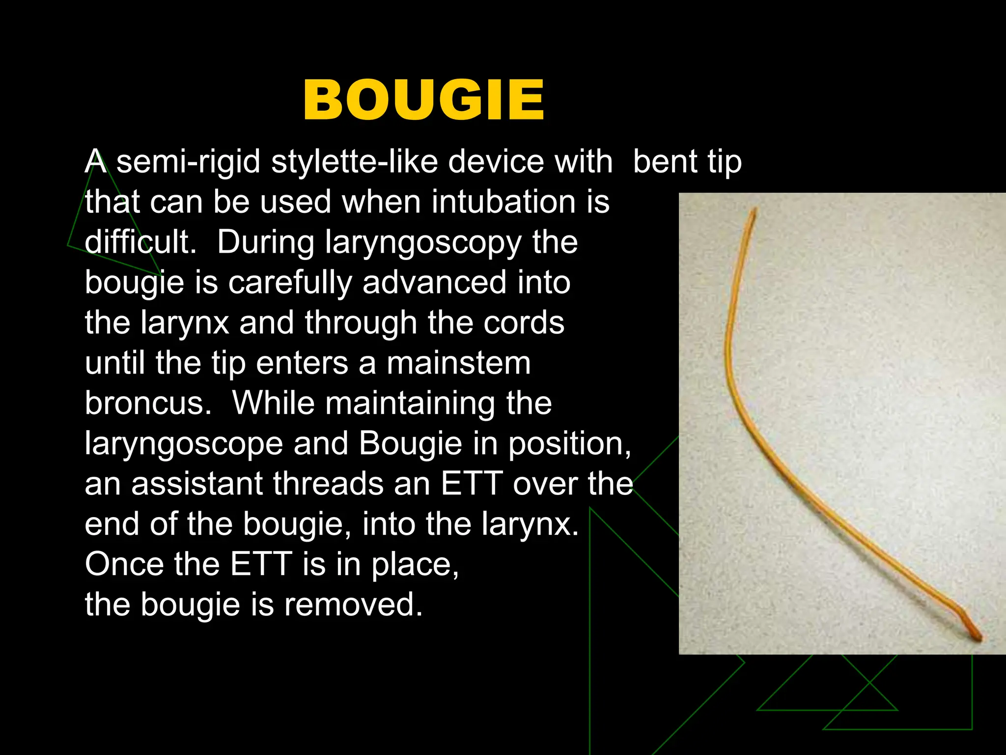
AIRWAY MANAGEMENT
OBJECTIVES
 Identify indications for intubation and prepare
the necessary equipment.
 Identify the advantages and disadvantages of
various devices for airway management.
 Identify difficult airway.
 Identify equipment for difficult airway and
know their use.
INDICATIONS OF
INTUBATION
 Cardiopulmonary Arrest
 Patient in coma
 Tachpnea/ Bradypnea
 Progressive cyanosis
 Surgical patients
 Airway protection from any cause
ADVANTAGES
 Provides an unobstructed airway
 Prevents aspiration of secretions into the
lungs
 Facilitates positive pressure ventilation
without gastric inflation
 Facilitates body positioning and movement
 May be utilized to deliver medication
 Narcan
 Atropine
 Epinephrine
 Lidocaine
DISADVANTAGES
 Needs advanced training to properly perform
the procedure
 Bypasses function of the nose to warm and
filter the inspired air
 Increased incidence of trauma due to neck
manipulation when spinal cord injury is
suspected
 May increase respiratory resistance
 Improper placement
INTUBATION ROLL
 Rigid Laryngoscopes
 Laryngoscope blades different sizes and
types
 ETT of various sizes
 Flexible Stylets
 Oral airways
 Exhaled CO2 detector
 ETT fixation device
 Lubricant gel
 Syringe
ENDOTRACHEAL TUBES
Types of endotracheal tube (ETT)
include oral or nasal, cuffed or un-
cuffed, preformed (eg RAE tube),
reinforced tubes, double-lumen tubes
and tracheostomy tubes. For human
use, tubes range in size from 2-10.5 mm
in internal diameter (ID).
 Endotracheal tubes are made from red rubber
and Polyvinylchloride. Those placed in a laser
field may be flexometallic.
REINFORCED ETT
Indications For Usage
 Patient's head is in extended
or flexed position
 Patient will be turned over
 Long-term cases
 Neurosurgical procedures
 Head and neck procedures
NASAL AND ORAL RAE
 NASAL
RAE TUBES II
 Preformed Endotracheal Tubes are designed
to conveniently position the anesthesia circuit
out of the surgical field for oral and
maxillofacial procedures.
 Oral Preformed shape directs tube downward,
to rest on patients chin
 Cuffed tubes available with Murphy Eye only
 Uncuffed tubes have two Murphy Eyes for
enhanced patient safety
 Bold marks at the center of bend with
distance to distal tip indicated
ENDOBRONCHIAL TUBE
Indications for
usage
 Thoracic surgery
 Broncho-spirometry
 Thoracoscopies
 Differential or
selective lung
ventilation
 Lung Lavage
ENDOBRONCHIAL TUBE
WITH CPAP SYSTEM
Indications For Usage
 Thoracic surgery
 Broncho-spirometry
 Thoracoscopies
 Differential or selective
lung ventilation
CONFIRMATION OF ETT
PLACEMENT
ETCO2 DETECTORS
 Single use to verify ETT placement
 Reliable carbon dioxide detectors help verify
ETT placement
 Responds quickly to exhaled CO2 with a
simple color change from purple to yellow
 Breath-to-breath response
 Constant visual feedback for up to 2 hours
Correct ET Tube Placement:
Capnography
Purpul Yellow
3-4 cm
Correct ET Tube
Placement
Correct ET Tube
Placement
Secure ET tube in place, note the number
Sedate patient with appropriate MAAS
Avoid accidental, or self extubation
SECURING THE AIRWAY
COMFIT™ ETT Holder
 The tapeless way to secure an ETT
 Completely adjustable
 Wide cotton-lined neckband minimizes skin
irritation, providing maximum patient
comfort
 Minimal plastic loop around the ET tube
allows access to the oral cavity
 Economical in two ways: low initial cost, no
frequent changing
 Latex-free product
COMFIT
EASY CAP II , PEDICAP
Easy Cap II Pedi-Cap
Weight over 15kg Weight 1kg-
15kg
Dead space25cc Dead space3
cc
Time 2 hours Time 2 hours
Tracheal Tube Cuff
Care
 These include bedside sphygmomanometers,
special aneroid cuff manometers, and
electronic cuff pressure devices.
 Ideally, most tubes seal at pressures between
14 and 20 mm Hg (19 to 27 cm H2O).
 Tracheal capillary pressure lies between 20
and 30 mm Hg
 Impairment in tracheal blood flow seen at 22
mm Hg and total obstruction seen at 37 mm
Hg
Sphygmomanometers
High Volume Low
Pressure Tubes
Minimum Leak Volume
Technique
 Air inflation of the tube cuff until the airflow
heard escaping around the cuff during
positive pressure breath ceases.
 Place a stethoscope over larynx. Indirectly
assesses inflation of cuff.
 Slowly withdraw air (in 0.1-mL increments)
until a small leak is heard on inspiration.
 Remove syringe tip, check inflation of pilot
balloon
SECRETION CLEARANCE
OPEN SUCTION SYSTEM
 Made of non-toxic PVC
 Available coded for size identification Closed
suction systems
CLOSED SUCTION SYSTEM
 (CSS) are increasingly replacing open suction
systems (OSS) to perform endotracheal toilet
in mechanically ventilated intensive care unit
patients.
Endotracheal or Tracheostomy
Tube Suctioning
Open Suctioning
Disconnection from the
ventilator
Not recommended when
PEEP >10
Closed Suctioning:
Facilitate continuous
mechanical ventilation and
oxygenation during the
suctioning.
Indicated when PEEP level
above 10cmH2O
Open Suctioning Technique
Closed Suctioning Technique
ETT WITH EVACUATION
LUMEN
INDICATIONS
For airway management by
oral/nasal intubation of the
trachea and for evacuation
or drainage of secretion from
the subglottic space
ADVANTAGES OF EVAC
 Helps decrease the rate of ventilator-
associated pneumonia (VAP) in the hospital
and to reduce VAP related costs
 Convenient and safe method for suctioning
accumulated secretions in the subglottic
space
 Large elliptical evacuation port located on
dorsal side proximal to cuff provides effective
evacuation
 Integral suction lumen allows continuous
suctioning without risking trauma to the vocal
cords as with manual catheter suctioning
ETT CARE
 Use of Gause @ the angles of mouth to
prevent damage to mucosa
 Moving ETT Q NOC from one to the other
side to avoid damage to mucosa
 Monitoring the correct position of ETT@ the
lip mark and positioning it properly
 Monitoring the ETT position on CXR from time
to time
 Regular suctioning through ETT
DIFFICULT AIRWAY
LET US SEE…
 What is a difficult airway ?
 The importance of difficult airway cart.
 Different modalities to be used in difficult
airways situations.
 Anticipate Difficult Airway.
 Be Prepared and have many back up plans.
WHAT IS A DIFFICULT
AIRWAY
 According to American Association of
Anesthesiologist, it is a clinical situation in
which a trained anesthesiologist experiences
difficulty with mask ventilation, tracheal
intubation or both.
 Requires more than 3 attempts or 10 min. to
intubate.
 Grade lll to lV in both Cormack and
Mallampadi Classifications.
PRE-INTUBATION
EVALUATION
Potentially difficult laryngoscopy includes:
 Less than 35 degree neck extension.
 Less than 7 cm distance between mandible
and the hyoid bone.
 Less than 12.5 cm sternomandibular distance
with head fully extended.
 Poorly visualized uvula.
 Short, thick neck.
 Receding mandible and protruding teeth.
MALLAMPADI
CLASSIFICATION
 Grade I: soft palate, uvula, tonsillar pillars
visible.
 Grade II: soft palate, uvula visible.
 Grade III: soft palate, base of uvula visible.
 Grade IV: soft palate not visible (100% Grade
lll or Grade lV view).
DIFFICULT AIRWAY
CART
Necessary equipment needed for an
anticipated or unexpected difficult airway
 LMAs
 Combitube
 Bougie
 Oral and nasopahryngeal airways
 Fast Track
 Cricothyrotomy kit
 Tube Exchangers
 Fiberoptic bronchoscope
INTUBATING STYLET
 A stylet for intubating an endotracheal tube is
like medico-surgical tube comprising of a
bendable metal rod sealed in a tubular plastic
sheath. The ends of the sheath are molded in
a smoothly rounded closed shape.
 Passed through an ETT, can be bend to give
ETT the shape of a hockey stick.
.
STYLET
ADVANTAGES
 Alow intubation of the trachea with minimal
visualization of the vocal cords.
 Easy to learn.
 Helps in stablizing the ETT for intubation
DISADVANTAGES
 May be incorrectly inserted and can damage
tracheal tissues.
VARIOUS STYLETS
 Shikani seeing stylet
 Bonfils fiberscope
 Machida Portable Stylet Fibersopce
 Video-Optical Intubation Stylet
 Aeroview
 Schroeder Stylet
 Nanoscope
 Many Others………..
LMA
 The Laryngeal Mask Airway is an
alternative airway device used for
anesthesia and airway support. It consists
of an inflatable silicone mask and rubber
connecting tube. It is inserted blindly into
the pharynx, forming a low-pressure seal
around the laryngeal inlet and permitting
gentle positive pressure ventilation. All
parts are latex-free.
LARYNGEAL MASK AIRWAY
LMA
INDICATIONS
 The Laryngeal Mask Airway is an
appropriate airway for short procedures
and in emergency situations.
 Can be used as rescue airway and
fiberoptic conduit when intubation is
difficult.
 Can be used for bronchoscopy in awake
patients.
LMA
CONTRAINDICATIONS
 Non-fasted patients
 Morbidly obese patients
 Pregnancy
 Obstructive or abnormal lesions of the
oropharynx
 Increased Airway resistance and decreased
lung compliance
VARIOUS SIZES OF LMA
MASK SIZE PATIENT
SIZE
WEIGHT CUFF
VOLUME
1 INFANT <6.5 KG 2-4 ML
2 CHILD 6.5-20 KG UP TO 10 ML
2 1/2 CHILD 20-30 KG UP TO 15 ML
3 SMALL
ADULT
>30 KG UP TO 20 ML
4 NORMAL
ADULT
UP TO 30 ML
LMA
Tips for Success:
 Begin with ASA I & II patients
 Learn and use standard insertion technique
 Use appropriate size and do NOT overinflate
 Maintain adequate anesthetic depth
 Remove when the patient opens mouth to
command
COMBITUBE
 Consists of two fused tubes with a 15 mm
connector at proximal end.
 Contains 2 cuffs, 100 cc proximal and 15 cc
distal.
 Distal lumen usually lies in esophagus so
the gas through blue tube will ventilate
Trachea.
 If Combitube enters trachea, ventilation is
through clear tube. Available in only one
disposable size for age> 15 years , height
>5ft.
COMBITUBE
COMBITUBE II
BOUGIE
A semi-rigid stylette-like device with bent tip
that can be used when intubation is
difficult. During laryngoscopy the
bougie is carefully advanced into
the larynx and through the cords
until the tip enters a mainstem
broncus. While maintaining the
laryngoscope and Bougie in position,
an assistant threads an ETT over the
end of the bougie, into the larynx.
Once the ETT is in place,
the bougie is removed.
ETT EXCHANGER
AIRWAY EXCHANGE
CATHETERS
SIZE (ID) LENGTH
2.5-4.0 56 cm
4.0-6.0 56 cm
6.0-8.5 81 cm
7.5-10.0 81 cm
ETT EXCHANGER
 Facilitates quick, efficient endotracheal tube
exchange or replacement without using a
laryngoscope
 Flexible material, frosted surface and depth
marks aid precise placement and minimize
drag
 Internal lumen allows for spontaneous
breathing during tube exchange
 Longer size allows exchange of the ETT
while exchanger is still in the trachea
 These devices allow insufflation of O2 and jet
ventilation.
ETT EXCHNAGER
ADVANTAGES
 Relatively short learning time
 Allow changing endotracheal tube with
guide still in the trachea e.g. in case of
ruptured ETT cuff
DISADVANTAGE
 Improper placement of ETT may still occur
with these devices if guide is not placed
completely in the trachea
CRICOTHYROTOMY
 Kits that allow introduction of some type of
tube into the trachea via cricothyrotomy .Most
of the kits are designed as temporary airway
and need to be replaced by a tracheostomy
tube after establishment of ventilation and
stabilization of patient
CRICOTHYROTOMY KIT
ADVANTAGES
 Rapid access to subglottic
area
Does not require visualization
of the larynx.
FLEXIBLE FIBEROPTIC
BRONCHOSCOPE
 The fibreoptic bronchoscope is constructed of
fibreoptic bundles and cables encased in a
slender, waterproof sheath from the handle to
the tip.
 The cable system permits manipulation of the
tip of the bronchoscope by adjustments @the
handle, the operating end of the device.
 Excellent visualization of the airway with
minimal homodynamic stress when properly
performed.
FIBEROPTIC
BRONCHOSCOPE
FIBEROPTIC II
Disadvantages
 Expensive
 Requires careful maintenance
 Presence of blood or secretion
 Impairs visualization.
COMPLICATIONS OF
INTUBATION
During intubation
 Esophageal intubation
 Endobronchial intubation
 Damage of tooth, lip, tongue, mucosa
 Increased B.P, HR, ICP, IOP
 Laryngospasm
 Unanticipated difficult airway
 Pt can code and die
COMPLICATIONS OF
INTUBATION
While ETT in place
 Unintentional extubation
 Endobroncial intubation
 Obstruction
 Mucosal inflammation and ulceration
 ETT malfunction
COMPLICATIONS OF
INTUBATION
Following extubation
 Edema and stenosis of glottic, subglottic and
trachesl regions
 Hoarse of voice due to vocal cord paralysis
 Laryngospasm
REFERENCES
 CLINICAL ANESTHESIOLOGY by G.Edward Morgan
and Maged S. Mikhail
 www.nellcor.com
 TEXTBOOK OF ADVANCED CARDIAC LIFE
SUPPORT
THANK YOU
BY
KANWAL SHAHZAD RRT

vdocuments.mx_airway-management-56ecf37df3080.ppt

  • 1.
  • 2.
    OBJECTIVES  Identify indicationsfor intubation and prepare the necessary equipment.  Identify the advantages and disadvantages of various devices for airway management.  Identify difficult airway.  Identify equipment for difficult airway and know their use.
  • 3.
    INDICATIONS OF INTUBATION  CardiopulmonaryArrest  Patient in coma  Tachpnea/ Bradypnea  Progressive cyanosis  Surgical patients  Airway protection from any cause
  • 4.
    ADVANTAGES  Provides anunobstructed airway  Prevents aspiration of secretions into the lungs  Facilitates positive pressure ventilation without gastric inflation  Facilitates body positioning and movement  May be utilized to deliver medication  Narcan  Atropine  Epinephrine  Lidocaine
  • 5.
    DISADVANTAGES  Needs advancedtraining to properly perform the procedure  Bypasses function of the nose to warm and filter the inspired air  Increased incidence of trauma due to neck manipulation when spinal cord injury is suspected  May increase respiratory resistance  Improper placement
  • 6.
    INTUBATION ROLL  RigidLaryngoscopes  Laryngoscope blades different sizes and types  ETT of various sizes  Flexible Stylets  Oral airways  Exhaled CO2 detector  ETT fixation device  Lubricant gel  Syringe
  • 7.
    ENDOTRACHEAL TUBES Types ofendotracheal tube (ETT) include oral or nasal, cuffed or un- cuffed, preformed (eg RAE tube), reinforced tubes, double-lumen tubes and tracheostomy tubes. For human use, tubes range in size from 2-10.5 mm in internal diameter (ID).
  • 9.
     Endotracheal tubesare made from red rubber and Polyvinylchloride. Those placed in a laser field may be flexometallic.
  • 10.
    REINFORCED ETT Indications ForUsage  Patient's head is in extended or flexed position  Patient will be turned over  Long-term cases  Neurosurgical procedures  Head and neck procedures
  • 11.
    NASAL AND ORALRAE  NASAL
  • 12.
    RAE TUBES II Preformed Endotracheal Tubes are designed to conveniently position the anesthesia circuit out of the surgical field for oral and maxillofacial procedures.  Oral Preformed shape directs tube downward, to rest on patients chin  Cuffed tubes available with Murphy Eye only  Uncuffed tubes have two Murphy Eyes for enhanced patient safety  Bold marks at the center of bend with distance to distal tip indicated
  • 13.
    ENDOBRONCHIAL TUBE Indications for usage Thoracic surgery  Broncho-spirometry  Thoracoscopies  Differential or selective lung ventilation  Lung Lavage
  • 14.
    ENDOBRONCHIAL TUBE WITH CPAPSYSTEM Indications For Usage  Thoracic surgery  Broncho-spirometry  Thoracoscopies  Differential or selective lung ventilation
  • 15.
  • 16.
    ETCO2 DETECTORS  Singleuse to verify ETT placement  Reliable carbon dioxide detectors help verify ETT placement  Responds quickly to exhaled CO2 with a simple color change from purple to yellow  Breath-to-breath response  Constant visual feedback for up to 2 hours
  • 17.
    Correct ET TubePlacement: Capnography Purpul Yellow
  • 18.
  • 19.
  • 20.
    Correct ET Tube Placement SecureET tube in place, note the number Sedate patient with appropriate MAAS Avoid accidental, or self extubation
  • 21.
    SECURING THE AIRWAY COMFIT™ETT Holder  The tapeless way to secure an ETT  Completely adjustable  Wide cotton-lined neckband minimizes skin irritation, providing maximum patient comfort  Minimal plastic loop around the ET tube allows access to the oral cavity  Economical in two ways: low initial cost, no frequent changing  Latex-free product
  • 22.
  • 23.
    EASY CAP II, PEDICAP Easy Cap II Pedi-Cap Weight over 15kg Weight 1kg- 15kg Dead space25cc Dead space3 cc Time 2 hours Time 2 hours
  • 24.
    Tracheal Tube Cuff Care These include bedside sphygmomanometers, special aneroid cuff manometers, and electronic cuff pressure devices.  Ideally, most tubes seal at pressures between 14 and 20 mm Hg (19 to 27 cm H2O).  Tracheal capillary pressure lies between 20 and 30 mm Hg  Impairment in tracheal blood flow seen at 22 mm Hg and total obstruction seen at 37 mm Hg
  • 25.
  • 26.
  • 27.
    Minimum Leak Volume Technique Air inflation of the tube cuff until the airflow heard escaping around the cuff during positive pressure breath ceases.  Place a stethoscope over larynx. Indirectly assesses inflation of cuff.  Slowly withdraw air (in 0.1-mL increments) until a small leak is heard on inspiration.  Remove syringe tip, check inflation of pilot balloon
  • 28.
    SECRETION CLEARANCE OPEN SUCTIONSYSTEM  Made of non-toxic PVC  Available coded for size identification Closed suction systems CLOSED SUCTION SYSTEM  (CSS) are increasingly replacing open suction systems (OSS) to perform endotracheal toilet in mechanically ventilated intensive care unit patients.
  • 29.
    Endotracheal or Tracheostomy TubeSuctioning Open Suctioning Disconnection from the ventilator Not recommended when PEEP >10 Closed Suctioning: Facilitate continuous mechanical ventilation and oxygenation during the suctioning. Indicated when PEEP level above 10cmH2O
  • 30.
  • 31.
  • 32.
    ETT WITH EVACUATION LUMEN INDICATIONS Forairway management by oral/nasal intubation of the trachea and for evacuation or drainage of secretion from the subglottic space
  • 33.
    ADVANTAGES OF EVAC Helps decrease the rate of ventilator- associated pneumonia (VAP) in the hospital and to reduce VAP related costs  Convenient and safe method for suctioning accumulated secretions in the subglottic space  Large elliptical evacuation port located on dorsal side proximal to cuff provides effective evacuation  Integral suction lumen allows continuous suctioning without risking trauma to the vocal cords as with manual catheter suctioning
  • 34.
    ETT CARE  Useof Gause @ the angles of mouth to prevent damage to mucosa  Moving ETT Q NOC from one to the other side to avoid damage to mucosa  Monitoring the correct position of ETT@ the lip mark and positioning it properly  Monitoring the ETT position on CXR from time to time  Regular suctioning through ETT
  • 35.
    DIFFICULT AIRWAY LET USSEE…  What is a difficult airway ?  The importance of difficult airway cart.  Different modalities to be used in difficult airways situations.  Anticipate Difficult Airway.  Be Prepared and have many back up plans.
  • 36.
    WHAT IS ADIFFICULT AIRWAY  According to American Association of Anesthesiologist, it is a clinical situation in which a trained anesthesiologist experiences difficulty with mask ventilation, tracheal intubation or both.  Requires more than 3 attempts or 10 min. to intubate.  Grade lll to lV in both Cormack and Mallampadi Classifications.
  • 37.
    PRE-INTUBATION EVALUATION Potentially difficult laryngoscopyincludes:  Less than 35 degree neck extension.  Less than 7 cm distance between mandible and the hyoid bone.  Less than 12.5 cm sternomandibular distance with head fully extended.  Poorly visualized uvula.  Short, thick neck.  Receding mandible and protruding teeth.
  • 38.
    MALLAMPADI CLASSIFICATION  Grade I:soft palate, uvula, tonsillar pillars visible.  Grade II: soft palate, uvula visible.  Grade III: soft palate, base of uvula visible.  Grade IV: soft palate not visible (100% Grade lll or Grade lV view).
  • 39.
    DIFFICULT AIRWAY CART Necessary equipmentneeded for an anticipated or unexpected difficult airway  LMAs  Combitube  Bougie  Oral and nasopahryngeal airways  Fast Track  Cricothyrotomy kit  Tube Exchangers  Fiberoptic bronchoscope
  • 40.
    INTUBATING STYLET  Astylet for intubating an endotracheal tube is like medico-surgical tube comprising of a bendable metal rod sealed in a tubular plastic sheath. The ends of the sheath are molded in a smoothly rounded closed shape.  Passed through an ETT, can be bend to give ETT the shape of a hockey stick. .
  • 41.
    STYLET ADVANTAGES  Alow intubationof the trachea with minimal visualization of the vocal cords.  Easy to learn.  Helps in stablizing the ETT for intubation DISADVANTAGES  May be incorrectly inserted and can damage tracheal tissues.
  • 42.
    VARIOUS STYLETS  Shikaniseeing stylet  Bonfils fiberscope  Machida Portable Stylet Fibersopce  Video-Optical Intubation Stylet  Aeroview  Schroeder Stylet  Nanoscope  Many Others………..
  • 43.
    LMA  The LaryngealMask Airway is an alternative airway device used for anesthesia and airway support. It consists of an inflatable silicone mask and rubber connecting tube. It is inserted blindly into the pharynx, forming a low-pressure seal around the laryngeal inlet and permitting gentle positive pressure ventilation. All parts are latex-free.
  • 44.
  • 45.
    LMA INDICATIONS  The LaryngealMask Airway is an appropriate airway for short procedures and in emergency situations.  Can be used as rescue airway and fiberoptic conduit when intubation is difficult.  Can be used for bronchoscopy in awake patients.
  • 46.
    LMA CONTRAINDICATIONS  Non-fasted patients Morbidly obese patients  Pregnancy  Obstructive or abnormal lesions of the oropharynx  Increased Airway resistance and decreased lung compliance
  • 47.
    VARIOUS SIZES OFLMA MASK SIZE PATIENT SIZE WEIGHT CUFF VOLUME 1 INFANT <6.5 KG 2-4 ML 2 CHILD 6.5-20 KG UP TO 10 ML 2 1/2 CHILD 20-30 KG UP TO 15 ML 3 SMALL ADULT >30 KG UP TO 20 ML 4 NORMAL ADULT UP TO 30 ML
  • 48.
    LMA Tips for Success: Begin with ASA I & II patients  Learn and use standard insertion technique  Use appropriate size and do NOT overinflate  Maintain adequate anesthetic depth  Remove when the patient opens mouth to command
  • 49.
    COMBITUBE  Consists oftwo fused tubes with a 15 mm connector at proximal end.  Contains 2 cuffs, 100 cc proximal and 15 cc distal.  Distal lumen usually lies in esophagus so the gas through blue tube will ventilate Trachea.  If Combitube enters trachea, ventilation is through clear tube. Available in only one disposable size for age> 15 years , height >5ft.
  • 50.
  • 51.
  • 52.
    BOUGIE A semi-rigid stylette-likedevice with bent tip that can be used when intubation is difficult. During laryngoscopy the bougie is carefully advanced into the larynx and through the cords until the tip enters a mainstem broncus. While maintaining the laryngoscope and Bougie in position, an assistant threads an ETT over the end of the bougie, into the larynx. Once the ETT is in place, the bougie is removed.
  • 53.
  • 54.
    AIRWAY EXCHANGE CATHETERS SIZE (ID)LENGTH 2.5-4.0 56 cm 4.0-6.0 56 cm 6.0-8.5 81 cm 7.5-10.0 81 cm
  • 55.
    ETT EXCHANGER  Facilitatesquick, efficient endotracheal tube exchange or replacement without using a laryngoscope  Flexible material, frosted surface and depth marks aid precise placement and minimize drag  Internal lumen allows for spontaneous breathing during tube exchange  Longer size allows exchange of the ETT while exchanger is still in the trachea  These devices allow insufflation of O2 and jet ventilation.
  • 56.
    ETT EXCHNAGER ADVANTAGES  Relativelyshort learning time  Allow changing endotracheal tube with guide still in the trachea e.g. in case of ruptured ETT cuff DISADVANTAGE  Improper placement of ETT may still occur with these devices if guide is not placed completely in the trachea
  • 57.
    CRICOTHYROTOMY  Kits thatallow introduction of some type of tube into the trachea via cricothyrotomy .Most of the kits are designed as temporary airway and need to be replaced by a tracheostomy tube after establishment of ventilation and stabilization of patient
  • 58.
    CRICOTHYROTOMY KIT ADVANTAGES  Rapidaccess to subglottic area Does not require visualization of the larynx.
  • 59.
    FLEXIBLE FIBEROPTIC BRONCHOSCOPE  Thefibreoptic bronchoscope is constructed of fibreoptic bundles and cables encased in a slender, waterproof sheath from the handle to the tip.  The cable system permits manipulation of the tip of the bronchoscope by adjustments @the handle, the operating end of the device.  Excellent visualization of the airway with minimal homodynamic stress when properly performed.
  • 60.
  • 61.
    FIBEROPTIC II Disadvantages  Expensive Requires careful maintenance  Presence of blood or secretion  Impairs visualization.
  • 62.
    COMPLICATIONS OF INTUBATION During intubation Esophageal intubation  Endobronchial intubation  Damage of tooth, lip, tongue, mucosa  Increased B.P, HR, ICP, IOP  Laryngospasm  Unanticipated difficult airway  Pt can code and die
  • 63.
    COMPLICATIONS OF INTUBATION While ETTin place  Unintentional extubation  Endobroncial intubation  Obstruction  Mucosal inflammation and ulceration  ETT malfunction
  • 64.
    COMPLICATIONS OF INTUBATION Following extubation Edema and stenosis of glottic, subglottic and trachesl regions  Hoarse of voice due to vocal cord paralysis  Laryngospasm
  • 65.
    REFERENCES  CLINICAL ANESTHESIOLOGYby G.Edward Morgan and Maged S. Mikhail  www.nellcor.com  TEXTBOOK OF ADVANCED CARDIAC LIFE SUPPORT
  • 66.