Airway Management
for the Trauma
Provider
SarahBeth Hartlage, MD MS
Assistant Professor
Department of Anesthesiology and Perioperative Medicine
University of Louisville
Disclosures
• No financial relationships to disclose
Objectives
• When and why to intubate
• Basics of airway management
• Recognizing difficult airways
• Emergency situations
WHY TO INTUBATE
Noninvasive Oxygen Therapy
Device FiO2
Room Air 21%
Nasal Cannula 1L 24%
NC 2L 28%
NC 4L 36%
NC 6L 44%
Simple Face Mask 6L 35%
FM 8L 40-45%
FM 10L 55-60%
Nonrebreather Mask 6L 60%
NRBM 8L 80%
NRBM 10L 95%
NRBM 15L ~100%
Venturi Mask 4-8L 24-40%
Venturi Mask 10-12L 40-50%
Noninvasive Ventilation
• CPAP/BiPAP
• Bag Valve Mask
Aids to Mask Ventilation
• Head Tilt / Chin Lift
• Oral Airway (OPA)
• Nasal Airway (Trumpet)
Indications for Endotracheal
Intubation
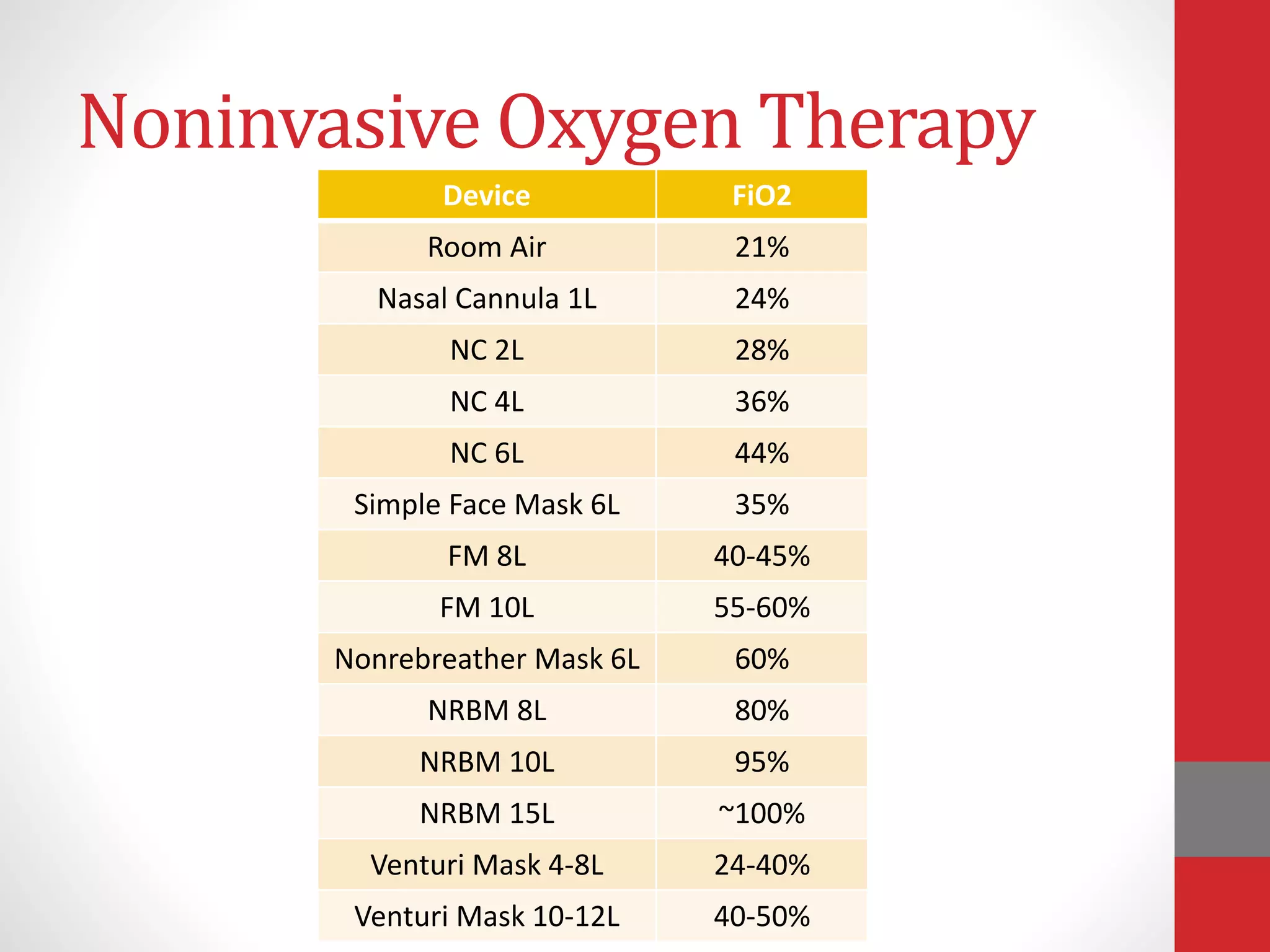
• Inability to oxygenate/ventilate adequately
• Via spontaneous respiration [respiratory failure]
• Via noninvasive ventilation [need for increased support]
• Inability to protect the airway
• GCS <8
• Hyporeflexia / areflexia
• Inability to maintain cardiac output [code situation]
Airway Anatomy
Mallampati Score
• Class I – complete visualization of
soft palate, uvula, anterior and
posterior tonsillar pillars
• Class II – complete visualization of
soft palate, uvula; partial
visualization of tonsillar pillars
• Class III – complete visualization of
soft palate; partial visualization of
uvula
• Class IV – no visualization of soft
palate; hard palate and tongue only
visible structures
Evaluate the airway with the patient sitting
upright, with head in neutral position.
HOW TO INTUBATE
7 P’s of Airway Management
• Planning
• Preparation
• Positioning
• Preoxygenation
• Paralysis
• Placement
• Proof
Planning Preparation Positioning Preoxygenation Paralysis Placement Proof
Time
0
T +1 T +3T -3T -5
Planning and Preparation
• Plan
• Evaluate the patient for necessity of intubation
• Examine the airway
• Equipment
• Suction, oxygen, monitors
• Bag valve mask
• Ancillary noninvasive devices – oral and nasal airways
• Laryngoscope handle and blade(s)
• Endotracheal tube(s) and stylet(s)
• Backup equipment – bougie, LMA, surgical airway kit
• Drugs
• Relevant personnel
• Physician, nurse, respiratory therapist
• Defined roles for team members
• Communication is key
Airway Tools - Basics
Airway Tools - Laryngoscope
Airway Tools – Endotracheal Tube
Airway Tools - Ancillary
Positioning
• “Sniffing Position” aligns oral / pharyngeal / laryngeal axes
• Requires alignment of the tragus with the sternal notch
• Typically the shoulders remain on the bed while the head is
lifted 3 or more inches
• Obese patients may require ramping of the shoulders and
significantly more elevation of the head
• Note that this is not simple “extension of the spine”
• Caution in patients with cervical spine injury or unknown
pathology
Flexion of cervical
spine
Extension of
atlantoaxial joint
Preoxygenation
• Used to “denitrogenate” the lungs and extend the safe apnea
period
• Safe apnea = time until saturation falls below ~90%
• 1-2 minutes if breathing room air
• Up to 8 minutes if fully preoxygenated
• Reduced in patients with decreased FRC (pregnancy, extremes of
age, obesity, ascites), increased O2 consumption (sepsis,
hypermetabolic state), shunt physiology, etc
• If adequate respiratory effort, may use FiO2 100% fo 3
minutes of tidal breathing OR 8 vital capacity breaths
• If patient unable to perform above, may “preoxygenate” with
positive pressure breaths
Preoxygenation
• Useful in the optimal situation – clearly not always the case
• Do the best you can
• Can also “preoxygenate” with bag-valve mask in some cases
Paralysis - A Brief Note on Drugs
• Many choices for medications
• Induction agent
• Midazolam 0.1-0.3mg/kg
• Fentanyl 2-10mcg/kg
• Propofol 1-2.5mg/kg
• Etomidate 0.3-0.4mg/kg
• Ketamine 1.5-2mg/kg
• Paralytic
• Succinylcholine 1mg/kg
• Rocuronium 1.2mg/kg
• Vecuronium 0.25mg/kg
Placement – Technique
• Position the patient
• Open the mouth – spread the molars with your right hand
• Insert the laryngoscope with your left hand
• Direct blade (Macintosh, Miller) – insert on right of mouth, sweep
tongue to left
• Indirect 60° video blade (Glide, D) – insert down center of tongue
• Remove right hand from mouth, may need for cricoid pressure
or other optimization of positioning
• Advance blade, visualize epiglottis
• Macintosh – advance anterior to epiglottis, into vallecula
• Miller – advance posterior to epiglottis
• Indirect – advance anterior to epiglottis, into vallecula
Placement – Technique
• Lift epiglottis to reveal vocal cords – lift up and away, never
back
• Miller blade – active lifting of epiglottis
• Macintosh, indirect video blades – passive lifting
• Use right hand to place endotracheal tube between vocal
cords
• After passing glottic opening, ask assistant to remove stylet
• Advance tube to desired depth
• Remove laryngoscope
• Inflate ETT cuff
• Ventilate
Proof of ETT Placement
• Fog in tube
• End Tidal CO2
• POC detector changes from purple  yellow when exposed to
CO2
• Continuous capnography will show ventilatory pattern
• Bilateral breath sounds
• Stable / increasing SpO2
• Tidal volumes / compliance
• Chest X Ray
Planning Preparation Positioning Preoxygenation Paralysis Placement Proof
Time
0
T +1 T +3T -3T -5
WHAT TO DO WITH A DIFFICULT AIRWAY
Difficult Airway
• Bread and butter for anesthesiologists, but also…
• One of the most common causes of lawsuits in closed claims
analysis
• Incidence of difficult intubation in OR 1.5-8%
• Incidence of difficult intubation out of OR as high as 30%
“The difficult airway is
anticipated; the failed airway is
experienced.”
Difficult Airway
“[…]the clinical situation in which a conventionally trained
Anesthesiologist experiences difficulty with facemask
ventilation, difficulty in supraglottic device ventilation, difficulty
in tracheal intubation or all three.”
- ASA definition
Inadequate
Ventilation
Esophageal
Intubation
Airway
Obstruction
Bronchospasm
Difficult
Intubation
Aspiration
Premature
Extubation
Other
% CLAIMS
Death
Brain Damage
Nerve Damage
MI / CVA
Miscellaneous
% COMPLICATIONS
Signs of Difficult Intubation
Testing
• Mallampati score ≥3
• Thyromental distance ≤5cm
• Upper lip bite test
• Class I – lower incisors reach
above vermillion border
• Class II – lower incisors reach
upper lip below vermillion
border
• Class III – lower incisors cannot
bite upper lip
• Each has poor sensitivity with
decent specificity; improved
utility when used together
Other Exam Findings
• Facial trauma, burns
• Obstruction / foreign
body
• Obesity
• Secretions, blood,
edema in airway
• Personal history of
difficult intubation
Signs of Difficult Mask Ventilation
• Beard
• Obesity
• Edentulous
• Elderly
• Sleep apnea, diagnosed or suspected
Emergency Surgical Airway
• Needle cricothyrotomy
• Open cricothyrotomy
• Open tracheostomy
Things to Remember in a
Difficult Airway Situation
• When in doubt, give a shout
• Call for help early
• The harder I practice, the luckier I get
• Your first experience shouldn’t be in an emergency
• Doing the same thing and expecting different
results is insane
• Do not keep repeating a failed technique
Nobody ever died from failure to
intubate, but patients die every day from
failure to oxygenate.
Airway Management for the Trauma Provider

Airway Management for the Trauma Provider

  • 1.
    Airway Management for theTrauma Provider SarahBeth Hartlage, MD MS Assistant Professor Department of Anesthesiology and Perioperative Medicine University of Louisville
  • 2.
    Disclosures • No financialrelationships to disclose
  • 3.
    Objectives • When andwhy to intubate • Basics of airway management • Recognizing difficult airways • Emergency situations
  • 4.
  • 5.
    Noninvasive Oxygen Therapy DeviceFiO2 Room Air 21% Nasal Cannula 1L 24% NC 2L 28% NC 4L 36% NC 6L 44% Simple Face Mask 6L 35% FM 8L 40-45% FM 10L 55-60% Nonrebreather Mask 6L 60% NRBM 8L 80% NRBM 10L 95% NRBM 15L ~100% Venturi Mask 4-8L 24-40% Venturi Mask 10-12L 40-50%
  • 6.
  • 7.
    Aids to MaskVentilation • Head Tilt / Chin Lift • Oral Airway (OPA) • Nasal Airway (Trumpet)
  • 8.
    Indications for Endotracheal Intubation •Inability to oxygenate/ventilate adequately • Via spontaneous respiration [respiratory failure] • Via noninvasive ventilation [need for increased support] • Inability to protect the airway • GCS <8 • Hyporeflexia / areflexia • Inability to maintain cardiac output [code situation]
  • 9.
  • 10.
    Mallampati Score • ClassI – complete visualization of soft palate, uvula, anterior and posterior tonsillar pillars • Class II – complete visualization of soft palate, uvula; partial visualization of tonsillar pillars • Class III – complete visualization of soft palate; partial visualization of uvula • Class IV – no visualization of soft palate; hard palate and tongue only visible structures Evaluate the airway with the patient sitting upright, with head in neutral position.
  • 11.
  • 12.
    7 P’s ofAirway Management • Planning • Preparation • Positioning • Preoxygenation • Paralysis • Placement • Proof
  • 13.
    Planning Preparation PositioningPreoxygenation Paralysis Placement Proof Time 0 T +1 T +3T -3T -5
  • 14.
    Planning and Preparation •Plan • Evaluate the patient for necessity of intubation • Examine the airway • Equipment • Suction, oxygen, monitors • Bag valve mask • Ancillary noninvasive devices – oral and nasal airways • Laryngoscope handle and blade(s) • Endotracheal tube(s) and stylet(s) • Backup equipment – bougie, LMA, surgical airway kit • Drugs • Relevant personnel • Physician, nurse, respiratory therapist • Defined roles for team members • Communication is key
  • 15.
  • 16.
    Airway Tools -Laryngoscope
  • 17.
    Airway Tools –Endotracheal Tube
  • 18.
    Airway Tools -Ancillary
  • 19.
    Positioning • “Sniffing Position”aligns oral / pharyngeal / laryngeal axes • Requires alignment of the tragus with the sternal notch • Typically the shoulders remain on the bed while the head is lifted 3 or more inches • Obese patients may require ramping of the shoulders and significantly more elevation of the head • Note that this is not simple “extension of the spine” • Caution in patients with cervical spine injury or unknown pathology
  • 20.
  • 22.
    Preoxygenation • Used to“denitrogenate” the lungs and extend the safe apnea period • Safe apnea = time until saturation falls below ~90% • 1-2 minutes if breathing room air • Up to 8 minutes if fully preoxygenated • Reduced in patients with decreased FRC (pregnancy, extremes of age, obesity, ascites), increased O2 consumption (sepsis, hypermetabolic state), shunt physiology, etc • If adequate respiratory effort, may use FiO2 100% fo 3 minutes of tidal breathing OR 8 vital capacity breaths • If patient unable to perform above, may “preoxygenate” with positive pressure breaths
  • 23.
    Preoxygenation • Useful inthe optimal situation – clearly not always the case • Do the best you can • Can also “preoxygenate” with bag-valve mask in some cases
  • 24.
    Paralysis - ABrief Note on Drugs • Many choices for medications • Induction agent • Midazolam 0.1-0.3mg/kg • Fentanyl 2-10mcg/kg • Propofol 1-2.5mg/kg • Etomidate 0.3-0.4mg/kg • Ketamine 1.5-2mg/kg • Paralytic • Succinylcholine 1mg/kg • Rocuronium 1.2mg/kg • Vecuronium 0.25mg/kg
  • 25.
    Placement – Technique •Position the patient • Open the mouth – spread the molars with your right hand • Insert the laryngoscope with your left hand • Direct blade (Macintosh, Miller) – insert on right of mouth, sweep tongue to left • Indirect 60° video blade (Glide, D) – insert down center of tongue • Remove right hand from mouth, may need for cricoid pressure or other optimization of positioning • Advance blade, visualize epiglottis • Macintosh – advance anterior to epiglottis, into vallecula • Miller – advance posterior to epiglottis • Indirect – advance anterior to epiglottis, into vallecula
  • 26.
    Placement – Technique •Lift epiglottis to reveal vocal cords – lift up and away, never back • Miller blade – active lifting of epiglottis • Macintosh, indirect video blades – passive lifting • Use right hand to place endotracheal tube between vocal cords • After passing glottic opening, ask assistant to remove stylet • Advance tube to desired depth • Remove laryngoscope • Inflate ETT cuff • Ventilate
  • 29.
    Proof of ETTPlacement • Fog in tube • End Tidal CO2 • POC detector changes from purple  yellow when exposed to CO2 • Continuous capnography will show ventilatory pattern • Bilateral breath sounds • Stable / increasing SpO2 • Tidal volumes / compliance • Chest X Ray
  • 30.
    Planning Preparation PositioningPreoxygenation Paralysis Placement Proof Time 0 T +1 T +3T -3T -5
  • 31.
    WHAT TO DOWITH A DIFFICULT AIRWAY
  • 32.
    Difficult Airway • Breadand butter for anesthesiologists, but also… • One of the most common causes of lawsuits in closed claims analysis • Incidence of difficult intubation in OR 1.5-8% • Incidence of difficult intubation out of OR as high as 30% “The difficult airway is anticipated; the failed airway is experienced.”
  • 33.
    Difficult Airway “[…]the clinicalsituation in which a conventionally trained Anesthesiologist experiences difficulty with facemask ventilation, difficulty in supraglottic device ventilation, difficulty in tracheal intubation or all three.” - ASA definition
  • 34.
  • 35.
    Signs of DifficultIntubation Testing • Mallampati score ≥3 • Thyromental distance ≤5cm • Upper lip bite test • Class I – lower incisors reach above vermillion border • Class II – lower incisors reach upper lip below vermillion border • Class III – lower incisors cannot bite upper lip • Each has poor sensitivity with decent specificity; improved utility when used together Other Exam Findings • Facial trauma, burns • Obstruction / foreign body • Obesity • Secretions, blood, edema in airway • Personal history of difficult intubation
  • 36.
    Signs of DifficultMask Ventilation • Beard • Obesity • Edentulous • Elderly • Sleep apnea, diagnosed or suspected
  • 39.
    Emergency Surgical Airway •Needle cricothyrotomy • Open cricothyrotomy • Open tracheostomy
  • 40.
    Things to Rememberin a Difficult Airway Situation • When in doubt, give a shout • Call for help early • The harder I practice, the luckier I get • Your first experience shouldn’t be in an emergency • Doing the same thing and expecting different results is insane • Do not keep repeating a failed technique
  • 41.
    Nobody ever diedfrom failure to intubate, but patients die every day from failure to oxygenate.