Downloaded 115 times


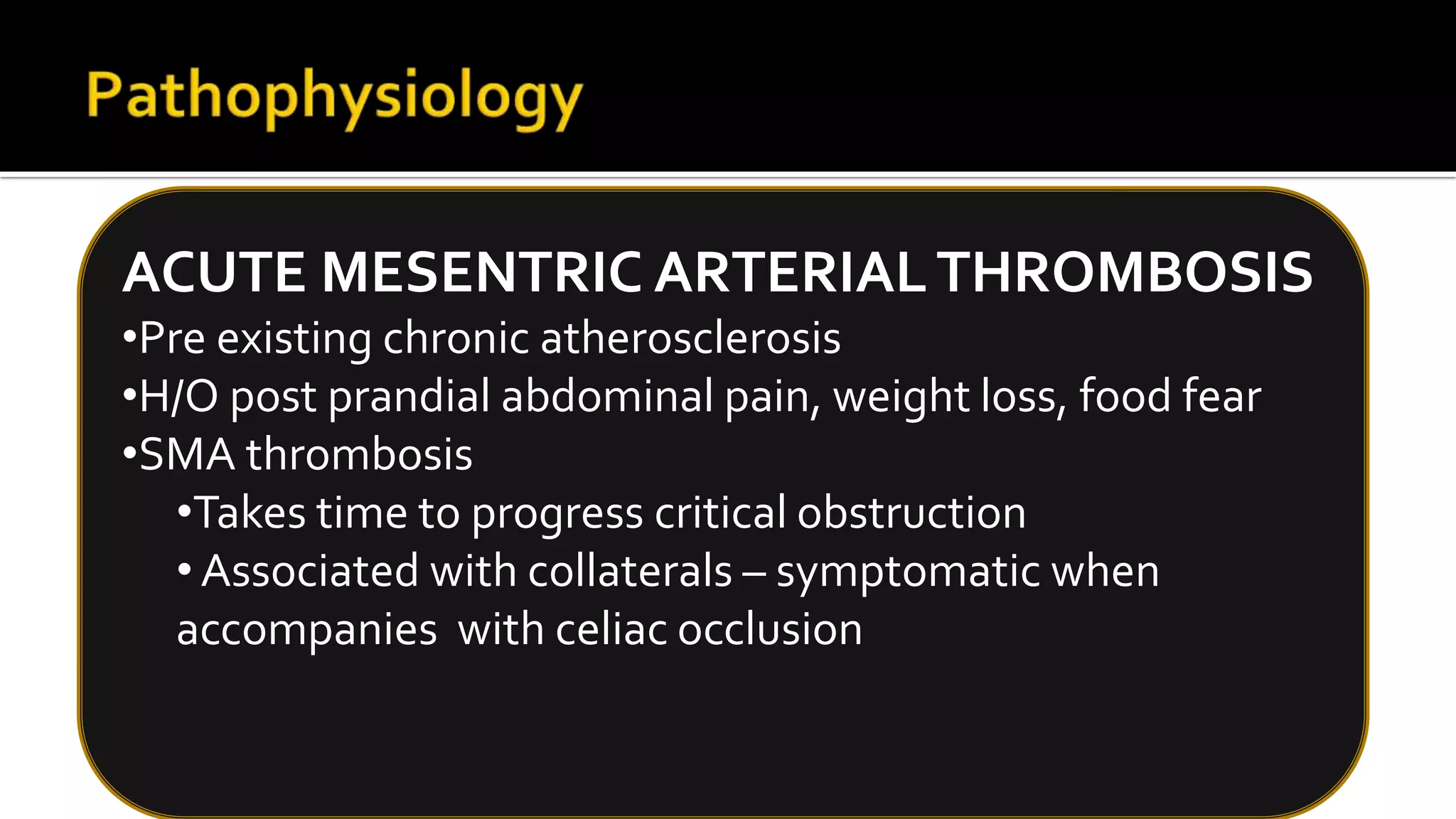


This document discusses acute mesenteric ischemia (AMI), a rare but life-threatening condition caused by sudden interruption of blood supply to the intestine. It can be caused by arterial embolism, thrombosis, or venous thrombosis. Clinical features include severe abdominal pain, nausea, vomiting, and bloody stools. Early diagnosis via CT angiography and prompt resuscitation, antibiotics, anticoagulation, and surgery are important, as delayed treatment can lead to bowel necrosis and death. Surgical management focuses on reestablishing blood flow and resecting non-viable bowel. Outcomes depend on how quickly treatment is initiated.


