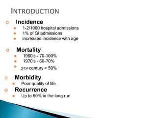
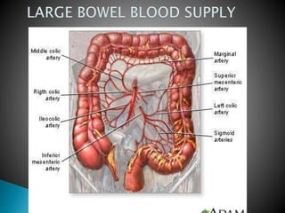
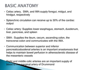
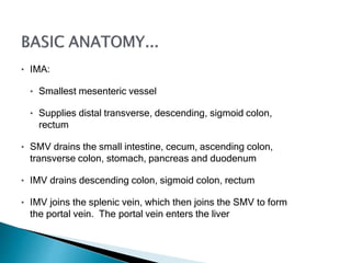
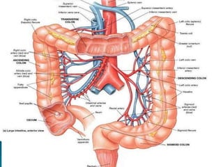
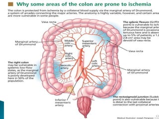
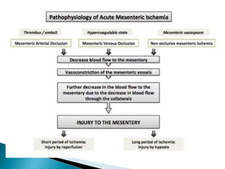
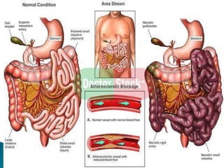
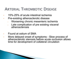
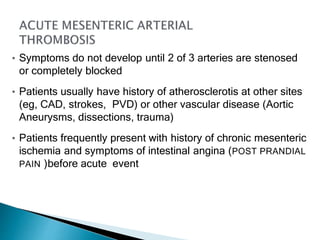
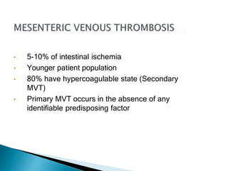
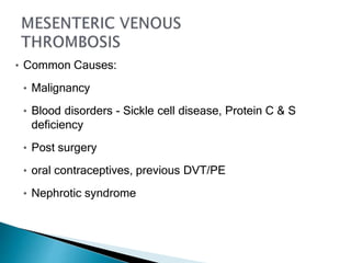
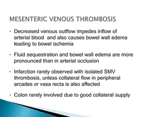
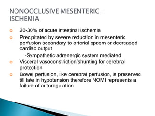
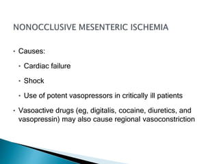
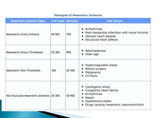
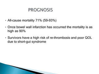
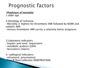
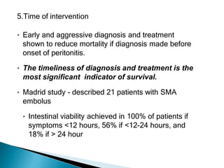
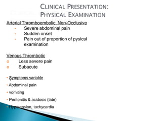
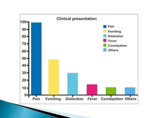
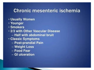
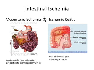
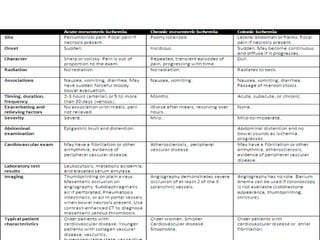
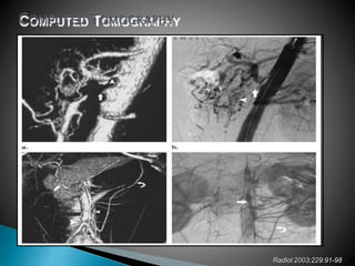
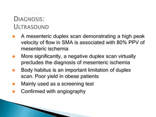
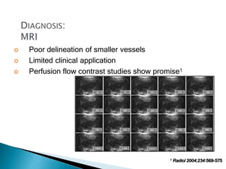
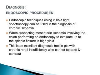
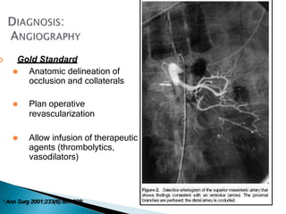
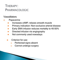
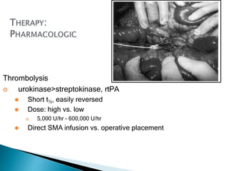
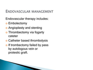
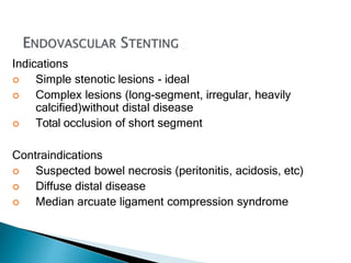
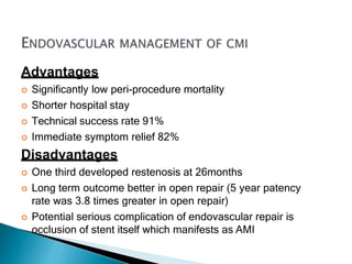
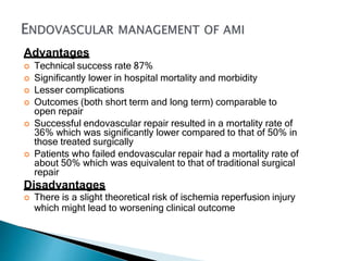
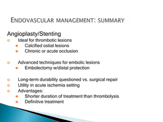
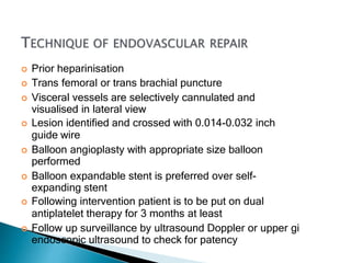
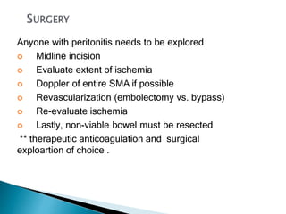
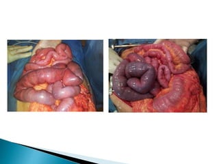
BY
RES 
Steven Nabil Fouad
--occlusion of the mesenteric vessels is regarded as
one of those conditions of which the diagnosis is
impossible, the prognosis hopeless, and the treatment
almost useless.
-- early dianosis result in better outcome
--Intestinal ischemia occurs when the splanchnic
perfusion fails to meet the metabolic demands of the
intestines resulting in ischemic tissue injury
--Occlusive or non-occlusive mechanism leads to
hypo perfusion of one or more mesenteric vessels
 Incidence
⚫ 1-2/1000 hospital admissions
⚫ 1% of GI admissions
⚫ increased incidence with age
 Mortality
⚫ 1960’s - 70-100%
⚫ 1970’s - 60-70%
⚫
21st century > 50%
 Morbidity
⚫ Poor quality of life
 Recurrence
⚫ Up to 60% in the long run
• Celiac artery, SMA, and IMA supply foregut, midgut, and
hindgut, respectively
• Splanchnic circulation can receive up to 30% of the cardiac
output
• Celiac artery: Supplies lower esophagus, stomach, duodenum,
liver, pancreas, and spleen
• SMA : Supplies the ileum, cecum, ascending colon, the
transverse colon and communicates with the IMA.
• Communication between superior and inferior
pancreaticoduodenal arteries is an important anastomosis that
helps to maintain bowel perfusion in atherosclerotic disease of
the mesenteric vessels
• Right and middle colic arteries are an important supply of
blood to the marginal artery of Drummond
• IMA:
• Smallest mesenteric vessel
• Supplies distal transverse, descending, sigmoid colon,
rectum
• SMV drains the small intestine, cecum, ascending colon,
transverse colon, stomach, pancreas and duodenum
• IMV drains descending colon, sigmoid colon, rectum
• IMV joins the splenic vein, which then joins the SMV to form
the portal vein. The portal vein enters the liver
 Abrupt Cessation of mesenteric blood flow
lead to malperfusion of bowel associated with
bowel necrosis.
Insufficient blood perfusion of small bowel or colon may
result from:
 Arterial Embolic Disease (50%)
 Arterial Thrombotic Disease (25%)
• Venous Thrombotic Disease (10%)
 Non-occlusive Mesenteric Ischemia
(20%)
• Injury severity is inversely proportional to mesenteric blood flow
• Number of vessels involved, mean arterial pressure, duration of
ischemia, and extent of collateral circulation all determine mesenteric
blood flow
• SM vessels are more frequently involved than the IM vessels (larger
diameter and better collaterals with inferior vessels)
• Damage may range from reversible ischemia to transmural infarction with
necrosis and perforation
• Arterial insufficiency causes tissue hypoxia, leading to bowel wall spasm
initially(vomiting or diarrhea)
• Injury complicated by reactive vasospasm in SMA after initial occlusion
• Mucosal sloughing may cause bleeding into the GIT
• Minimal abdominal tenderness is present despite symptoms of
intense visceral pain
• If ischemia persists disruption of mucosal barrier occurs and
bacteria, toxins, and vasoactive substances are released into the
systemic circulation
• This can cause septic shock, cardiac failure, or multi-organ failure
before bowel necrosis actually occurs
• With worsening hypoxic damage the bowel wall becomes edematous
and cyanotic
• Bowel necrosis occurs in 8-12 hours from onset of symptoms
• Transmural necrosis leads to peritonitis and indicates grave
prognosis
 Majority of cases (>50%): SMA occlusion
 Location: origin of middle colic artery (ischemia from
proximal jejunam to splenic flexure)
 Occlusion is sudden and no time to develop
compensatory increase in collateral flow
 Ischemia is more severe in SMA occlusion
 Celiac and IMA occlusion usually less severe and is
tolerated
 Most have underlying stenosis as well
 Embolic sources: cardiac (80%), aortic plaques
 Typical causes:
 Mural thrombi - MI
 Atrial thrombi - mitral stenosis and AF, vegetative
endocarditis
 Aortic thrombi - mycotic aneurysm, thrombi at sites of
atheromatous plaques, sites of vascular aortic prosthetic
grafts interposed between heart and SMA
• 15%-25% of acute intestinal ischemia
• Pre-existing atherosclerotic disease
— Worsening chronic mesenteric ischemia
— Late complication of pre existing visceral
atherosclerosis
• Found at ostium of SMA
• More delayed onset of symptoms - Slow process of
atherosclerotic stenosis before acute occlusion allows
time for development of collateral circulation
• Symptoms do not develop until 2 of 3 arteries are stenosed
or completely blocked
• Patients usually have history of atherosclerotis at other sites
(eg, CAD, strokes, PVD) or other vascular disease (Aortic
Aneurysms, dissections, trauma)
• Patients frequently present with history of chronic mesenteric
ischemia and symptoms of intestinal angina (POST PRANDIAL
PAIN )before acute event
• 5-10% of intestinal ischemia
• Younger patient population
• 80% have hypercoagulable state (Secondary
MVT)
• Primary MVT occurs in the absence of any
identifiable predisposing factor
• Common Causes:
• Malignancy
• Blood disorders - Sickle cell disease, Protein C & S
deficiency
• Post surgery
• oral contraceptives, previous DVT/PE
• Nephrotic syndrome
• Decreased venous outflow impedes inflow of
arterial blood and also causes bowel wall edema
leading to bowel ischemia
• Fluid sequestration and bowel wall edema are more
pronounced than in arterial occlusion
• Infarction rarely observed with isolated SMV
thrombosis, unless collateral flow in peripheral
arcades or vasa recta is also affected
• Colon rarely involved due to good collateral supply
 20-30% of acute intestinal ischemia
 Precipitated by severe reduction in mesenteric
perfusion secondary to arterial spasm or decreased
cardiac output
-Sympathetic adrenergic system mediated
 Visceral vasoconstriction/shunting for cerebral
protection
 Bowel perfusion, like cerebral perfusion, is preserved
till late in hypotension therefore NOMI represents a
failure of autoregulation
• Causes:
• Cardiac failure
• Shock
• Use of potent vasopressors in critically ill patients
• Vasoactive drugs (eg, digitalis, cocaine, diuretics, and
vasopressin) may also cause regional vasoconstriction
• All-cause mortality 71% (59-93%)
• Once bowel wall infarction has occurred the mortality is as
high as 90%
• Survivors have a high risk of re-thrombosis and poor QOL
due to short-gut syndrome
*Predictors of mortality:
1.older age
2.Atieology of ischemia;
-Mortality is highest for thrombotic AMI followed by NOMI and
embolic AMI.
-Venous thrombotic AMI carries a relatively better prognosis
3.Labaratory indicators;
-hepatic and renal impairment
-metabolic acidosis (LDH)
-lecucytosis (sepsis)
4. radilogical indicators;
-intramural pneumatosis
-free IP fluid collection (PERFPRATION)
5.Time of intervention
• Early and aggressive diagnosis and treatment
shown to reduce mortality if diagnosis made before
onset of peritonitis.
• The timeliness of diagnosis and treatment is the
most significant indicator of survival.
• Madrid study - described 21 patients with SMA
embolus
• Intestinal viability achieved in 100% of patients if
symptoms <12 hours, 56% if <12-24 hours, and
18% if > 24 hour
Arterial Thromboembolic, Non-Occlusive
• Severe abdominal pain
• Sudden onset
• Pain out of proportion of pysical
examination
Venous Thrombotic
 Less severe pain
 Subacute
• Symptoms variable
• Abdominal pain
• vomiting
• Peritonitis & acidosis (late)
• Hypotension, tachycardia
Limited clinical utility
 CBC
 PC &INR
 LIVER FUNCTION
 KIDNEY FUNCTION
 ELECTROLYTE
 arterial blood gases
 D.dimer ,CRP
 Other markers : LDH, TNF
1 Eur J Surg 1994;160:381-4
2 Br J Surg 1986;73:219-21
3 Dig Dis Sci 1991;36:1589-93
4 Br J Surg 1982;69:S52-3
Non-Invasive Imaging
 Plain X-ray
Computed Tomography
(helical/angiography)
 Ultrasound
 MRI/MRA
Invasive
 Endoscopic procedures
 Angiography
Plain Films
 pneumatosis
 portal venous gas
 thumbprinting →
 Findings late, associated with high mortality
Sensitivity: 96%
Specificity: 94%
Criteria
• pneumatosis
• venous gas
•SMA/celiac/IMA occlusion w/distal
disease
• arterial embolism
OR
•bowel wall thickening + one of
following:
– lack of bowel wall
enhancement
– solid organ infarction
– venous thrombosis
1 Radiol 2003;229:91-98
Ct Angio with 3D reconstruction
is a highly sensitive test for
intestinal ischemia
Radiol 2003;229:91-98
⚫ A mesenteric duplex scan demonstrating a high peak
velocity of flow in SMA is associated with 80% PPV of
mesenteric ischemia
⚫ More significantly, a negative duplex scan virtually
precludes the diagnosis of mesenteric ischemia
⚫ Body habitus is an important limitation of duplex
scan. Poor yield in obese patients
⚫ Mainly used as a screening test
⚫ Confirmed with angiography
 Poor delineation of smaller vessels
 Limited clinical application
 Perfusion flow contrast studies show promise1
1 Radiol 2004;234:569-575
ENDOSCOPIC PROCEDURES
 Endoscopic techniques using visible light
spectroscopy can be used in the diagnosis of
chronic ischemia
 When suspecting mesenteric ischemia involving the
colon performing an endoscopy to evaluate up to
the splenic flexure is high yield
 This is an excellent diagnostic tool in pts with
chronic renal insufficiency who cannot tolerate iv
contrast
 Gold Standard
⚫ Anatomic delineation of
occlusion and collaterals
⚫ Plan operative
revascularization
⚫ Allow infusion of therapeutic
agents (thrombolytics,
vasodilators)
1 Ann Surg 2001;233(6):801-808
 Diagnosis
 Supportive Care
 Restoring Blood Flow
 Resection of non-viable gut
 Second-Look
Supportive measures
 IV resuscitation
 Optimize cardiac status
 Broad-spectrum antibiotics (no data)
 Nasogastric decompression
 Correction of electrolyte imbalances
Anticoagulation
 Heparin IV
⚫ Prevents clot propagation
⚫ Systemic vs. intra-arterial
⚫ Restart 48 hrs after any surgical intervention
 Warfarin
⚫ Prevents clot propagation
⚫ Give for 6-12 mos if no clotting disorder (no data)
1 Surg Gynecol Obstet 1981;153:561-569
2 Vascular Emergencies. 1982;553-561
Vasodilators
 Papaverine
⚫ Increases cAMP, relaxes smooth muscle
⚫ Primary indication: Non-occlusive arterial disease
⚫ Early SMA infusion reduces mortality to 40-50%
⚫ Directed infusion via angiography
⚫ Not commonly used nowadays
⚫ Criterion for use:
 Peritoneal signs absent
 Cannot undergo surgery
Thrombolysis
 urokinase>streptokinase, rtPA
⚫ Short t½, easily reversed
⚫ Dose: high vs. low
 5,000 U/hr - 600,000 U/hr
⚫ Direct SMA infusion vs. operative placement
Thrombolysis
 Duration: minutes – 48 hrs1
⚫ Risk of bowel necrosis if treatment delayed
⚫ Treat to re-establish flow vs. complete dissolution
⚫ > 48 hrs-Greater risk of bleeding
Discontinue if
 Worsening abdominal symptoms without evidence of
thrombolysis
 Bleeding
 No angiographic improvement
1 JVIR 2005;16:317-329
 Criterion for use:
⚫ Embolic/thrombotic disease
⚫ Poor operative candidates
⚫ No contraindications to fibrinolytics
⚫ No bowel infarction (no peritonitis/acidosis)
 Expansion of use to all patients without bowel infarction and
without contraindications to thrombolysis
Endovascular therapy includes:
 Embolectomy
 Angioplasty and stenting
 Thrombectomy via fogarty
cateter
 Catheter based thrombolysis
 If trombectomy failed by pass
by autologous vein or
protestic graft.
Indications
 Simple stenotic lesions - ideal
 Complex lesions (long-segment, irregular, heavily
calcified)without distal disease
 Total occlusion of short segment
Contraindications
 Suspected bowel necrosis (peritonitis, acidosis, etc)
 Diffuse distal disease
 Median arcuate ligament compression syndrome
Advantages
 Significantly low peri-procedure mortality
 Shorter hospital stay
 Technical success rate 91%
 Immediate symptom relief 82%
Disadvantages
 One third developed restenosis at 26months
 Long term outcome better in open repair (5 year patency
rate was 3.8 times greater in open repair)
 Potential serious complication of endovascular repair is
occlusion of stent itself which manifests as AMI
Indications
 Prohibitive operative risk
 No clinical signs of peritoneal inflammation
 Those with no autologous vessel available for graft
even with contaminated peritoneal cavity
Advantages
 Technical success rate 87%
 Significantly lower in hospital mortality and morbidity
 Lesser complications
 Outcomes (both short term and long term) comparable to
open repair
 Successful endovascular repair resulted in a mortality rate of
36% which was significantly lower compared to that of 50% in
those treated surgically
 Patients who failed endovascular repair had a mortality rate of
about 50% which was equivalent to that of traditional surgical
repair
Disadvantages
 There is a slight theoretical risk of ischemia reperfusion injury
which might lead to worsening clinical outcome
Angioplasty/Stenting
 Ideal for thrombotic lesions
⚫ Calcified ostial lesions
⚫ Chronic or acute occlusion
 Advanced techniques for embolic lesions
⚫ Embolectomy w/distal protection
 Long-term durability questioned vs. surgical repair
 Utility in acute ischemia setting
 Advantages:
⚫ Shorter duration of treatment than thrombolysis
⚫ Definitive treatment
 Prior heparinisation
 Trans femoral or trans brachial puncture
 Visceral vessels are selectively cannulated and
visualised in lateral view
 Lesion identified and crossed with 0.014-0.032 inch
guide wire
 Balloon angioplasty with appropriate size balloon
performed
 Balloon expandable stent is preferred over self-
expanding stent
 Following intervention patient is to be put on dual
antiplatelet therapy for 3 months at least
 Follow up surveillance by ultrasound Doppler or upper gi
endoscopic ultrasound to check for patency
Anyone with peritonitis needs to be explored
 Midline incision
 Evaluate extent of ischemia
 Doppler of entire SMA if possible
 Revascularization (embolectomy vs. bypass)
 Re-evaluate ischemia
 Lastly, non-viable bowel must be resected
** therapeutic anticoagulation and surgical
exploartion of choice .
 Mesenteric ischemia is diagnosis which is often missed
and even today it carries a relatively grim prognosis.
 Mesenteric ischemia is to be suspected in any patient
presenting with pain abdomen out of proportion to the
physical findings.
 The timeliness of diagnosis and treatment is vitally
important.
 Surgical procedures have been the mainstay of
treatment over the years but the advent of endovascular
treatment options offer great promise in reducing the
mortality and morbidity without compromising on the
efficacy of treatment.
]
1. Nuzzo, A.; Guedj, K.; Curac, S.; Hercend, C.; Bendavid, C.; Gault, N.; Tran-Dinh, A.; Ronot,
M.; Nicoletti, A.; Bouhnik, Y.; et al.Accuracy of citrulline, I-FABP and d-lactate in the
diagnosis of acute mesenteric ischemia. Sci. Rep. 2021, 11, 18929. [CrossRef]
2. Björck, M.; Koelemay, M.; Acosta, S.; Bastos Goncalves, F.; Kölbel, T.; Kolkman, J.J.; Lees, T.;
Lefevre, J.H.; Menyhei, G.; Oderich,G.; et al. Editor’s Choice—Management of the Diseases of
Mesenteric Arteries and Veins: Clinical Practice Guidelines of theEuropean Society of Vascular
Surgery (ESVS). Eur. J. Vasc. Endovasc. Surg. 2017, 53, 460–510. [CrossRef]
3.Kougias, P.; Lau, D.; El Sayed, H.F.; Zhou, W.; Huynh, T.T.; Lin, P.H. Determinants of mortality
and treatment outcome followingsurgical interventions for acute mesenteric ischemia. J. Vasc.
Surg. 2007, 46, 467–474. [CrossRef]
4. Acosta-Merida, M.A.; Marchena-Gomez, J.; Hemmersbach-Miller, M.; Roque-Castellano, C.;
Hernandez-Romero, J.M. Identification of risk factors for perioperative mortality in acute
mesenteric ischemia. World J. Surg. 2006, 30, 1579–1585. [CrossRef]
5. Bala, M.; Kashuk, J.; Moore, E.E.; Kluger, Y.; Biffl, W.; Gomes, C.A.; Ben-Ishay, O.; Rubinstein, C.;
Balogh, Z.J.; Civil, I.; et al.
Acute mesenteric ischemia: Guidelines of the World Society of Emergency Surgery. World J. Emerg.
Surg. 2017, 12, 38. [CrossRef]
[PubMed]
6. Dahlke, M.H.; Asshoff, L.; Popp, F.C.; Feuerbach, S.; Lang, S.A.; Renner, P.;
Slowik, P.; Stoeltzing, O.; Schlitt, H.J.; Piso, P.
Mesenteric ischemia—Outcome after surgical therapy in 83 patients. Dig. Surg.
2008, 25, 213–219. [CrossRef] [PubMed]
7. Tilsed, J.V.T.; Casamassima, A.; Kurihara, H.; Mariani, D.; Martinez, I.; Pereira,
J.; Ponchietti, L.; Shamiyeh, A.; al-Ayoubi, F.;
Barco, L.A.B.; et al. ESTES guidelines: Acute mesenteric ischaemia. Eur. J. Trauma
Emerg. Surg. 2016, 42, 253–270. [CrossRef]
8. Huang, H.H.; Chang, Y.C.; Yen, D.H.; Kao, W.F.; Chen, J.D.; Wang, L.M.; Huang
C.I.; Lee, C.H. Clinical factors and outcomes in
patients with acute mesenteric ischemia in the emergency department. J. Chin.
Med. Assoc. 2005, 68, 299–306. [CrossRef]
9. Adaba, F.; Askari, A.; Dastur, J.; Patel, A.; Gabe, S.M.; Vaizey, C.J.; Faiz, O.;
Nightingale, J.M.D.; Warusavitarne, J. Mortality after
acute primary mesenteric infarction: A systematic review and meta-analysis of
observational studies. Colorectal Dis. 2015, 17,
566–577. [CrossRef]
10. Pedersoli, F.; Schönau, K.; Schulze-Hagen, M.; Keil, S.; Isfort, P.; Gombert, A.;
Alizai, P.H.; Kuhl, C.K.; Bruners, P.; Zimmermann,
M. Endovascular Revascularization with Stent Implantation in Patients with Acute
Mesenteric Ischemia due to Acute Arterial
Thrombosis: Clinical Outcome and Predictive Factors. Cardiovasc. Interv. Radiol.
2021, 44, 1030–1038. [CrossRef]
11. Cudnik, M.T.; Darbha, S.; Jones, J.; Macedo, J.; Stockton, S.W.; Hiestand, B.C.
The diagnosis of acute mesenteric ischemia: A
systematic review and meta-analysis. Acad. Emerg. Med. 2013, 20, 1087–1100.
[CrossRef]
12. Treskes, N.; Persoon, A.M.; van Zanten, A.R.H. Diagnostic accuracy of novel
serological biomarkers to detect acute mesenteric
ischemia: A systematic review and meta-analysis. Intern. Emerg. Med. 2017, 12,
821–836. [CrossRef]
13. Leone, M.; Bechis, C.; Baumstarck, K.; Ouattara, A.; Collange, O.; Augustin, P.;
Annane, D.; Arbelot, C.; Asehnoune, K.; Baldési,
O.; et al. Outcome of acute mesenteric ischemia in the intensive care unit: A
retrospective, multicenter study of 780 cases. Intensive
Care Med. 2015, 41, 667–676. [CrossRef]
AMI final 2A.pptx

AMI final 2A.pptx

  • 1.
  • 2.
    --occlusion of themesenteric vessels is regarded as one of those conditions of which the diagnosis is impossible, the prognosis hopeless, and the treatment almost useless. -- early dianosis result in better outcome --Intestinal ischemia occurs when the splanchnic perfusion fails to meet the metabolic demands of the intestines resulting in ischemic tissue injury --Occlusive or non-occlusive mechanism leads to hypo perfusion of one or more mesenteric vessels
  • 3.
     Incidence ⚫ 1-2/1000hospital admissions ⚫ 1% of GI admissions ⚫ increased incidence with age  Mortality ⚫ 1960’s - 70-100% ⚫ 1970’s - 60-70% ⚫ 21st century > 50%  Morbidity ⚫ Poor quality of life  Recurrence ⚫ Up to 60% in the long run
  • 5.
    • Celiac artery,SMA, and IMA supply foregut, midgut, and hindgut, respectively • Splanchnic circulation can receive up to 30% of the cardiac output • Celiac artery: Supplies lower esophagus, stomach, duodenum, liver, pancreas, and spleen • SMA : Supplies the ileum, cecum, ascending colon, the transverse colon and communicates with the IMA. • Communication between superior and inferior pancreaticoduodenal arteries is an important anastomosis that helps to maintain bowel perfusion in atherosclerotic disease of the mesenteric vessels • Right and middle colic arteries are an important supply of blood to the marginal artery of Drummond
  • 6.
    • IMA: • Smallestmesenteric vessel • Supplies distal transverse, descending, sigmoid colon, rectum • SMV drains the small intestine, cecum, ascending colon, transverse colon, stomach, pancreas and duodenum • IMV drains descending colon, sigmoid colon, rectum • IMV joins the splenic vein, which then joins the SMV to form the portal vein. The portal vein enters the liver
  • 9.
     Abrupt Cessationof mesenteric blood flow lead to malperfusion of bowel associated with bowel necrosis.
  • 10.
    Insufficient blood perfusionof small bowel or colon may result from:  Arterial Embolic Disease (50%)  Arterial Thrombotic Disease (25%) • Venous Thrombotic Disease (10%)  Non-occlusive Mesenteric Ischemia (20%)
  • 11.
    • Injury severityis inversely proportional to mesenteric blood flow • Number of vessels involved, mean arterial pressure, duration of ischemia, and extent of collateral circulation all determine mesenteric blood flow • SM vessels are more frequently involved than the IM vessels (larger diameter and better collaterals with inferior vessels) • Damage may range from reversible ischemia to transmural infarction with necrosis and perforation • Arterial insufficiency causes tissue hypoxia, leading to bowel wall spasm initially(vomiting or diarrhea) • Injury complicated by reactive vasospasm in SMA after initial occlusion • Mucosal sloughing may cause bleeding into the GIT
  • 12.
    • Minimal abdominaltenderness is present despite symptoms of intense visceral pain • If ischemia persists disruption of mucosal barrier occurs and bacteria, toxins, and vasoactive substances are released into the systemic circulation • This can cause septic shock, cardiac failure, or multi-organ failure before bowel necrosis actually occurs • With worsening hypoxic damage the bowel wall becomes edematous and cyanotic • Bowel necrosis occurs in 8-12 hours from onset of symptoms • Transmural necrosis leads to peritonitis and indicates grave prognosis
  • 15.
     Majority ofcases (>50%): SMA occlusion  Location: origin of middle colic artery (ischemia from proximal jejunam to splenic flexure)  Occlusion is sudden and no time to develop compensatory increase in collateral flow  Ischemia is more severe in SMA occlusion  Celiac and IMA occlusion usually less severe and is tolerated  Most have underlying stenosis as well
  • 16.
     Embolic sources:cardiac (80%), aortic plaques  Typical causes:  Mural thrombi - MI  Atrial thrombi - mitral stenosis and AF, vegetative endocarditis  Aortic thrombi - mycotic aneurysm, thrombi at sites of atheromatous plaques, sites of vascular aortic prosthetic grafts interposed between heart and SMA
  • 17.
    • 15%-25% ofacute intestinal ischemia • Pre-existing atherosclerotic disease — Worsening chronic mesenteric ischemia — Late complication of pre existing visceral atherosclerosis • Found at ostium of SMA • More delayed onset of symptoms - Slow process of atherosclerotic stenosis before acute occlusion allows time for development of collateral circulation
  • 18.
    • Symptoms donot develop until 2 of 3 arteries are stenosed or completely blocked • Patients usually have history of atherosclerotis at other sites (eg, CAD, strokes, PVD) or other vascular disease (Aortic Aneurysms, dissections, trauma) • Patients frequently present with history of chronic mesenteric ischemia and symptoms of intestinal angina (POST PRANDIAL PAIN )before acute event
  • 19.
    • 5-10% ofintestinal ischemia • Younger patient population • 80% have hypercoagulable state (Secondary MVT) • Primary MVT occurs in the absence of any identifiable predisposing factor
  • 20.
    • Common Causes: •Malignancy • Blood disorders - Sickle cell disease, Protein C & S deficiency • Post surgery • oral contraceptives, previous DVT/PE • Nephrotic syndrome
  • 21.
    • Decreased venousoutflow impedes inflow of arterial blood and also causes bowel wall edema leading to bowel ischemia • Fluid sequestration and bowel wall edema are more pronounced than in arterial occlusion • Infarction rarely observed with isolated SMV thrombosis, unless collateral flow in peripheral arcades or vasa recta is also affected • Colon rarely involved due to good collateral supply
  • 22.
     20-30% ofacute intestinal ischemia  Precipitated by severe reduction in mesenteric perfusion secondary to arterial spasm or decreased cardiac output -Sympathetic adrenergic system mediated  Visceral vasoconstriction/shunting for cerebral protection  Bowel perfusion, like cerebral perfusion, is preserved till late in hypotension therefore NOMI represents a failure of autoregulation
  • 23.
    • Causes: • Cardiacfailure • Shock • Use of potent vasopressors in critically ill patients • Vasoactive drugs (eg, digitalis, cocaine, diuretics, and vasopressin) may also cause regional vasoconstriction
  • 25.
    • All-cause mortality71% (59-93%) • Once bowel wall infarction has occurred the mortality is as high as 90% • Survivors have a high risk of re-thrombosis and poor QOL due to short-gut syndrome
  • 26.
    *Predictors of mortality: 1.olderage 2.Atieology of ischemia; -Mortality is highest for thrombotic AMI followed by NOMI and embolic AMI. -Venous thrombotic AMI carries a relatively better prognosis 3.Labaratory indicators; -hepatic and renal impairment -metabolic acidosis (LDH) -lecucytosis (sepsis) 4. radilogical indicators; -intramural pneumatosis -free IP fluid collection (PERFPRATION)
  • 27.
    5.Time of intervention •Early and aggressive diagnosis and treatment shown to reduce mortality if diagnosis made before onset of peritonitis. • The timeliness of diagnosis and treatment is the most significant indicator of survival. • Madrid study - described 21 patients with SMA embolus • Intestinal viability achieved in 100% of patients if symptoms <12 hours, 56% if <12-24 hours, and 18% if > 24 hour
  • 28.
    Arterial Thromboembolic, Non-Occlusive •Severe abdominal pain • Sudden onset • Pain out of proportion of pysical examination Venous Thrombotic  Less severe pain  Subacute • Symptoms variable • Abdominal pain • vomiting • Peritonitis & acidosis (late) • Hypotension, tachycardia
  • 33.
    Limited clinical utility CBC  PC &INR  LIVER FUNCTION  KIDNEY FUNCTION  ELECTROLYTE  arterial blood gases  D.dimer ,CRP  Other markers : LDH, TNF 1 Eur J Surg 1994;160:381-4 2 Br J Surg 1986;73:219-21 3 Dig Dis Sci 1991;36:1589-93 4 Br J Surg 1982;69:S52-3
  • 34.
    Non-Invasive Imaging  PlainX-ray Computed Tomography (helical/angiography)  Ultrasound  MRI/MRA Invasive  Endoscopic procedures  Angiography
  • 35.
    Plain Films  pneumatosis portal venous gas  thumbprinting →  Findings late, associated with high mortality
  • 36.
    Sensitivity: 96% Specificity: 94% Criteria •pneumatosis • venous gas •SMA/celiac/IMA occlusion w/distal disease • arterial embolism OR •bowel wall thickening + one of following: – lack of bowel wall enhancement – solid organ infarction – venous thrombosis 1 Radiol 2003;229:91-98 Ct Angio with 3D reconstruction is a highly sensitive test for intestinal ischemia
  • 37.
  • 38.
    ⚫ A mesentericduplex scan demonstrating a high peak velocity of flow in SMA is associated with 80% PPV of mesenteric ischemia ⚫ More significantly, a negative duplex scan virtually precludes the diagnosis of mesenteric ischemia ⚫ Body habitus is an important limitation of duplex scan. Poor yield in obese patients ⚫ Mainly used as a screening test ⚫ Confirmed with angiography
  • 39.
     Poor delineationof smaller vessels  Limited clinical application  Perfusion flow contrast studies show promise1 1 Radiol 2004;234:569-575
  • 40.
    ENDOSCOPIC PROCEDURES  Endoscopictechniques using visible light spectroscopy can be used in the diagnosis of chronic ischemia  When suspecting mesenteric ischemia involving the colon performing an endoscopy to evaluate up to the splenic flexure is high yield  This is an excellent diagnostic tool in pts with chronic renal insufficiency who cannot tolerate iv contrast
  • 41.
     Gold Standard ⚫Anatomic delineation of occlusion and collaterals ⚫ Plan operative revascularization ⚫ Allow infusion of therapeutic agents (thrombolytics, vasodilators) 1 Ann Surg 2001;233(6):801-808
  • 42.
     Diagnosis  SupportiveCare  Restoring Blood Flow  Resection of non-viable gut  Second-Look
  • 43.
    Supportive measures  IVresuscitation  Optimize cardiac status  Broad-spectrum antibiotics (no data)  Nasogastric decompression  Correction of electrolyte imbalances
  • 44.
    Anticoagulation  Heparin IV ⚫Prevents clot propagation ⚫ Systemic vs. intra-arterial ⚫ Restart 48 hrs after any surgical intervention  Warfarin ⚫ Prevents clot propagation ⚫ Give for 6-12 mos if no clotting disorder (no data) 1 Surg Gynecol Obstet 1981;153:561-569 2 Vascular Emergencies. 1982;553-561
  • 45.
    Vasodilators  Papaverine ⚫ IncreasescAMP, relaxes smooth muscle ⚫ Primary indication: Non-occlusive arterial disease ⚫ Early SMA infusion reduces mortality to 40-50% ⚫ Directed infusion via angiography ⚫ Not commonly used nowadays ⚫ Criterion for use:  Peritoneal signs absent  Cannot undergo surgery
  • 46.
    Thrombolysis  urokinase>streptokinase, rtPA ⚫Short t½, easily reversed ⚫ Dose: high vs. low  5,000 U/hr - 600,000 U/hr ⚫ Direct SMA infusion vs. operative placement
  • 47.
    Thrombolysis  Duration: minutes– 48 hrs1 ⚫ Risk of bowel necrosis if treatment delayed ⚫ Treat to re-establish flow vs. complete dissolution ⚫ > 48 hrs-Greater risk of bleeding Discontinue if  Worsening abdominal symptoms without evidence of thrombolysis  Bleeding  No angiographic improvement 1 JVIR 2005;16:317-329
  • 48.
     Criterion foruse: ⚫ Embolic/thrombotic disease ⚫ Poor operative candidates ⚫ No contraindications to fibrinolytics ⚫ No bowel infarction (no peritonitis/acidosis)  Expansion of use to all patients without bowel infarction and without contraindications to thrombolysis
  • 49.
    Endovascular therapy includes: Embolectomy  Angioplasty and stenting  Thrombectomy via fogarty cateter  Catheter based thrombolysis  If trombectomy failed by pass by autologous vein or protestic graft.
  • 50.
    Indications  Simple stenoticlesions - ideal  Complex lesions (long-segment, irregular, heavily calcified)without distal disease  Total occlusion of short segment Contraindications  Suspected bowel necrosis (peritonitis, acidosis, etc)  Diffuse distal disease  Median arcuate ligament compression syndrome
  • 51.
    Advantages  Significantly lowperi-procedure mortality  Shorter hospital stay  Technical success rate 91%  Immediate symptom relief 82% Disadvantages  One third developed restenosis at 26months  Long term outcome better in open repair (5 year patency rate was 3.8 times greater in open repair)  Potential serious complication of endovascular repair is occlusion of stent itself which manifests as AMI
  • 52.
    Indications  Prohibitive operativerisk  No clinical signs of peritoneal inflammation  Those with no autologous vessel available for graft even with contaminated peritoneal cavity
  • 53.
    Advantages  Technical successrate 87%  Significantly lower in hospital mortality and morbidity  Lesser complications  Outcomes (both short term and long term) comparable to open repair  Successful endovascular repair resulted in a mortality rate of 36% which was significantly lower compared to that of 50% in those treated surgically  Patients who failed endovascular repair had a mortality rate of about 50% which was equivalent to that of traditional surgical repair Disadvantages  There is a slight theoretical risk of ischemia reperfusion injury which might lead to worsening clinical outcome
  • 54.
    Angioplasty/Stenting  Ideal forthrombotic lesions ⚫ Calcified ostial lesions ⚫ Chronic or acute occlusion  Advanced techniques for embolic lesions ⚫ Embolectomy w/distal protection  Long-term durability questioned vs. surgical repair  Utility in acute ischemia setting  Advantages: ⚫ Shorter duration of treatment than thrombolysis ⚫ Definitive treatment
  • 55.
     Prior heparinisation Trans femoral or trans brachial puncture  Visceral vessels are selectively cannulated and visualised in lateral view  Lesion identified and crossed with 0.014-0.032 inch guide wire  Balloon angioplasty with appropriate size balloon performed  Balloon expandable stent is preferred over self- expanding stent  Following intervention patient is to be put on dual antiplatelet therapy for 3 months at least  Follow up surveillance by ultrasound Doppler or upper gi endoscopic ultrasound to check for patency
  • 56.
    Anyone with peritonitisneeds to be explored  Midline incision  Evaluate extent of ischemia  Doppler of entire SMA if possible  Revascularization (embolectomy vs. bypass)  Re-evaluate ischemia  Lastly, non-viable bowel must be resected ** therapeutic anticoagulation and surgical exploartion of choice .
  • 58.
     Mesenteric ischemiais diagnosis which is often missed and even today it carries a relatively grim prognosis.  Mesenteric ischemia is to be suspected in any patient presenting with pain abdomen out of proportion to the physical findings.  The timeliness of diagnosis and treatment is vitally important.  Surgical procedures have been the mainstay of treatment over the years but the advent of endovascular treatment options offer great promise in reducing the mortality and morbidity without compromising on the efficacy of treatment.
  • 59.
    ] 1. Nuzzo, A.;Guedj, K.; Curac, S.; Hercend, C.; Bendavid, C.; Gault, N.; Tran-Dinh, A.; Ronot, M.; Nicoletti, A.; Bouhnik, Y.; et al.Accuracy of citrulline, I-FABP and d-lactate in the diagnosis of acute mesenteric ischemia. Sci. Rep. 2021, 11, 18929. [CrossRef] 2. Björck, M.; Koelemay, M.; Acosta, S.; Bastos Goncalves, F.; Kölbel, T.; Kolkman, J.J.; Lees, T.; Lefevre, J.H.; Menyhei, G.; Oderich,G.; et al. Editor’s Choice—Management of the Diseases of Mesenteric Arteries and Veins: Clinical Practice Guidelines of theEuropean Society of Vascular Surgery (ESVS). Eur. J. Vasc. Endovasc. Surg. 2017, 53, 460–510. [CrossRef] 3.Kougias, P.; Lau, D.; El Sayed, H.F.; Zhou, W.; Huynh, T.T.; Lin, P.H. Determinants of mortality and treatment outcome followingsurgical interventions for acute mesenteric ischemia. J. Vasc. Surg. 2007, 46, 467–474. [CrossRef] 4. Acosta-Merida, M.A.; Marchena-Gomez, J.; Hemmersbach-Miller, M.; Roque-Castellano, C.; Hernandez-Romero, J.M. Identification of risk factors for perioperative mortality in acute mesenteric ischemia. World J. Surg. 2006, 30, 1579–1585. [CrossRef] 5. Bala, M.; Kashuk, J.; Moore, E.E.; Kluger, Y.; Biffl, W.; Gomes, C.A.; Ben-Ishay, O.; Rubinstein, C.; Balogh, Z.J.; Civil, I.; et al. Acute mesenteric ischemia: Guidelines of the World Society of Emergency Surgery. World J. Emerg. Surg. 2017, 12, 38. [CrossRef] [PubMed]
  • 60.
    6. Dahlke, M.H.;Asshoff, L.; Popp, F.C.; Feuerbach, S.; Lang, S.A.; Renner, P.; Slowik, P.; Stoeltzing, O.; Schlitt, H.J.; Piso, P. Mesenteric ischemia—Outcome after surgical therapy in 83 patients. Dig. Surg. 2008, 25, 213–219. [CrossRef] [PubMed] 7. Tilsed, J.V.T.; Casamassima, A.; Kurihara, H.; Mariani, D.; Martinez, I.; Pereira, J.; Ponchietti, L.; Shamiyeh, A.; al-Ayoubi, F.; Barco, L.A.B.; et al. ESTES guidelines: Acute mesenteric ischaemia. Eur. J. Trauma Emerg. Surg. 2016, 42, 253–270. [CrossRef] 8. Huang, H.H.; Chang, Y.C.; Yen, D.H.; Kao, W.F.; Chen, J.D.; Wang, L.M.; Huang C.I.; Lee, C.H. Clinical factors and outcomes in patients with acute mesenteric ischemia in the emergency department. J. Chin. Med. Assoc. 2005, 68, 299–306. [CrossRef] 9. Adaba, F.; Askari, A.; Dastur, J.; Patel, A.; Gabe, S.M.; Vaizey, C.J.; Faiz, O.; Nightingale, J.M.D.; Warusavitarne, J. Mortality after acute primary mesenteric infarction: A systematic review and meta-analysis of observational studies. Colorectal Dis. 2015, 17, 566–577. [CrossRef]
  • 61.
    10. Pedersoli, F.;Schönau, K.; Schulze-Hagen, M.; Keil, S.; Isfort, P.; Gombert, A.; Alizai, P.H.; Kuhl, C.K.; Bruners, P.; Zimmermann, M. Endovascular Revascularization with Stent Implantation in Patients with Acute Mesenteric Ischemia due to Acute Arterial Thrombosis: Clinical Outcome and Predictive Factors. Cardiovasc. Interv. Radiol. 2021, 44, 1030–1038. [CrossRef] 11. Cudnik, M.T.; Darbha, S.; Jones, J.; Macedo, J.; Stockton, S.W.; Hiestand, B.C. The diagnosis of acute mesenteric ischemia: A systematic review and meta-analysis. Acad. Emerg. Med. 2013, 20, 1087–1100. [CrossRef] 12. Treskes, N.; Persoon, A.M.; van Zanten, A.R.H. Diagnostic accuracy of novel serological biomarkers to detect acute mesenteric ischemia: A systematic review and meta-analysis. Intern. Emerg. Med. 2017, 12, 821–836. [CrossRef] 13. Leone, M.; Bechis, C.; Baumstarck, K.; Ouattara, A.; Collange, O.; Augustin, P.; Annane, D.; Arbelot, C.; Asehnoune, K.; Baldési, O.; et al. Outcome of acute mesenteric ischemia in the intensive care unit: A retrospective, multicenter study of 780 cases. Intensive Care Med. 2015, 41, 667–676. [CrossRef]