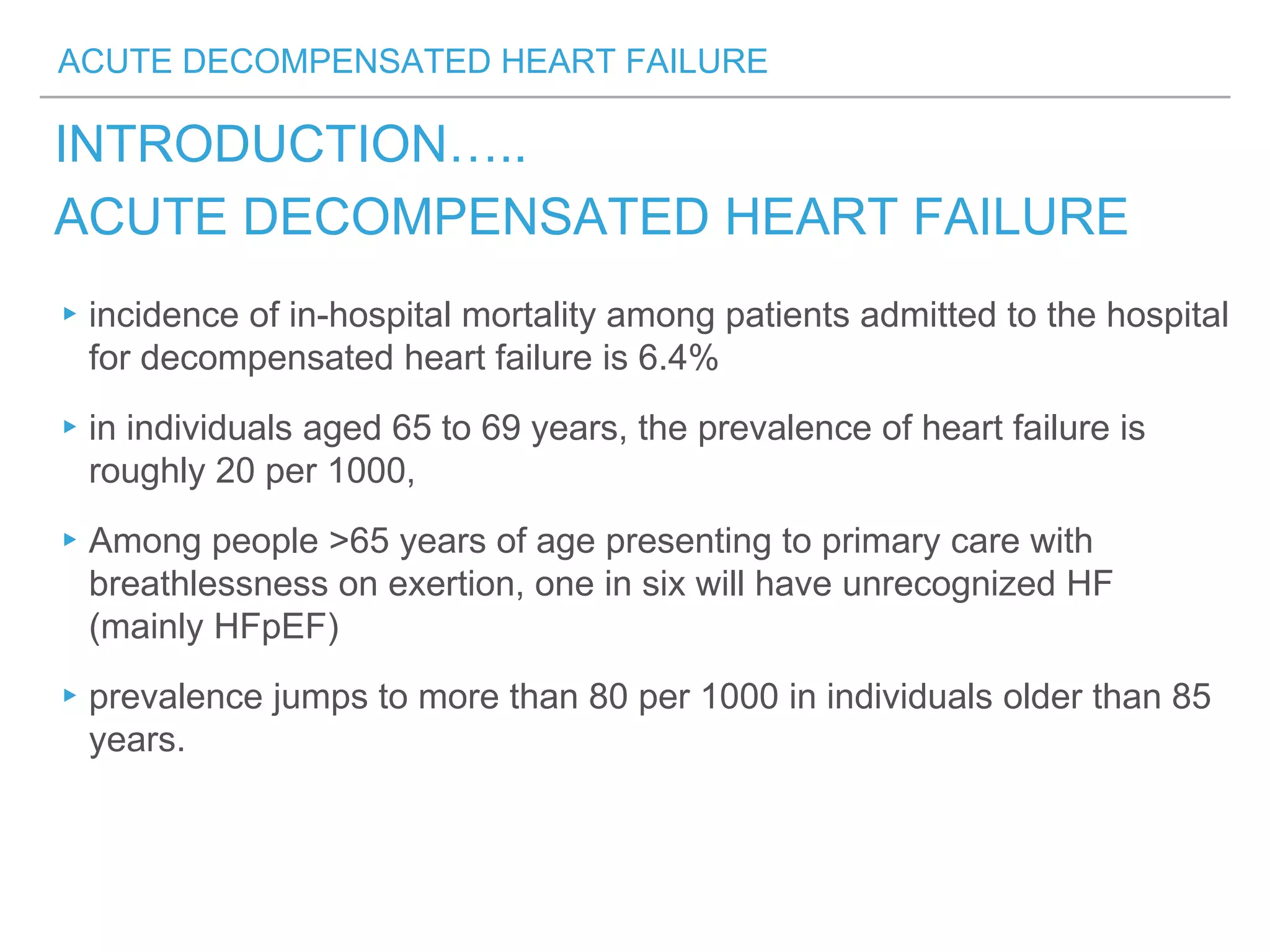
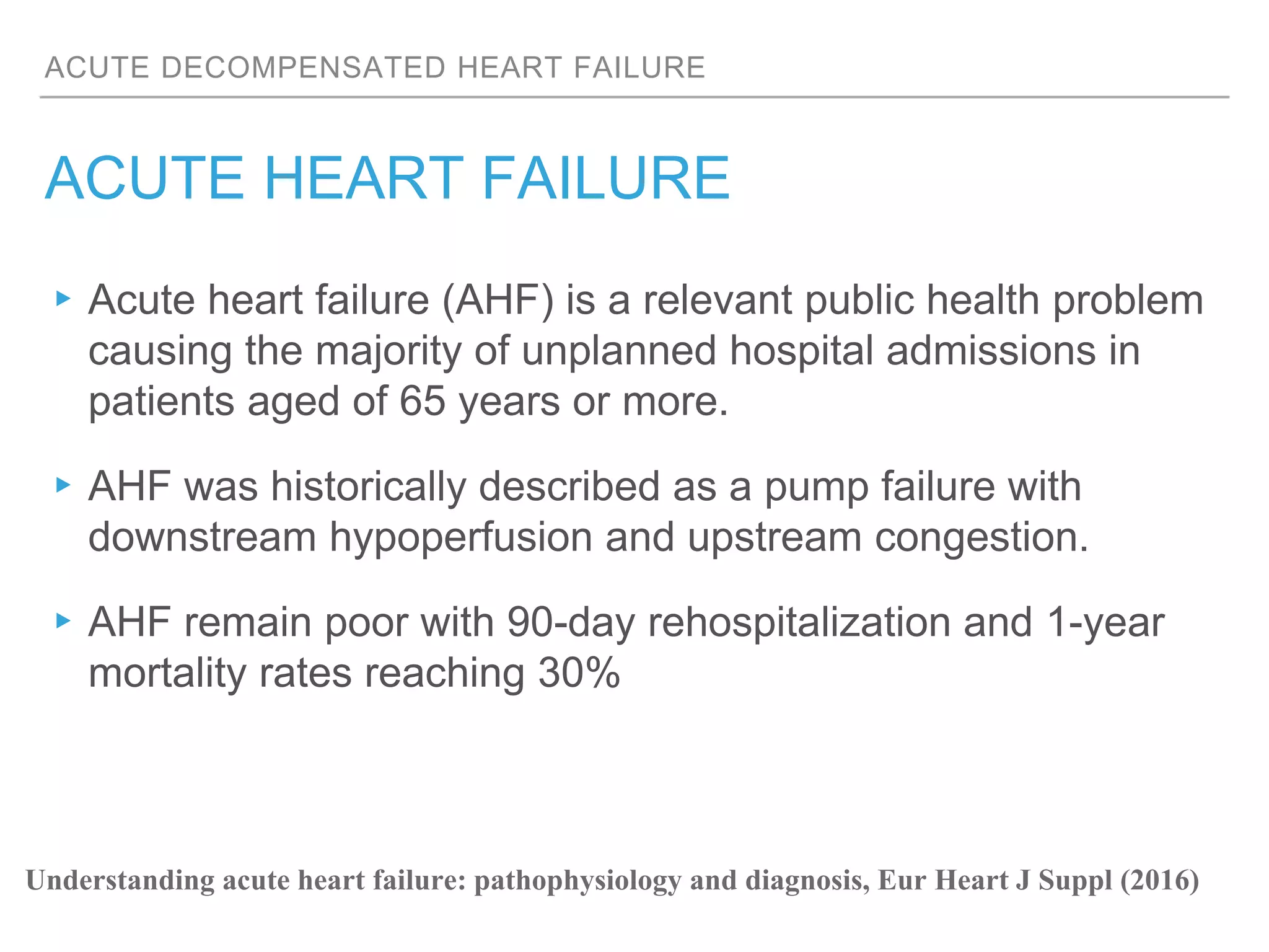
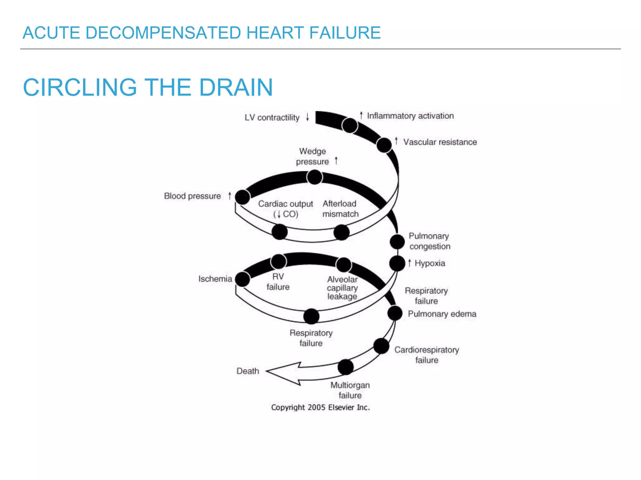
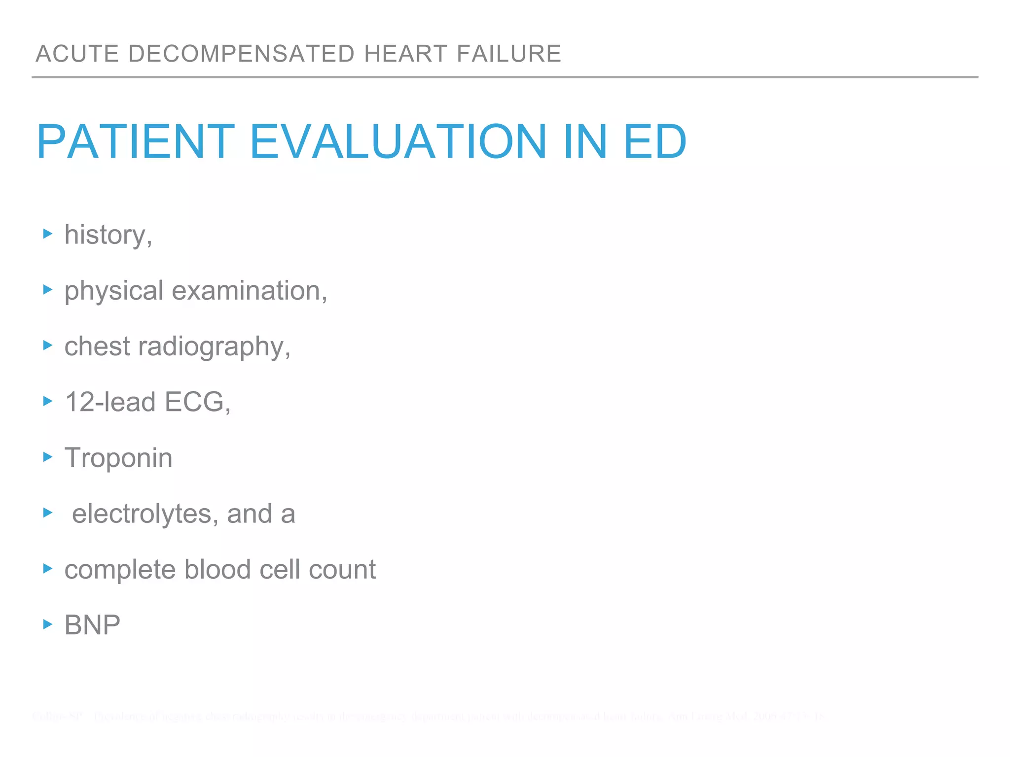
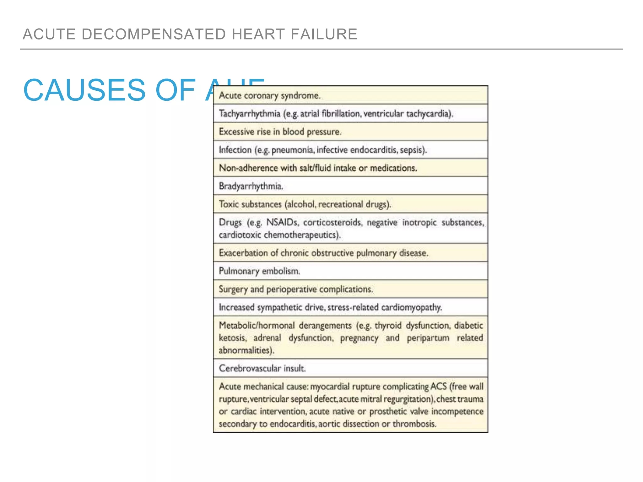
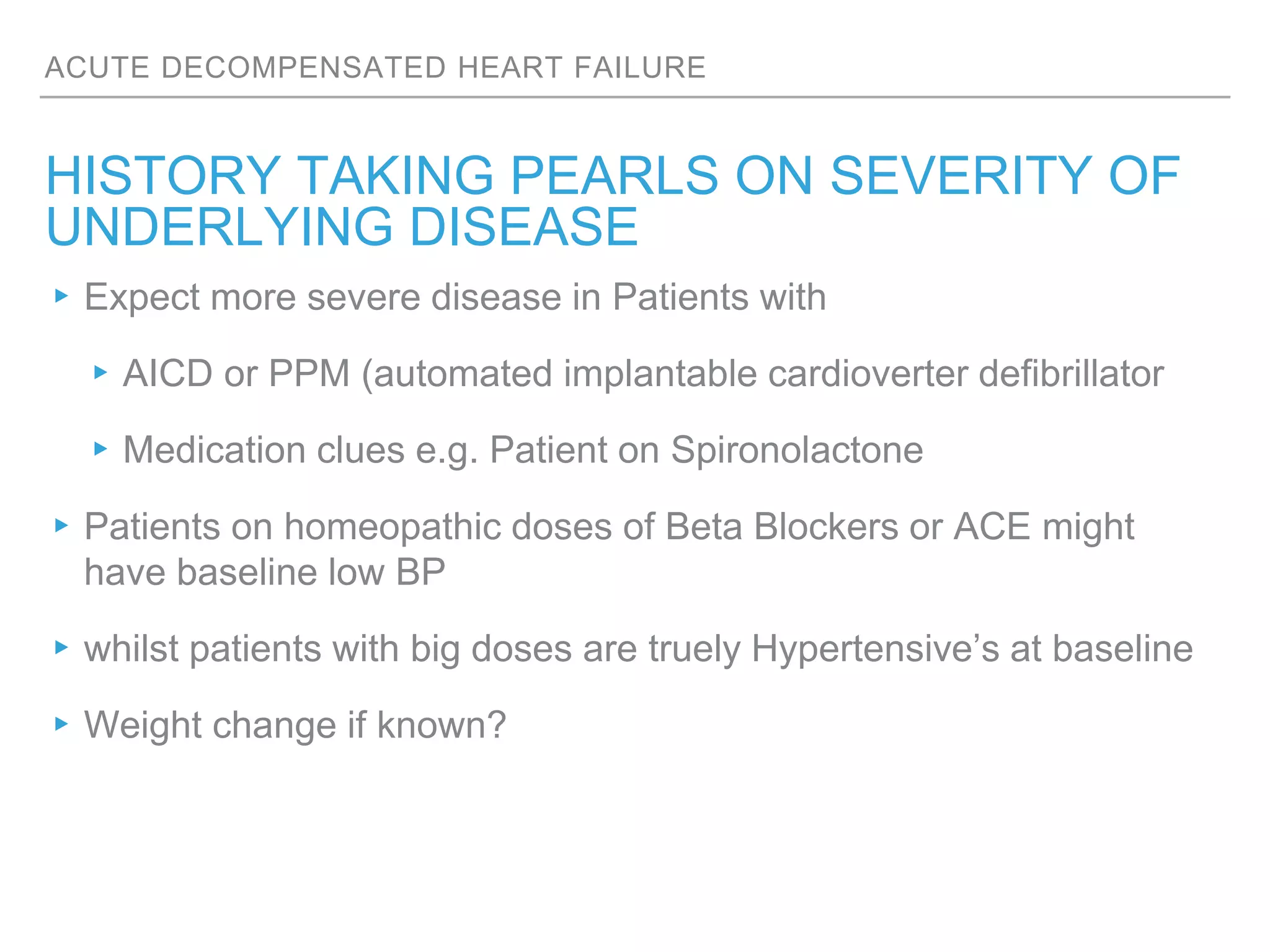
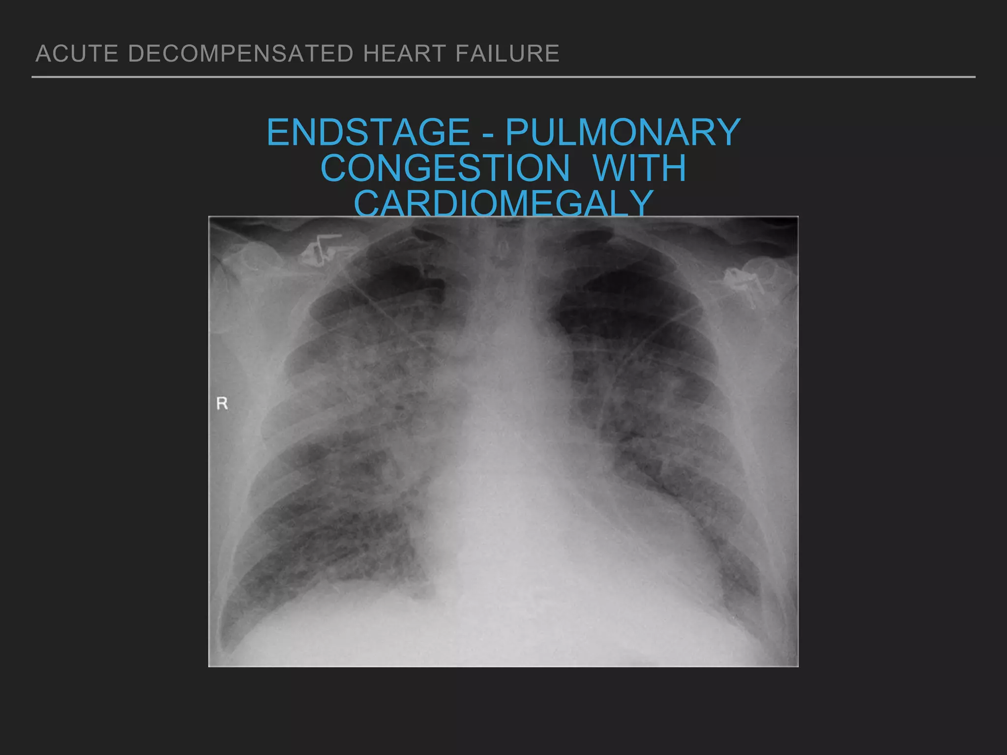
An overweight middle-aged man presented with acute shortness of breath, elevated blood pressure, coarse lung sounds, low oxygen saturation, and leg swelling. His initial evaluation found signs of severe congestive heart failure including pulmonary edema. Further assessment is needed to determine the underlying cause, guide treatment, and classify the type and severity of heart failure.
definition of heart failure, classification of heart failure, risk factors for heart failure, clinical features, general physical examination findings in heart failure
This is a comprehensive approach to a hypertensive patient presenting to the emergency department.
Discussing:-
- Hypertensive emergency
- Hypertensive Urgency
- Hypertensive Crisis
- Hypertensive encephalopathy and retinopathy
- Accelerated Hypertension
- Malignant hypertension
Acute kidney injury (AKI) is a potentially life-threatening
syndrome that occurs primarily in hospitalized patients
and frequently complicates the course of critically ill
patient.
Acute Kidney Injury is is (abrupt) reduction in kidney functions as evidence by changed in laboratory values; serum creatinine, blood urea nitrogen(BUN)and urine output
The presentation covers definitions, identification, Treatment goals, Special situations, Practice points, and cardinal pharmacotherapy. Session presented in NBE learning session
Note of management acute heart failure
Initially I just read the ESC 2016 guideline and was interested in choosing inotropic agents. I started to reads books and looked for more information.
Thyroid and its pathology (Hypothyroidism).Vikas Reddy
GREEK :- THYREOS – SHIELD ; EIDOS – FORM
1.LOCATION:- Anterior to trachea in between the cricoid cartilage and the suprasternal notch.
2.SHAPE:- It has 2 lobes connected with an isthmus, each lobe in turn has two poles.
3.Weighs around 10-20 gm, highly vascular and soft in consistency.
4. 4 Parathyroid glands which secrete PTH are located posterior to each pole of thyroid
The RLN traverse the lateral border of thyroid gland and must be identified during thyroid surgery to avoid injury and vocal cord paralysis.
Develops from the floor of primitive pharynx during the 3rd week of gestation.
Fetal cells in which developmental transcription factors TTF-1,TTF-2 & PAX-8 are expressed selectively form the thyroid gland ,secondly they result in induction of thyroid specific genes
Tg,TPO,NIS,TSH-R.
Mutations-THYROID AGENESIS & DYSHORMONOGENESIS(CONG. HYPOTHYROIDISM).
The developing gland migrates along the thyroglossal duct to reach its final location in the neck.
LINGUAL THYROID AND THYROGLOSSAL DUCT CYST.
Thyroid hormone synthesis begins at about 11 weeks of gestation.
Until 11 week of gestation and even later, it is the maternal thyroid hormones which cross the placenta to reach the fetus and aid its development.
Therefore a child born to a hypothyroid mother would suffer from features of congenital hypothyroidism.
Secondly if the mother has TSH-R blocking antibodies or has received anti thyroid therapy during pregnancy, might lead to transient congenital hypothyroidism.
Hyperthyroidism, Reference: Hyperthyroid, Harrison's Principles of Internal Medicine, Soheil Elahi, Islamic Azad University of Medicine- International Branch (IAUM-int)
Prof. Mridul M. Panditrao has added another of his very important, useful and in vogue topic to his collection. This is his well acclaimed andwell received faculty lecture at recently concluded International conference on Pain... ISSPCON 2014, at Mumbai/ Bombay from 6th to 9th Feb. 2014.
Periodontal Treatment of Medically Compromised Patients [Autosaved].pptxANIL KUMAR
The world's population is estimated to be over 7.7 billion. [1] Within this mass of humanity is a
substantial number of people who are elderly; the graying of the world's population is predicted to
produce millions of individuals with systemic medical conditions that can affect oral health and
dental treatment. The dental management of these medically compromised patients can be
problematic in terms of oral complications, dental therapy, and emergency care
Final presentation for my institutional APPE. I presented a full breakdown Cardiac Amyloidosis including clinical presentation, diagnosis and treatment options.
- Video recording of this lecture in English language: https://youtu.be/kqbnxVAZs-0
- Video recording of this lecture in Arabic language: https://youtu.be/SINlygW1Mpc
- Link to download the book free: https://nephrotube.blogspot.com/p/nephrotube-nephrology-books.html
- Link to NephroTube website: www.NephroTube.com
- Link to NephroTube social media accounts: https://nephrotube.blogspot.com/p/join-nephrotube-on-social-media.html
Tom Selleck Health: A Comprehensive Look at the Iconic Actor’s Wellness Journeygreendigital
Tom Selleck, an enduring figure in Hollywood. has captivated audiences for decades with his rugged charm, iconic moustache. and memorable roles in television and film. From his breakout role as Thomas Magnum in Magnum P.I. to his current portrayal of Frank Reagan in Blue Bloods. Selleck's career has spanned over 50 years. But beyond his professional achievements. fans have often been curious about Tom Selleck Health. especially as he has aged in the public eye.
Follow us on: Pinterest
Introduction
Many have been interested in Tom Selleck health. not only because of his enduring presence on screen but also because of the challenges. and lifestyle choices he has faced and made over the years. This article delves into the various aspects of Tom Selleck health. exploring his fitness regimen, diet, mental health. and the challenges he has encountered as he ages. We'll look at how he maintains his well-being. the health issues he has faced, and his approach to ageing .
Early Life and Career
Childhood and Athletic Beginnings
Tom Selleck was born on January 29, 1945, in Detroit, Michigan, and grew up in Sherman Oaks, California. From an early age, he was involved in sports, particularly basketball. which played a significant role in his physical development. His athletic pursuits continued into college. where he attended the University of Southern California (USC) on a basketball scholarship. This early involvement in sports laid a strong foundation for his physical health and disciplined lifestyle.
Transition to Acting
Selleck's transition from an athlete to an actor came with its physical demands. His first significant role in "Magnum P.I." required him to perform various stunts and maintain a fit appearance. This role, which he played from 1980 to 1988. necessitated a rigorous fitness routine to meet the show's demands. setting the stage for his long-term commitment to health and wellness.
Fitness Regimen
Workout Routine
Tom Selleck health and fitness regimen has evolved. adapting to his changing roles and age. During his "Magnum, P.I." days. Selleck's workouts were intense and focused on building and maintaining muscle mass. His routine included weightlifting, cardiovascular exercises. and specific training for the stunts he performed on the show.
Selleck adjusted his fitness routine as he aged to suit his body's needs. Today, his workouts focus on maintaining flexibility, strength, and cardiovascular health. He incorporates low-impact exercises such as swimming, walking, and light weightlifting. This balanced approach helps him stay fit without putting undue strain on his joints and muscles.
Importance of Flexibility and Mobility
In recent years, Selleck has emphasized the importance of flexibility and mobility in his fitness regimen. Understanding the natural decline in muscle mass and joint flexibility with age. he includes stretching and yoga in his routine. These practices help prevent injuries, improve posture, and maintain mobilit
These lecture slides, by Dr Sidra Arshad, offer a quick overview of the physiological basis of a normal electrocardiogram.
Learning objectives:
1. Define an electrocardiogram (ECG) and electrocardiography
2. Describe how dipoles generated by the heart produce the waveforms of the ECG
3. Describe the components of a normal electrocardiogram of a typical bipolar lead (limb II)
4. Differentiate between intervals and segments
5. Enlist some common indications for obtaining an ECG
6. Describe the flow of current around the heart during the cardiac cycle
7. Discuss the placement and polarity of the leads of electrocardiograph
8. Describe the normal electrocardiograms recorded from the limb leads and explain the physiological basis of the different records that are obtained
9. Define mean electrical vector (axis) of the heart and give the normal range
10. Define the mean QRS vector
11. Describe the axes of leads (hexagonal reference system)
12. Comprehend the vectorial analysis of the normal ECG
13. Determine the mean electrical axis of the ventricular QRS and appreciate the mean axis deviation
14. Explain the concepts of current of injury, J point, and their significance
Study Resources:
1. Chapter 11, Guyton and Hall Textbook of Medical Physiology, 14th edition
2. Chapter 9, Human Physiology - From Cells to Systems, Lauralee Sherwood, 9th edition
3. Chapter 29, Ganong’s Review of Medical Physiology, 26th edition
4. Electrocardiogram, StatPearls - https://www.ncbi.nlm.nih.gov/books/NBK549803/
5. ECG in Medical Practice by ABM Abdullah, 4th edition
6. Chapter 3, Cardiology Explained, https://www.ncbi.nlm.nih.gov/books/NBK2214/
7. ECG Basics, http://www.nataliescasebook.com/tag/e-c-g-basics
- Video recording of this lecture in English language: https://youtu.be/lK81BzxMqdo
- Video recording of this lecture in Arabic language: https://youtu.be/Ve4P0COk9OI
- Link to download the book free: https://nephrotube.blogspot.com/p/nephrotube-nephrology-books.html
- Link to NephroTube website: www.NephroTube.com
- Link to NephroTube social media accounts: https://nephrotube.blogspot.com/p/join-nephrotube-on-social-media.html
Adv. biopharm. APPLICATION OF PHARMACOKINETICS : TARGETED DRUG DELIVERY SYSTEMSAkankshaAshtankar
MIP 201T & MPH 202T
ADVANCED BIOPHARMACEUTICS & PHARMACOKINETICS : UNIT 5
APPLICATION OF PHARMACOKINETICS : TARGETED DRUG DELIVERY SYSTEMS By - AKANKSHA ASHTANKAR
263778731218 Abortion Clinic /Pills In Harare ,sisternakatoto
263778731218 Abortion Clinic /Pills In Harare ,ABORTION WOMEN’S CLINIC +27730423979 IN women clinic we believe that every woman should be able to make choices in her pregnancy. Our job is to provide compassionate care, safety,affordable and confidential services. That’s why we have won the trust from all generations of women all over the world. we use non surgical method(Abortion pills) to terminate…Dr.LISA +27730423979women Clinic is committed to providing the highest quality of obstetrical and gynecological care to women of all ages. Our dedicated staff aim to treat each patient and her health concerns with compassion and respect.Our dedicated group ABORTION WOMEN’S CLINIC +27730423979 IN women clinic we believe that every woman should be able to make choices in her pregnancy. Our job is to provide compassionate care, safety,affordable and confidential services. That’s why we have won the trust from all generations of women all over the world. we use non surgical method(Abortion pills) to terminate…Dr.LISA +27730423979women Clinic is committed to providing the highest quality of obstetrical and gynecological care to women of all ages. Our dedicated staff aim to treat each patient and her health concerns with compassion and respect.Our dedicated group of receptionists, nurses, and physicians have worked together as a teamof receptionists, nurses, and physicians have worked together as a team wwww.lisywomensclinic.co.za/
Ozempic: Preoperative Management of Patients on GLP-1 Receptor Agonists Saeid Safari
Preoperative Management of Patients on GLP-1 Receptor Agonists like Ozempic and Semiglutide
ASA GUIDELINE
NYSORA Guideline
2 Case Reports of Gastric Ultrasound
New Drug Discovery and Development .....NEHA GUPTA
The "New Drug Discovery and Development" process involves the identification, design, testing, and manufacturing of novel pharmaceutical compounds with the aim of introducing new and improved treatments for various medical conditions. This comprehensive endeavor encompasses various stages, including target identification, preclinical studies, clinical trials, regulatory approval, and post-market surveillance. It involves multidisciplinary collaboration among scientists, researchers, clinicians, regulatory experts, and pharmaceutical companies to bring innovative therapies to market and address unmet medical needs.
The Gram stain is a fundamental technique in microbiology used to classify bacteria based on their cell wall structure. It provides a quick and simple method to distinguish between Gram-positive and Gram-negative bacteria, which have different susceptibilities to antibiotics
Best Ayurvedic medicine for Gas and IndigestionSwastikAyurveda
Here is the updated list of Top Best Ayurvedic medicine for Gas and Indigestion and those are Gas-O-Go Syp for Dyspepsia | Lavizyme Syrup for Acidity | Yumzyme Hepatoprotective Capsules etc
2. ACUTE DECOMPENSATED HEART FAILURE
CLINICAL VIGNETTE - MY PATIENT FROM
SATURDAY NIGHT
Its 6:32 am and you are the Registrar in charge of the Saturday night shift at Charlie’s.
You are tired after battling through Amphetamine and ETOH inebriated Patients, its quiet,
….too quiet!
“Its quite quiet” the Intern says…… and 2 minutes later SJA rushes a patient into T2.
The patient is an overweight middle-aged man who is also struggling
to breathe. SJA report that his blood pressure was 220/130 mm Hg at the scene.
Shifty immediately attaches the patient to the cardiac monitor and obtains vital signs.
His blood pressure is now 240/140 mm Hg. HR is 140. You listen to his lungs and notice coarse, wet breath
sounds. Your patient is tachypnoeic, sweaty, leaning forward in bed, and saturating 72% on room air. His oxygen
saturation improves to 88% on a 100% nonrebreather mask.
His legs are oedematous, and he has marked conversational dyspnea(single words).
Respiratory arrest seems certain unless appropriate action is taken, and you wonder what to do…..
3. ACUTE DECOMPENSATED HEART FAILURE
INTRODUCTION…..
ACUTE DECOMPENSATED HEART FAILURE
▸incidence of in-hospital mortality among patients admitted to the hospital
for decompensated heart failure is 6.4%
▸in individuals aged 65 to 69 years, the prevalence of heart failure is
roughly 20 per 1000,
▸Among people >65 years of age presenting to primary care with
breathlessness on exertion, one in six will have unrecognized HF
(mainly HFpEF)
▸prevalence jumps to more than 80 per 1000 in individuals older than 85
years.
4. ACUTE DECOMPENSATED HEART FAILURE
ACUTE HEART FAILURE
▸Acute heart failure (AHF) is a relevant public health problem
causing the majority of unplanned hospital admissions in
patients aged of 65 years or more.
▸AHF was historically described as a pump failure with
downstream hypoperfusion and upstream congestion.
▸AHF remain poor with 90-day rehospitalization and 1-year
mortality rates reaching 30%
Understanding acute heart failure: pathophysiology and diagnosis, Eur Heart J Suppl (2016)
5. EPIDEMIOLOGY OF HEART
FAILURE (IN THE US)
MORE DEATHS FROM
HEART FAILURE THAN
FROM ALL FORMS OF
CANCER COMBINED
550,000 NEW CASES/YEAR
4.7 MILLION SYMPTOMATIC
PATIENTS;
ESTIMATED 10 MILLION IN
2037
*Rich M. J Am Geriatric Soc. 1997;45:968–974.
American Heart Association. 2001 Heart and Stroke
Statistical Update. 2000.
3.5
4.7
10
0
2
4
6
8
10
12
1991 2000 2037*
HeartFailurePatientsinUS
(Millions)
6. ACUTE DECOMPENSATED HEART FAILURE
NOT ALL HEART FAILURE IS THE
SAME.
- Heart failure with preserved ejection fraction (HFpEF)
- and heart failure with reduced ejection fraction (HFrEF)
- may present with either hypertension or hypotension
- represent distinct underlying pathophysiologies that
require different approaches in treatment.
- HFpEF and HFrEF are equal in terms of occurrence,
morbidity, and mortality.
Karrowni W, Chatterjee K. Diastolic heart failure: the current understanding and approach for management with focus on intensive care unit patients. J Intensive Care Med. 2014;29(3):119-127. (Review article)
7. ACUTE DECOMPENSATED HEART FAILURE
SYSTOLIC AND DIASTOLIC HEART FAILURE
Heart failure occurs when the heart is either unable-
-to Pump blood during systole (HFrEF) or
-to Fill with blood during diastole (HFpEF)
Systolic heart failure results in heart failure with reduced ejection fraction. Diastolic heart failure results in heart failure with preserved ejection fractio
8. ACUTE DECOMPENSATED HEART FAILURE
HEART FAILURE WITH PRESERVED
EJECTION FRACTIONHFpEF is defined as heart failure with ejection fraction that is either borderline (41%-
49%) or normal (> 50%).
Diastolic dysfunction
is characterised by elevated left ventricular filling pressures with impaired myocardial
relaxation.
responds differently to traditional heart failure therapies that were intended for the
traditional treatment of HFrEF
9. ACUTE DECOMPENSATED HEART FAILURE
ETIOLOGY AND PATHOPHYSIOLOGY
▸Injury to Myocardium
▸secondary to a number of causes that develop slowly over time (uncontrolled
hypertension, diabetes) or
▸more suddenly (eg, coronary ischaemia, STEMI)
▸Regardless of the cause, myocardial injury results in
▸structural-abnormalities of the ventricular wall that impair systolic
contraction or diastolic relaxation
▸electrophysiological, ->impaired conduction,(QRS widening on ECG),
which can lead to conduction blocks and re-entrant dysrhythmias and
▸biochemical remodeling -> impaired myocyte functionality and
increased risk of dysrhythmia
10. ACUTE DECOMPENSATED HEART FAILURE
THE ISSUE IS…
▸Regardless of the underlying aetiology, patients presenting
with AHF appear very similar to each other:
▸Sodium and Fluid retention,
▸excess Fluid backing up into the lungs, abdomen, and
extremities.
▸The result is fatigue, peripheral oedema, and dyspnea that
is often worse with exertion
11. ACUTE DECOMPENSATED HEART FAILURE
PATHOPHYSIOLOGY OF ACUTE HEART FAILURE
▸Acute heart failure is defined as new-onset or worsening of
symptoms and signs of HF
▸AHF typically includes symptoms or signs related to
congestion and volume overload rather than to
hypoperfusion
▸level of congestion and the number of congested organs
have prognostic relevance in HF patients
Gheorghiade M. European Society of Cardiology, European Society of Intensive Care
Medicine. Assessing and grading congestion in acute heart failure: a scientific statement
from the acute heart failure committee of the heart failure association of the European
Society of Cardiology and endorsed by the European Society of Intensive Care
Medicine. Eur J Heart Fail 2010
13. ACUTE DECOMPENSATED HEART FAILURE
PATIENT EVALUATION IN ED
▸history,
▸physical examination,
▸chest radiography,
▸12-lead ECG,
▸Troponin
▸ electrolytes, and a
▸complete blood cell count
▸BNP
Collins SP, . Prevalence of negative chest radiography results in the emergency department patient with decompensated heart failure. Ann Emerg Med. 2006;47:13–18.
14. ACUTE DECOMPENSATED HEART FAILURE
DIFFERENTIAL DIAGNOSIS
▸look for alternative diagnoses
▸do not miss reversible causes and
▸other life threatening causes
17. ACUTE DECOMPENSATED HEART FAILURE
HISTORY TAKING PEARLS ON SEVERITY OF
UNDERLYING DISEASE
▸Expect more severe disease in Patients with
▸AICD or PPM (automated implantable cardioverter defibrillator
▸Medication clues e.g. Patient on Spironolactone
▸Patients on homeopathic doses of Beta Blockers or ACE might
have baseline low BP
▸whilst patients with big doses are truely Hypertensive’s at baseline
▸Weight change if known?
18. ACUTE DECOMPENSATED HEART FAILURE
AHF - SIGNS AND SYMPTOMS
▸Exertional dyspnea and/or dyspnea at rest
▸Orthopnea
▸Acute pulmonary oedema
▸Chest pain/pressure and palpitations
▸Tachycardia
▸Fatigue and weakness
▸Nocturia and oliguria
▸Anorexia, weight loss, nausea
▸Exophthalmos and/or visible pulsation of eyes
▸Distention of neck veins
▸Weak, rapid, and thready pulse
▸Rales, wheezing
▸S 3 gallop and/or pulsus alternans
▸Increased intensity of P 2 heart sound
▸Hepatojugular reflux
▸Ascites, hepatomegaly, and/or anasarca
▸Central or peripheral cyanosis, pallor
▸Big Ticket items:
▸Patient Position
▸Hypertensive/Hypotensive
▸Distention of neck veins - Surrogate
for Right Heart pressure
▸Auscultation —>Rales, wheezing -
worse rales>worse AHD
▸New Heart Murmur / Distant Heart
sound
▸S 3 gallop - Diagnostic for AHD
▸uni/bilateral Leg swelling
19. ACUTE DECOMPENSATED HEART FAILURE
ASSESSMENT OF HF PROBABILITY
▸1. Clinical history:
▸History of CAD (MI, revascularization)
▸History of arterial hypertension
▸Exposition to cardiotoxic drug/radiation
▸Use of diuretics
▸Orthopnoea / paroxysmal nocturnal dyspnoea
▸2. Physical examination:
▸Rales
▸Bilateral ankle oedema
▸Heart murmur
▸Jugular venous dilatation
▸Laterally displaced/broadened apical beat
▸3. ECG:
▸Any abnormality
21. ACUTE DECOMPENSATED HEART FAILURE
ESC HEART FAILURE GUIDELINES
▸Use transthoracic echocardiography in patients with
suspected or established HF for the assessment of
myocardial structure and function along with the
measurement of LVEF to establish the diagnosis of
▸HF with reduced (HFrEF, LVEF<40%),
▸mid-range (HFmrEF, LVEF: 40-49%) or
▸preserved ejection fraction (HFpEF, LVEF≥50%)
23. ACUTE DECOMPENSATED HEART FAILURE
FORRESTER HAEMODYNAMIC SUBSETS -
AHF IN NUMBERS
Subset Description
I: Warm and dry (normal) PCWP 15–18 mmHg and CI >2.2 L/min/m2
II: Warm and wet (congestion) PCWP >18 mmHg and CI >2.2 L/min/m2
III: Cold and dry (hypoperfusion) PCWP 15–18 mmHg and CI <2.2 L/min/m2
IV: Cold and wet (congestion and
hypoperfusion)
PCWP >18 mmHg and CI <2.2 L/min/m2
Forrester Hemodynamic Subsets
CI: cardiac index; PCWP: pulmonary capillary wedge pressure.
Forrester JS, Diamond G, Chatterjee K, et al. Medical therapy of acute myocardial infarction by application of hemodynamic subsets. N Engl J Med. 1976;295:1356–1362.
25. ACUTE DECOMPENSATED HEART FAILURE
ECG - INFERIOR STEMI WITH RV INFARCTION
ST elevation in V1 – the only standard ECG lead that looks directly at the right ventricle.
ST elevation in lead III > lead II – because lead III is more “rightward facing” than lead II and hence
more sensitive to the injury current produced by the right ventricle.
Right ventricular infarction complicates up to 40% of inferior STEMIs.
Patients with RV infarction are very preload sensitive (due to poor RV contractility) and can develop
severe hypotension in response to nitrates or other preload-reducing agents.
Hypotension in right ventricular infarction is treated with cautious fluid loading, and nitrates are
contraindicated.
27. ACUTE DECOMPENSATED HEART FAILURE
RADIOLOGICAL FEATURES OF ACUTE
PULMONARY OEDEMA
▸There are distinct radiological phases.
▸Interstitial phase is the fluid filling up spaces between acini;
▸the alveolar phase is where that fluid floods the air spaces.
▸Interstitial pulmonary oedema may develop first.
▸Small pulmonary vessels lose their definition
▸Peribronchial cuffing occurs:
▸Kerley B lines
▸Thickening of fissures
▸Pleural effusions
▸Bat Wing" oedema
▸15% Normal CXR
29. ACUTE DECOMPENSATED HEART FAILURE
MILD PULMONARY CONGESTION WITH
CEPHALISATIONAP chest radiograph demonstrates cephalisation (upper lobe vascular redistribution) in stage I CCF where
there is elevation of the left atrial pressure 10-15 mmHg. Normal left atrial pressure is 5-10 mmHg.
http://www.radiologyassistant.nl/en/p4c132f36513d4
30. ACUTE DECOMPENSATED HEART FAILURE
MORE ADVANCED CONGESTION - KERLEY B
LINES
Stage II - Interstitial oedema
Stage II of CHF is characterised by fluid leakage into the interlobular and peribronchial interstitium as a result of the i
When fluid leaks into the peripheral interlobular septa it is seen as Kerley B or septal lines.
Kerley-B lines are seen as peripheral short 1-2 cm horizontal lines near the costophrenic angles.
These lines run perpendicular to the pleura.
31. ACUTE DECOMPENSATED HEART FAILURE
EVEN MORE ADVANCED CONGESTION
When fluid leaks into the peribronchovascular interstitium it is seen as thickening of the bronchial walls (peribronchial
cuffing) and as loss of definition of these vessels (perihilar haze).
On the left a patient with heart failure.
There is an increase in the caliber of the pulmonary vessels and they have lost their definition because they are
34. ACUTE DECOMPENSATED HEART FAILURE
ECHOCARDIOGRAPHY
▸ Primary imaging modality to
evaluate a patient’s cardiac
function and evaluate for
either systolic or diastolic
dysfunction
▸ only part of the initial ED
examination where systolic
and diastolic dysfunction can
be distinguished
▸ approximation of LVEF
▸ visual assessment that looks
for the general quality of the
“heart’s squeeze”
35. ACUTE DECOMPENSATED HEART FAILURE
ECHOCARDIOGRAPHY
▸ A more precise way to examine left
ventricular ejection fraction is via E-point
septal separation (EPSS),
▸ measures the smallest distance between
the tip of the mitral leaflet and the
interventricular septum during diastole.
▸ This distance is assessed using M-mode,
with the indicator overlying the tip of the
mitral lea et.
▸ The larger this distance, the lower the
ejection fraction. EPSS > 7 mm is
indicative of poor left ventricular
Secko MA, Lazar JM, Salciccioli LA, et al. Can junior emer- gency physicians use E-point septal
separation to accurately estimate left ventricular function in acutely dyspneic pa- tients? Acad Emerg
Normal
Normal
Abnormal
M-Mode
36. ACUTE DECOMPENSATED HEART FAILURE
PULMONARY
ULTRASOUND - B LINES
Multiple B-lines from a case of cardiogenic pulmonary oedema.
When a similar pattern is visualised on multiple locations in the anterior and lateral chest, it
is diagnostic of the interstitial syndrome.
Interstitial
Pulmonary fluid is identifiable on ultrasound as vertical hyper-echoic lines that arise from,
and run perpendicular to, the pleura.
These lines extend into the lung parenchyma and are referred to as B-lines.
The presence of 3 or more B-lines in at least 2 bilateral lung zones is indicative of
pulmonary oedema.
The greater the number of zones demonstrating B-lines, the higher the likelihood of ADHF
Combined with BNP - High Dx accuracy!
Liteplo AS, et al. Emergency thoracic ultrasound in the differentiation of the etiology of shortness of breath (ETUDES): sonographic B-lines and N-terminal pro-brain-type natriuretic peptide in diagnosing congestive heart failure. Acad
38. ACUTE DECOMPENSATED HEART FAILURE
BNP
▸BNP Cutoff of 100 ng/l,
▸BNP had a sensitivity 90%, specificity 76%, negative predictive 79%, and
positive predictive value of 89%.
▸In this capacity, BNP is highly useful to exclude AHF
▸negative likelihood ratio of BNP at 100 pg/mL is 0.13 -LR
▸< 100 ng/l strongly suggestive against AHF
▸>400ng/l suggestive of AHF exacerbation
• However may be falsely elevated in:
• Renal disease, atrial fibrillation, pulmonary HTN
• May be falsely low in:
• Obese patients, HFPEF
• High BNP increased Mortality in men
1. Maisel AS, et all . Rapid measurement of B-type natriuretic peptide in the emergency diagnosis of heart failure. N Engl J Med. 2002;347:161–167.
39. ACUTE DECOMPENSATED HEART FAILURE
TROPONIN
▸Useful for risk stratification
▸? underlying primary cause of AHF (cardiac strain,
ischaemia, infarction)
▸elevated trop = increased in hospital mortality 8% versus 2.7
% if Neg Trop
▸increased re-hospitalisation rate
▸increased risk at death at 90 days
40. ACUTE DECOMPENSATED HEART FAILURE
FBC, U/E,S + SUPPLEMENTAL TESTS
▸Anaemia- HB under 8 (in IHD under 10)- improve Oxygen
carrying capacity by transfusion
▸Sodium and Renal function (AKI?-end organ damage)
▸Liver function (end organ damage)
▸Thyroid function (cause of failure?)
▸Calcium - low? improves cardiac contractility
▸amyloidosis, pheochromocytoma etc as rare causes.
41. ACUTE DECOMPENSATED HEART FAILURE
TREATMENT AIMS
▸Decrease left ventricular diastolic pressure, by decreasing
systemic vascular resistance and improving systolic and
diastolic functional reserve.
▸Promote coronary blood flow.
▸Correct acute respiratory failure.
▸In-hospital mortality for APO is up to 12%, with one-year
mortality up to 40% !
42. ACUTE DECOMPENSATED HEART FAILURE
MANAGEMENT OF PRELOAD
▸ Diuretics
▸ Fluid restriction
▸ Venodilators
▸ Aldosterone agonists
▸ Beta-blockers
▸ Maintenance of sinus rhythm and atrial systolic contribution
▸ Pacing to maintain AV synchrony
45. ACUTE DECOMPENSATED HEART FAILURE
DIAGNOSIS AND INITIAL PROGNOSTIC EVALUATION - BE A CHAMP
Jessup, M. et al. (2016) 2016 ESC and
ACC/AHA/HFSA heart failure guideline
update — what is new and why is it
important?
Nat. Rev. Cardiol.
doi:10.1038/nrcardio.2016.134
46. ACUTE DECOMPENSATED HEART FAILURE
IDENTIFICATION OF PRECIPITANTS/CAUSES LEADING TO DECOMPENSATION
THAT NEEDS URGENT MANAGEMENT
▸ Acute coronary syndrome.
▸ Coexistence of these two clinical conditions (ACS and AHF) always identifies a very-high-risk group where an immediate (i.e. <2 h from hospital admission in patients
with NSTEMI, analogous to STEMI management)
▸ invasive strategy with intent to perform revascularization is recommended, irrespective of ECG or biomarker findings.
▸ Hypertensive emergency.
▸ AHF precipitated by rapid and excessive increase in arterial blood pressure typically manifests as acute pulmonary oedema. Aggressive blood pressure reduction (in
the range of 25% during the first few hours and cautiously thereafter) with i.v. vasodilators in combination with loop diuretics is recommended.
▸ Rapid arrhythmias or severe bradycardia/conduction disturbance.
▸ Severe rhythm disturbances in patients with AHF and unstable conditions should be corrected urgently with medical therapy, electrical cardioversion or temporary
pacing
▸ Electrical cardioversion is recommended if an atrial or ventricular arrhythmia is thought to be contributing to the patient's haemodynamic compromise in order to
restore sinus rhythm and improve the patient's clinical condition.
▸ Acute mechanical cause underlying AHF.
▸ mechanical complication of ACS (free wall rupture, ventricular septal defect, acute mitral regurgitation),
▸ chest trauma or cardiac intervention
▸ Echocardiography is essential for diagnosis, and treatment typically requires circulatory support with surgical or percutaneous intervention.
▸ • Acute pulmonary embolism. When acute pulmonary embolism is confirmed as the cause of shock or hypotension, immediate specific treatment is recommended with
primary reperfusion either with thrombolysis, catheter-based approach or surgical embolectomy.
48. ACUTE DECOMPENSATED HEART FAILURE
5 DISTINCT TREATMENT GROUPS
▸ Acute heart failure syndrome (AHFS) spectrum can be divided into 5 groups as
regards therapeutic management:
▸Dyspnoea + /- congestion with elevated systolic blood pressure (SBP)>140
mmHg, usually with abrupt onset APO (most frequent type)
▸Dyspnoea + /- congestion with normal SBP 100-140 mmHg, usually with gradual
onset predominant systemic oedema and milder APO
▸Dyspnoea + /- congestion with low SBP <100 mmHg, with predominant
cardiogenic shock or end-stage cardiac failure (most fatal type)
▸Dyspnoea + /- congestion with signs of ACS such as chest pain
▸Isolated RV failure usually without APO
Mebazza A, Gheoghiade M, Pina I et al. Practical recommendations for pre- hospital and early in-hospital management of patients presenting with acute heart failure. Crit Care Med 2008;36:S129-39.
49. ACUTE DECOMPENSATED HEART FAILURE
CLINICAL CLASSIFICATION
Classification of patients presenting with acutely decompensated heart failur
Yancy C W et al. Circulation. 2013;128:e240-e327
50. ACUTE DECOMPENSATED HEART FAILURE
PRACTICAL RECOMMENDATION
Alexandre Mebazaa, MD, PhD et all “Practical recommendations
for prehospital and early inhospital management of patients
presenting with acute heart failure
syndromes” Crit Care Med 2008 Vol. 36, No. 1
52. ACUTE DECOMPENSATED HEART FAILURE
VASODILATORS
▸ Safety of high dose initial GTN:
▸ 800 mcg(2x0,4mg sl tab) GTN sl if BP>180mmHg
▸ 1200 mcg(3x0.4mg sl Tab) for BP>200 mmHg
▸ only 3/75 incidence of Hypotension
▸ Initial GTN dose iv 50-100mcg/min up to 400mcg/min for 2 min max with Physician
in attendance
▸ Vasodilators should be used with extreme caution in patients with significant mitral
or aortic stenosis.
Clemency BM, Thompson JJ, Tundo GN, et al.
Prehospital Disaster Medicine high-dose
sublingual nitroglycerin rarely causes hypoten- sion.
Prehosp Disaster Med. 2013;28(5):477-481.
(Retrospective cohort study; 75 patients)
53. ACUTE DECOMPENSATED HEART FAILURE
VASODILATORS
‣ No robust evidence confirming their beneficial effects.
‣ They have dual benefit by
‣ decreasing venous tone (to optimize preload)
‣ decreasing arterial tone (decrease afterload).
‣ they may also increase stroke volume.
‣ Vasodilators are especially useful in patients with hypertensive AHF, whereas in SBP <90 mmHg (or with s
54. ACUTE DECOMPENSATED HEART FAILURE
INOTROPIC AGENTS
▸ Digoxin - no improvement over placebo in one (older) study
▸ Dopamine was compared with norepinephrine in the treatment of various shock patients.
▸ Increased Mortality
▸ A subgroup analysis suggested that norepinephrine would have fewer side effects and lower mortality
▸ Norepinephrine 1st choice but increasing Oxygen demand in Heart
De Backer D , Biston P, Devriendt J, Madl C, Chochrad D, Aldecoa C, Brasseur A, Defrance
P, Gottignies P, Vincent J-L. Comparison of dopamine and norepinephrine in the treatment of
shock. N Engl J Med 2010;362:779–789.
56. ACUTE DECOMPENSATED HEART FAILURE
DIGOXIN
▸Digoxin is if at all indicated in patients with AF and rapid
ventricular rate (>110 bpm)
▸boluses of 0.25–0.5 mg i.v. if not used previously
57. ACUTE DECOMPENSATED HEART FAILURE
MANAGEMENT OF PATIENTS WITH CARDIOGENIC SHOCK
▸Pharmacologic therapy aims to improve organ perfusion
▸by increasing cardiac output and blood pressure.
▸fluid challenge,
▸inotropic agent (Dobutamine)and a
▸vasopressor (noradrenaline) as needed.
▸immediate Coronary Angiogram recommended
58. ACUTE DECOMPENSATED HEART FAILURE
DIURETICS
▸Diuretics are a cornerstone in the treatment of patients with AHF and signs of fluid overload and
congestion.
▸Diuretics increase renal salt and water excretion and have some vasodilatory effect.
▸In patients with AHF and signs of hypoperfusion, diuretics should be avoided before adequate perfusion
is attained.
▸The initial approach to congestion management involves i.v. diuretics with the addition of vasodilators for
dyspnoea relief if blood pressure allows.
▸Options include dual nephron blockade by loop diuretics (i.e. furosemide or torasemide) with thiazide
diuretics or natriuretic doses of MRAs
▸Administration of furosemide at 2.5 times the previous oral dose resulted in greater improvement in
dyspnoea, larger weight change and fluid loss at the cost of transient worsening in renal function.
▸patients with new-onset AHF or those with chronic HF without a history of renal failure and previous use
of diuretics may respond to i.v. boluses of 20–40 mg
59. ACUTE DECOMPENSATED HEART FAILURE
FURUSEMIDE
Felker GM , Lee KL, Bull DA, Redfield MM, Stevenson LW, Goldsmith SR, LeWinter MM,
Deswal A, Rouleau JL, Ofili EO, Anstrom KJ, Hernandez AF, McNulty SE, Velazquez EJ,
Kfoury AG, Chen HH, Givertz MM, Semigran MJ, Bart BA, Mascette AM, Braunwald E,
O'Connor CM. Diuretic strategies in patients with acute decompensated heart failure. N Engl J
Med 2011;364:797–805.
▸In Patient with Evidence of Fluid overload Diuretics to be started
early in ED stay
▸In the ‘high-dose’ arm of the DOSE study, administration of
furosemide at 2.5 times the previous oral dose resulted in
greater improvement in dyspnoea, larger weight change and
fluid loss at the cost of transient worsening in renal function
▸i.v. Furusemide should be given at least the same or larger
the daily po dose to improve outcomes.
▸starting does for Furusemide in de move patients 20-40mg iv
60. ACUTE DECOMPENSATED HEART FAILURE
OXYGEN
▸In AHF, oxygen should not be used routinely in non-
hypoxaemic patients, as it causes vasoconstriction and a
reduction in cardiac output.
▸ In COPD, hyperoxygenation may increase ventilation–
perfusion mismatch, suppressing ventilation and leading to
hypercapnia.
▸During oxygen therapy, acid–base balance and
transcutaneous SpO2 should be monitored.
Park JH , Balmain S, Berry C, Morton JJ, McMurray JJV. Potentially detrimental
cardiovascular effects of oxygen in patients with chronic left ventricular systolic dysfunction.
Heart 2010;96:533–538.
61. ACUTE DECOMPENSATED HEART FAILURE
OPIATES…RATHER USE BENZO’S
▸Opiates relieve dyspnoea and anxiety.
▸In AHF, routine use of opiates is not recommended and they may
only be cautiously considered in patients with severe dyspnoea,
mostly with pulmonary oedema.
▸Dose-dependent side effects include nausea, hypotension,
bradycardia and respiratory depression (potentially increasing the
need for invasive ventilation).
▸There are controversies regarding the potentially elevated
mortality risk in patients receiving morphine
Peacock WF , Hollander JE, Diercks DB, Lopatin M, Fonarow G, Emerman CL. Morphine and
outcomes in acute decompensated heart failure: an ADHERE analysis. Emerg Med
J 2008;25:205–209.
62. ACUTE DECOMPENSATED HEART FAILURE
LEVOSIMENDAN
▸“Calcium-sensitiser”
▸works by increasing myocardial contractility
▸by sensitising the cardiac myocytes to calcium
▸ In the REVIVE study levosimendan, when added to standard therapy,
resulted in a more rapid symptomatic improvement when compared to
placebo with standard therapy;
▸increased risk of hypotension and dysrhythmias associated with its
administration
Packer M, Colucci W, Fisher L, et al. Effect of Levosimendan on the
short term clinical course of patients with acutely decompensated heart
failure. JACC Heart Fail2013;1(2):103- 111. (Randomized controlled trial;
700 patients
63. ACUTE DECOMPENSATED HEART FAILURE
VENTILATORY ASSISTANCE
▸Non-invasive ventilation (NIV) refers to CPAP; or bilevel positive airway pressure
(BiPAP) non-invasive pressure support ventilation (NIPSV), where IPAP – EPAP
(≡PEEP) reflects the amount of pressure support delivered.
▸CPAP reduces mortality (RR 0.64) and need to intubate (RR 0.44), with no effect
on incidence of new MI. BiPAP reduces need to intubate (RR 0.54), but not
mortality or new MI. Thus CPAP preferred in APO due to AMI / ischaemia.
▸Note 3CPO trial findings showed negative effect of NIV compared to standard
medical therapy alone, but may be explained by sickest patients were excluded,
low overall rates of intubation, ischaemia and mortality (i.e. their patients were
different), and considerable treatment group crossover after first 2 hours.
64. ACUTE DECOMPENSATED HEART FAILURE
NIV
▸Non-invasive positive pressure ventilation includes both CPAP and bi-level positive
pressure ventilation (PPV). Bi-level PPV also allows inspiratory pressure support that
improves minute ventilation and is especially useful in patients with hypercapnia, most
typically COPD patients.
▸Non-invasive positive pressure ventilation includes both CPAP and bi-level positive
pressure ventilation (PPV). Bi-level PPV also allows inspiratory pressure support that
improves minute ventilation and is especially useful in patients with hypercapnia, most
typically COPD patients.
▸Non-invasive positive pressure ventilation reduces respiratory distress and may decrease
intubation and mortality rate
▸In one study (Emerg Med J 2004; 21:155-161) survival to hospital discharge was improved
with CPAP (10 mm/Hg) over BiPap (Ipap 15 Epap 5) and conventional therapy
Vital FMR , Ladeira MT, Atallah AN. Non-invasive positive pressure ventilation (CPAP or
bilevel NPPV) for cardiogenic pulmonary oedema. Cochrane Database Syst
Rev 2013;5:CD005351.
65. ACUTE DECOMPENSATED HEART FAILURE
EARLY REVASCULARISATION THERAPY IN
ISCHAEMIC ECG
▸The SHOCK Trial showed a 67% relative improvement in
long-term survival, measured at 6 years, for patients
managed with rapid revascularization.
▸Role of revascularization is not clear for patients presenting
with failure without obvious acute ischemia.
Hochman JS, Sleeper LA, Webb JG, et al. Early revasculariza- tion and long-
term survival in cardiogenic shock complicat- ing acute myocardial infarction.
JAMA. 2006;295(21):2511- 2515. (Randomized controlled trial; 302 patients)
66. ACUTE DECOMPENSATED HEART FAILURE
ULTRAFILTRATION
▸Ultrafiltration is similar to hemodialysis; however, it focuses on
fluid removal rather than solute exchange.
▸can be accomplished through a smaller-diameter catheter than
hemodialysis, but it generally requires a peripherally inserted
central catheter (PICC) line
▸The UNLOAD trial evaluated ultrafiltration versus IV diuretic
therapy in patients with functioning kidneys, and it demonstrated
that ultrafiltration removes a larger volume of fluid and is
associated with a greater reduction in 90-day resource utilisation
compared to diuretic therapy.
68. ACUTE DECOMPENSATED HEART FAILURE
INITIAL MANAGEMENT OF A PATIENT WITH
ACUTE HEART FAILURE
Jessup, M. et al. (2016) 2016 ESC and ACC/AHA/HFSA heart failure guideline update — what is new and why is it important?
Nat. Rev. Cardiol. doi:10.1038/nrcardio.2016.134
Modified from Ponikowski, P. et al. 2016 ESC guidelines for the diagnosis and treatment of acute and chronic heart
failure: the Task Force for the diagnosis and treatment of acute and chronic heart failure of the European Society
of Cardiology (ESC). Developed with the special contribution of the Heart Failure Association (HFA) of the ESC.
Eur. Heart J. http://dx.doi.org/10.1093/eurheartj/ehw128 (2016), with permission from Oxford University Press and the ESC
69. ACUTE DECOMPENSATED HEART FAILURE
MECHANICAL RESCUE DEVICES AND
CHEATS
▸Increase cardiac output by unnatural means:
▸LVAD
▸ECMO
▸Increase the pacemaker rate (60-90 increase of CO by 30%)
▸Decrease the organism's demand for cardiac output
▸Hypothermia
▸Paralysis/sedation
71. TEXT
OPIATES
▸Opiates relieve dyspnoea and anxiety.
▸In AHF, routine use of opiates is not recommended
▸only be cautiously considered in patients with severe dyspnoea, mostly
with pulmonary oedema.
▸Dose-dependent side effects
▸nausea, hypotension, bradycardia and respiratory depression
(potentially increasing the need for invasive ventilation).
▸There are controversies regarding the potentially elevated mortality
risk in patients receiving morphine
Iakobishvili et all, Use of intravenous morphine for acute decompensated heart failure in patients with and without acute coronary syndromes.
Acute Card Care 2011;13:76–80.
PeacockWF.ett all.Morphine and outcomes in acute decompensated heart failure: an ADHERE analysis.Emerg Med J
2008;25:205–209.
72. ACUTE DECOMPENSATED HEART FAILURE
DEVICE THERAPY - RRT
▸Renal Replacement Therapy - routine use of ultrafiltration is not
recommended
▸Criteria for initiation of renal replacement therapy in patients with refractory
volume overload:
▸oliguria unresponsive to fluid resuscitation measures,
▸severe hyperkalaemia (K+ >6.5 mmol/L),
▸severe acidaemia (pH <7.2),
▸serum urea level >25 mmol/L (150 mg/dL) and
▸serum creatinine >300 µmol/L (>3.4 mg/dL)
73. ACUTE DECOMPENSATED HEART FAILURE
DEVICE THERAPY - MECHANICAL ASSIST
DEVICES
▸Intra-aortic balloon pump
▸in cardiac shock
▸otherwise no good evidence
▸IABP did not improve outcomes in patients
suffering from AMI and cardiogenic shock -
(IABP-SHOCK II trial)
▸Left Ventricular assist devices (LVAD)
▸(MCS) may be used as a ‘bridge to
decision’ or longer term in selected
patients
Thiele H. et all.Intra-aortic balloon counterpulsation in acute myocardial infarction complicated by cardiogenic shock (IABP-SHOCK II): final 12 month results of a randomised, open-label trial. Lancet 2013 38
74. ACUTE DECOMPENSATED HEART FAILURE
OTHER INTERVENTIONS
▸In patients with AHF and pleural effusion, pleurocentesis
with fluid evacuation may be considered if feasible in order
to alleviate dyspnoea.
▸In ascites, ascitic paracentesis with fluid evacuation may be
considered in order to alleviate symptoms.
▸reduction in intra-abdominal pressure,
▸partially normalizes the transrenal pressure gradient, thus
improving renal filtration.
75. ACUTE DECOMPENSATED HEART FAILURE
DISPOSITION
▸The criteria for ICU/CCU admission include any of the following:
▸ need for intubation (or already intubated)
▸ signs/symptoms of hypoperfusion
▸ oxygen saturation (SpO2) <90% (despite supplemental
oxygen)
▸ use of accessory muscles for breathing, respiratory rate
>25/min
▸ heart rate <40 or >130 bpm, SBP <90 mmHg.
76. ACUTE DECOMPENSATED HEART FAILURE
CLINICAL VIGNETTE - OUTCOME OF YOUR
PATIENT
▸The middle-aged man with hypertensive decompensated heart
failure with acute pulmonary oedema, was started immediately
on BiPAP to support his breathing, and he responded well.
Bedside pulmonary ultrasound showed B-lines, confirming the
diagnosis of pulmonary oedema. He was started on a high-dose
nitroglycerin drip, which resulted in a significant improvement in
his respiratory symptoms. He received IV diuresis and was
admitted to the ICU for further management.
78. USEFUL SCRIPTURE
▸ EBMEDICINE Acute Decompensated Heart Failure: New Strategies
for Improving Outcomes May 2017
▸ 2016 ESC Guidelines for the diagnosis and treatment of acute and
chronic heart failure: The Task Force for the diagnosis and treatment
of acute and chronic heart failure of the European Society of
Cardiology (ESC)Eur Heart J (2016) 37 (27): 2129-2200
Editor's Notes
Acute heart failure is defined as new-onset or worsening of symptoms and signs of HF,5 often requiring rapid escalation of therapy and hospital admission. The clinical presentation of AHF typically includes symptoms or signs related to congestion and volume overload rather than to hypoperfusion.7 Since congestion plays a central role for the vast majority of AHF cases, understanding of the underlying pathophysiological mechanisms related to congestion is essential for treating AHF patients.8 More importantly, the level of congestion and the number of congested organs have prognostic relevance in HF patients
The evaluation of the patient in the ED with possible AHFS includes history, physical examination, chest radiography, 12-lead ECG, cardiac troponin testing (I or T), electrolytes, and a complete blood cell count. The chest radiograph remains a cornerstone for diagnostic testing, but can lack signs of congestion in over 15% of patients, thus limiting its ability as a screening tool.24 I
It is recommended that initial diagnosis of AHF should be based on a thorough history assessing symptoms, prior cardiovascular history and potential cardiac and non-cardiac precipitants, as well as on the assessment of signs/symptoms of congestion and/or hypoperfusion by physical examination and further confirmed by appropriate additional investigations such as ECG, chest X-ray, laboratory assessment (with specific biomarkers) and echocardiography.
AHF may present as a first occurrence (de novo) or, more frequently, as a consequence of acute decompensation of chronic HF, and may be caused by primary cardiac dysfunction or precipitated by extrinsic factors, often in patients with chronic HF. Acute myocardial dysfunction (ischaemic, inflammatory or toxic), acute valve insufficiency or pericardial tamponade are among the most frequent acute primary cardiac causes of AHF.
Clinical classification can be based on bedside physical examination in order to detect the presence of clinical symptoms/signs of congestion (‘wet’ vs. ‘dry’ if present vs. absent) and/or peripheral hypoperfusion (‘cold’ vs. ‘warm’ if present vs. absent) (Figure 12.1).514,515 The combination of these options identifies four groups: warm and wet (well perfused and congested) —most commonly present; cold and wet (hypoperfused and congested); cold and dry (hypoperfused without congestion); and warm and dry (compensated, well perfused without congestion). This classification may be helpful to guide therapy in the initial phase and carries prognostic information.510,5
Radiological features of acute pulmonary oedema
There are two distinct radiological phases. The interstitial phase is the fluid filling up spaces between acini; the alveolar phase is where that fluid floods the air spaces. Both phases tend to appear at the same time in ICU patients, at least in the context of fluid overload and heart failure.
• Interstitial pulmonary oedema may develop first.
• Small pulmonary vessels lose their definition on the radiograph; blurring occurs. This can be mistaken for something
• Peribronchial cuffing occurs: the blood vessels which follow the bronchi become engorged, and thus causes a "cuffed" appearance. These cuffs are usually the most prominent around the hila.
• Kerley B lines may appear - these are linear shadows created by collections of septal fluid; they tend to be most prominent near the peripheries of the lungs, because the fissures are seen side-on.
• Thickening of fissures also occurs for the same reason; think of them as really big Kerley lines.
• Pleural effusions may form
• "Bat Wing" oedema is alveolar oedema in a non-gravity-dependent distribution, and is usually seen in hyper-acute heart failure, eg. if there is acute mitral incompetence due to papillary muscle rupture.
The natriuretic peptides b-type natriuretic peptide (BNP) and N-terminal (NT)-proBNP have demonstrated diagnostic utility in this patient population when clinical uncertainty remains after initial history, physical examination, and chest radiography. These biomarkers are generated from a prohormone released from cardiac myocytes in response to ventricular dilatation and pressure overload.25,–,27 After release from the cardiac myocyte, the prohormone proBNP is cleaved into BNP, which is metabolically active, and NT-proBNP, which is metabolically inactive. Both BNP and NT-proBNP are elevated in AHFS and the magnitude of marker elevation is correlated with severity
of illness.
Using a cutoff of 100 pg/mL, BNP had a sensitivity, specificity, negative predictive, and positive predictive value of 90%, 76%, 79%, and 89%, respectiv ely.In this capacity, BNP is highly useful to exclude AHFS
The natriuretic peptides are particularly good at ruling out AHFS; the negative likelihood ratio of BNP at 100 pg/mL is 0.13
Identification of precipitants/causes leading to decompensation that needs urgent management
The next step should comprise the identification of major precipitants/causes leading to decompensation, which should be managed urgently to avoid further deterioration (Figure 12.2). These include the following: Identification of acute aetiologies/precipitants with subsequent initiation of specific treatments should be done within the immediate phase of AHF management (initial 60–120 min) (Figure 12.2).
• Acute coronary syndrome. Patients presenting with ACS should be managed according to the ESC guidelines on non-ST elevation ACS (NSTE-ACS) and STEMI.114,535 Coexistence of these two clinical conditions (ACS and AHF) always identifies a very-high-risk group where an immediate (i.e. <2 h from hospital admission in patients with NSTEMI, analogous to STEMI management) invasive strategy with intent to perform revascularization is recommended, irrespective of ECG or biomarker findings.114,535 See below for patients presenting with persistent haemodynamic instability due to mechanical ACS complication.
• Hypertensive emergency. AHF precipitated by rapid and excessive increase in arterial blood pressure typically manifests as acute pulmonary oedema. A prompt reduction in blood pressure should be considered as a primary therapeutic target and initiated as soon as possible. Aggressive blood pressure reduction (in the range of 25% during the first few hours and cautiously thereafter) with i.v. vasodilators in combination with loop diuretics is recommended.317,536,537
• Rapid arrhythmias or severe bradycardia/conduction disturbance. Severe rhythm disturbances in patients with AHF and unstable conditions should be corrected urgently with medical therapy, electrical cardioversion or temporary pacing260,316,389 (see also Section 10.1 for AF management).Electrical cardioversion is recommended if an atrial or ventricular arrhythmia is thought to be contributing to the patient's haemodynamic compromise in order to restore sinus rhythm and improve the patient's clinical condition.Patients with AHF and incessant ventricular arrhythmias present a challenging scenario, as arrhythmias and haemodynamic instability operate in a vicious circle, perpetuating each other. In selected cases, immediate angiography (with resultant revascularization, if needed) and electrophysiological testing with radiofrequency ablation may be considered.260
• Acute mechanical cause underlying AHF. This may present as a mechanical complication of ACS (free wall rupture, ventricular septal defect, acute mitral regurgitation), chest trauma or cardiac intervention, or as acute native or prosthetic valve incompetence secondary to endocarditis, aortic dissection or thrombosis and comprise rare causes of obstruction (e.g. cardiac tumours). Echocardiography is essential for diagnosis, and treatment typically requires circulatory support with surgical or percutaneous intervention.
• Acute pulmonary embolism. When acute pulmonary embolism is confirmed as the cause of shock or hypotension, immediate specific treatment is recommended with primary reperfusion either with thrombolysis, catheter-based approach or surgical embolectomy.526 Patients presenting with acute pulmonary embolism should be managed according to the appropriate guidelines.
A large number of overlapping classifications of AHF based on different criteria have been proposed.510–513 In practice the most useful classifications are those based on clinical presentation at admission, allowing clinicians to identify patients at high risk of complications and to direct management at specific targets, which creates a pathway for personalized care in the AHF setting. In most cases, patients with AHF present with either preserved (90–140 mmHg) or elevated (>140 mmHg; hypertensive AHF) systolic blood pressure (SBP). Only 5–8% of all patients present with low SBP (i.e. <90 mmHg; hypotensive AHF), which is associated with poor prognosis, particularly when hypoperfusion is also present
Clinical classification can be based on bedside physical examination in order to detect the presence of clinical symptoms/signs of congestion (‘wet’ vs. ‘dry’ if present vs. absent) and/or peripheral hypoperfusion (‘cold’ vs. ‘warm’ if present vs. absent) (Figure 12.1).514,515 The combination of these options identifies four groups: warm and wet (well perfused and congested) —most commonly present; cold and wet (hypoperfused and congested); cold and dry (hypoperfused without congestion); and warm and dry (compensated, well perfused without congestion). This classification may be helpful to guide therapy in the initial phase and carries prognostic information.510,5
Vasodilators
Intravenous vasodilators (Table 12.4) are the second most often used agents in AHF for symptomatic relief; however, there is no robust evidence confirming their beneficial effects.
They have dual benefit by decreasing venous tone (to optimize preload) and arterial tone (decrease afterload). Consequently, they may also increase stroke volume. Vasodilators are especially useful in patients with hypertensive AHF, whereas in those with SBP <90 mmHg (or with symptomatic hypotension) they should be avoided. Dosing should be carefully controlled to avoid excessive decreases in blood pressure, which is related to poor outcome. Vasodilators should be used with caution in patients with significant mitral or aortic stenosis.
Use of an inotrope (Table 12.5) should be reserved for patients with a severe reduction in cardiac output resulting in compromised vital organ perfusion, which occurs most often in hypotensive AHF. Inotropic agents are not recommended in cases of hypotensive AHF where the underlying cause is hypovolaemia or other potentially correctable factors before elimination of these causes. Levosimendan is preferable over dobutamine to reverse the effect of beta-blockade if beta-blockade is thought to be contributing to hypoperfusion.572 However, levosimendan is a vasodilator, thus it is not suitable for treatment of patients with hypotension (SBP <85 mmHg) or cardiogenic shock unless in combination with other inotropes or vasopressors.559,573,574 Inotropes, especially those with adrenergic mechanisms, can cause sinus tachycardia and may induce myocardial ischaemia and arrhythmias, thus ECG monitoring is required. There is long-standing concern that they may increase mortality, which derives from studies in which intermittent or continuous infusions of inotropes were given.559–563,575 In any case, inotropes have to be used with caution starting from rather low doses and up-titrating with close monitoring.
Digoxin is mostly indicated in patients with AF and rapid ventricular rate (>110 bpm) and given in boluses of 0.25–0.5 mg i.v. if not used previously (0.0625–0.125 mg may be an adequate dose in patients with moderate to severe renal dysfunction). However, in patients with co-morbidities or other factors affecting digoxin metabolism (including other drugs) and/or the elderly, the maintenance dose may be difficult to estimate theoretically, and in this situation it should be established empirically, based on the measurements of digoxin concentration in peripheral blood.
Cardiogenic shock is defined as hypotension (SBP <90 mmHg) despite adequate filling status with signs of hypoperfusion (Table 12.2). The pathogenetic scenarios of cardiogenic shock range from low-output advanced end-stage chronic HF to acute-onset de novo cardiogenic shock most often caused by STEMI, but also by various aetiologies other than ACS. A patient in cardiogenic shock should undergo immediate comprehensive assessment. ECG and echocardiography are required immediately in all patients with suspected cardiogenic shock. In patients with cardiogenic shock complicating ACS, an immediate coronary angiography is recommended (within 2 h from hospital admission) with an intent to perform coronary revascularization.
As a vasopressor, norepinephrine is recommended when mean arterial pressure needs pharmacologic support. Dobutamine is the most commonly used adrenergic inotrope. Levosimendan may also be used in combination with a vasopressor.
Diuretics are a cornerstone in the treatment of patients with AHF and signs of fluid overload and congestion. Diuretics increase renal salt and water excretion and have some vasodilatory effect. In patients with AHF and signs of hypoperfusion, diuretics should be avoided before adequate perfusion is attained.
The initial approach to congestion management involves i.v. diuretics with the addition of vasodilators for dyspnoea relief if blood pressure allows. To enhance diuresis or overcome diuretic resistance, options include dual nephron blockade by loop diuretics (i.e. furosemide or torasemide) with thiazide diuretics or natriuretic doses of MRAs.570,571 However, this combination requires careful monitoring to avoid hypokalaemia, renal dysfunction and hypovolaemia.
Data defining optimal dosing, timing and method of delivery are incomplete. In the ‘high-dose’ arm of the DOSE study, administration of furosemide at 2.5 times the previous oral dose resulted in greater improvement in dyspnoea, larger weight change and fluid loss at the cost of transient worsening in renal function.548 In AHF, i.v. furosemide is the most commonly used first-line diuretic. The dose should be limited to the smallest amount to provide adequate clinical effect and modified according to previous renal function and previous dose of diuretics. The initial i.v. dose should be at least equal to the pre-existing oral dose used at home. Consequently, patients with new-onset AHF or those with chronic HF without a history of renal failure and previous use of diuretics may respond to i.v. boluses of 20–40 mg, whereas those with previous use of diuretics usually require higher doses. A bolus of 10–20 mg i.v. torasemide may be considered as an alternative.
Opiates relieve dyspnoea and anxiety. In AHF, routine use of opiates is not recommended and they may only be cautiously considered in patients with severe dyspnoea, mostly with pulmonary oedema. Dose-dependent side effects include nausea, hypotension, bradycardia and respiratory depression (potentially increasing the need for invasive ventilation). There are controversies regarding the potentially elevated mortality risk in patients receiving morphine
Renal replacement therapy
Ultrafiltration involves the removal of plasma water across a semipermeable membrane in response to a transmembrane pressure gradient. There is no evidence favouring ultrafiltration over loop diuretics as first-line therapy in patients with AHF.571,578 At the present time, routine use of ultrafiltration is not recommended and should be confined to patients who fail to respond to diuretic-based strategies.
The following criteria may indicate the need for initiation of renal replacement therapy in patients with refractory volume overload: oliguria unresponsive to fluid resuscitation measures, severe hyperkalaemia (K+ >6.5 mmol/L), severe acidaemia (pH <7.2), serum urea level >25 mmol/L (150 mg/dL) and serum creatinine >300 µmol/L (>3.4 mg/dL).
Mechanical assist devices
Intra-aortic balloon pump
The conventional indications for an intra-aortic balloon pump (IABP) are to support the circulation before surgical correction of specific acute mechanical problems (e.g. interventricular septal rupture and acute mitral regurgitation), during severe acute myocarditis and in selected patients with acute myocardial ischaemia or infarction before, during and after percutaneous or surgical revascularization. There is no good evidence that an IABP is of benefit in other causes of cardiogenic shock (for details see below).
Ventricular assist devices
Ventricular assist devices and other forms of mechanical circulatory support (MCS) may be used as a ‘bridge to decision’ or longer term in selected patients (see Section 13).
Other interventions
In patients with AHF and pleural effusion, pleurocentesis with fluid evacuation may be considered if feasible in order to alleviate dyspnoea.
In patients with ascites, ascitic paracentesis with fluid evacuation may be considered in order to alleviate symptoms. This procedure, through reduction in intra-abdominal pressure, may also partially normalize the transrenal pressure gradient, thus improving renal filtration.581
The conventional indications for an intra-aortic balloon pump (IABP) are to support the circulation before surgical correction of specific acute mechanical problems (e.g. interventricular septal rupture and acute mitral regurgitation), during severe acute myocarditis and in selected patients with acute myocardial ischaemia or infarction before, during and after percutaneous or surgical revascularization. There is no good evidence that an IABP is of benefit in other causes of cardiogenic shock (for details see below).
Other interventions
In patients with AHF and pleural effusion, pleurocentesis with fluid evacuation may be considered if feasible in order to alleviate dyspnoea.
In patients with ascites, ascitic paracentesis with fluid evacuation may be considered in order to alleviate symptoms. This procedure, through reduction in intra-abdominal pressure, may also partially normalize the transrenal pressure gradient, thus improving renal filtration.