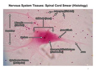
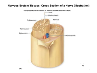
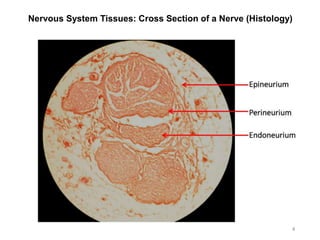
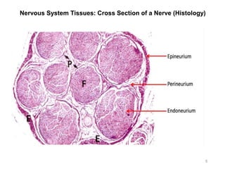
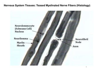
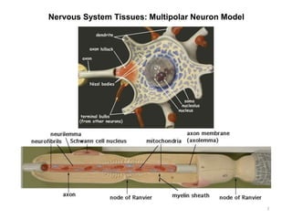
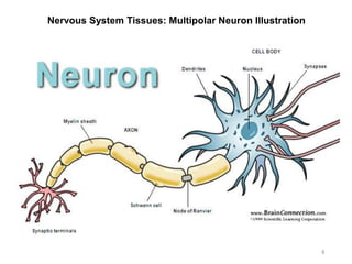
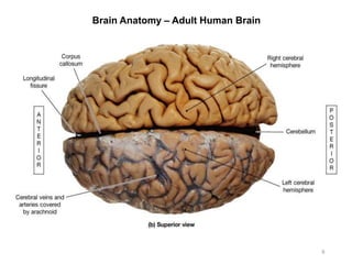
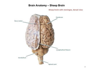
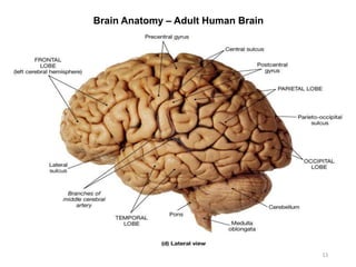
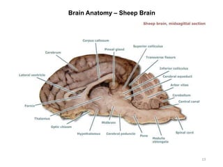
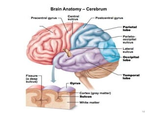
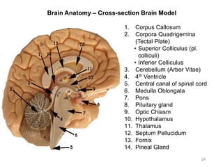
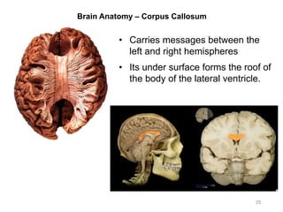
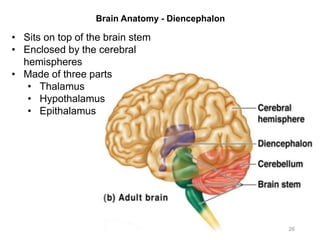
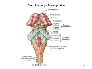
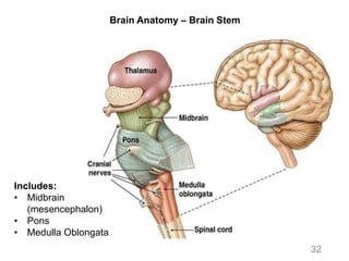
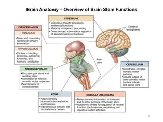
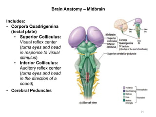
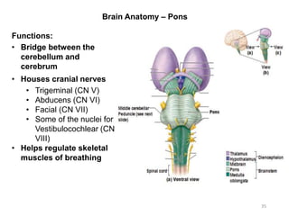
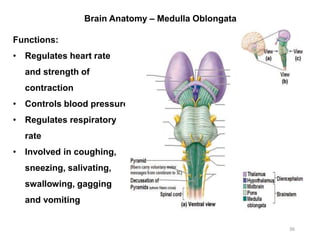
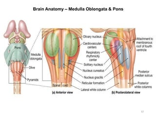
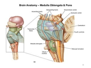
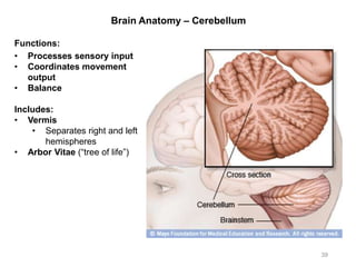
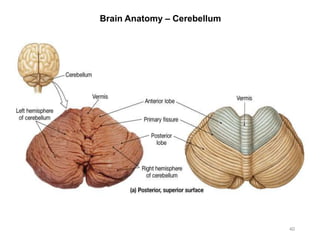
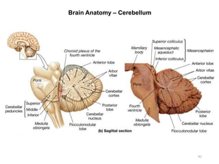
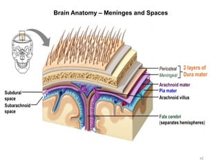
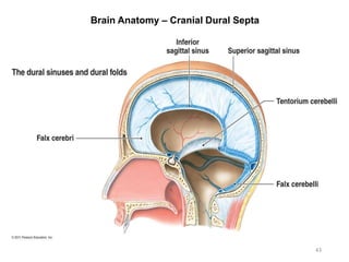
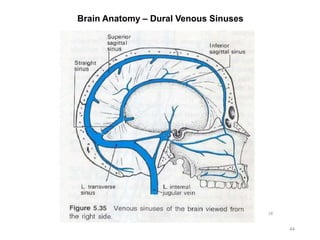
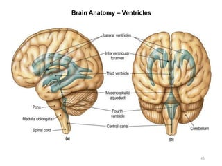
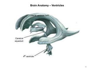
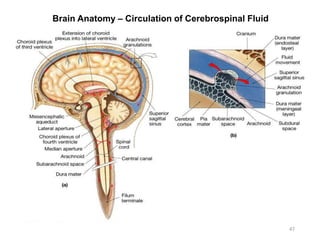
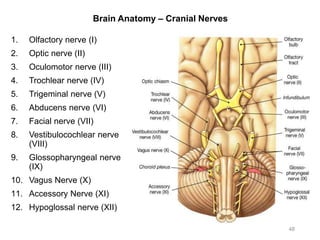
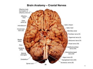
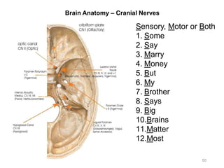
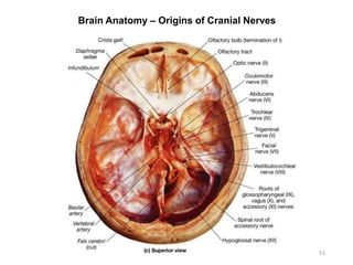
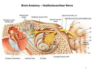
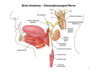
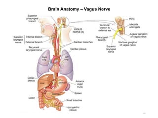
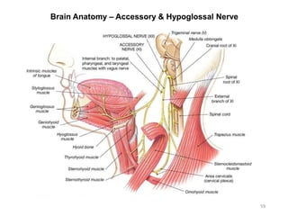
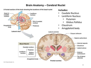
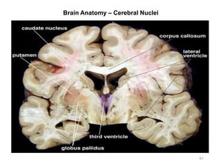
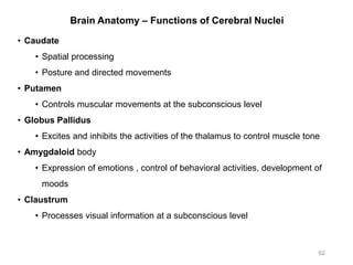
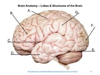
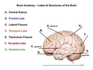
Activity 7:
Nervous System Histology, Brain, &
Cranial Nerves
Chapters 14 & 15 – McKinley et al., Human Anatomy, 4e.
Objectives:
• Histology: Identify structures indicated on three different
slides or images of nervous system tissue. Some of these are
also visible on the classroom model of a neuron.
• Human brain: Identify listed structures of the human brain on
classroom models, the cranial meninges, and structures
involved in cerebrospinal fluid circulation.
• Human brain: Identify the 12 pairs of cranial nerves by name
and number on a model and on the sheep brain.
• Dissect a sheep brain and identify structures listed.
1Compilation: Mohammad Tomaraei & Cristin Fail
Nervous System Tissues: Spinal Cord Smear (Histology)
2
Nervous System Tissues: Cross Section of a Nerve (Illustration)
3
Nervous System Tissues: Cross Section of a Nerve (Histology)
4
Nervous System Tissues: Cross Section of a Nerve (Histology)
5
Nervous System Tissues: Teased Myelinated Nerve Fibers (Histology)
6
Nervous System Tissues: Multipolar Neuron Model
7
Nervous System Tissues: Multipolar Neuron Illustration
8
Brain Anatomy – Adult Human Brain
9
Brain Anatomy – Sheep Brain
10
Brain Anatomy – Adult Human Brain
11
Brain Anatomy – Sheep Brain
12
Brain Anatomy – Sheep Brain
13
Brain Anatomy – Cerebrum
14
Brain Anatomy – Cerebrum
15
• Cerebral hemispheres are divided by the longitudinal fissure into left
and right sides
• Central sulcus divides the frontal lobe from parietal lobes
Brain Anatomy – Lobes of the Brain
16
Brain Anatomy – White Matter of Cerebrum
17
Brain Anatomy – Cerebral Hemispheres
18
Brain Anatomy – Hemispheric Lateralization
19
Brain Anatomy – Lobes of the Brain
20
Brain Anatomy – Lobes of the Brain & their Functions
21
Frontal Lobe Parietal Lobe
Occipital
Lobe
Temporal
Lobe
Brain
Stem
Cerebellum
• Primary Motor
Cortex
(Precentral
Gyrus)
• Voluntary motor
functions
• Concentration
• Verbal
communication
• Decision making
• Planning
• Personality
• Primary
Somatosensory
Cortex
(Postcentral
Gyrus)
• Sensory
functions (Pain,
heat and other
sensations)
• Comprehension
of language
• Primary
Visual
Cortex
• Processing
visual
information
• Storing
visual
memories
• Understanding
Speech
• Interpretation
and storage of
auditory and
olfactory
sensations
• Breathing
• Swallowing
• Heart rate
Includes
Midbrain,
Pons, and
Medulla
Oblongata
• Coordination
• Balance
• Stores
memories of
previously
learned
movement
patterns
Brain Anatomy – Primary Somatosensory Cortex
22
The amount of cortex devoted to any given body region is
proportional to how richly innervated that region is.
Brain Anatomy – Homunculus Model
23
Which model is more realistic?
Brain Anatomy – Cross-section Brain Model
24
1. Corpus Callosum
2. Corpora Quadrigemina
(Tectal Plate)
• Superior Colliculus (pl.
colliculi)
• Inferior Colliculus
3. Cerebellum (Arbor Vitae)
4. 4th Ventricle
5. Central canal of spinal cord
6. Medulla Oblongata
7. Pons
8. Pituitary gland
9. Optic Chiasm
10. Hypothalamus
11. Thalamus
12. Septum Pellucidum
13. Fornix
14. Pineal Gland
Brain Anatomy – Corpus Callosum
25
• Carries messages between the
left and right hemispheres
• Its under surface forms the roof of
the body of the lateral ventricle.
Brain Anatomy - Diencephalon
• Sits on top of the brain stem
• Enclosed by the cerebral
hemispheres
• Made of three parts
• Thalamus
• Hypothalamus
• Epithalamus
26
Brain Anatomy - Diencephalon
27
Brain Anatomy – Epithalamus & Pineal Gland
• Pineal gland is an
endocrine gland that
secretes the hormone
Melatonin.
• Melatonin helps
regulate day-night
cycles known as the
body’s Circadian
Rhythm.
28
Brain Anatomy – Thalamus & the Interthalamic Adhesion
• The Interthalamic
Adhesion is
located midsagitally
and connects the
left and right
thalamic bodies
29
Brain Anatomy – Hypothalamus
30
Includes:
• Mammillary
Body
• Infundibulum
• Pituitary Gland
• Optic Chiasm
• Optic Tracts
• 3rd Ventricle
Brain Anatomy – Functions of Hypothalamus
31
• Master control of the autonomic nervous system
• Heart rate, blood pressure, digestive activities and respiration
• Master control of the endocrine system
• Metabolism, growth, stress responses and reproduction
• Regulation of body temperature
• Body’s “thermostat”, detects altered blood temperatures, heat or cool
the body: shivering/sweating
• Control of emotional behavior
• Emotional responses: pleasure, aggression, fear, rage, contentment
and sex drive
• Control of food intake
• Monitors levels of glucose and amino acids in the blood and
produces sensations of hunger
• Control of water intake
• Monitors blood solute concentration
• Regulation of sleep-wake rhythms
• The Suprachiasmatic Nucleus directs the Pineal gland to secrete
melatonin, regulating circadian rhythms.
Brain Anatomy – Brain Stem
32
Includes:
• Midbrain
(mesencephalon)
• Pons
• Medulla Oblongata
Brain Anatomy – Overview of Brain Stem Functions
33
Brain Anatomy – Midbrain
34
Includes:
• Corpora Quadrigemina
(tectal plate)
• Superior Colliculus:
Visual reflex center
(turns eyes and head
in response to visual
stimulus)
• Inferior Colliculus:
Auditory reflex center
(turns eyes and head
in the direction of a
sound)
• Cerebral Peduncles
Brain Anatomy – Pons
35
Functions:
• Bridge between the
cerebellum and
cerebrum
• Houses cranial nerves
• Trigeminal (CN V)
• Abducens (CN VI)
• Facial (CN VII)
• Some of the nuclei for
Vestibulocochlear (CN
VIII)
• Helps regulate skeletal
muscles of breathing
Brain Anatomy – Medulla Oblongata
36
Functions:
• Regulates heart rate
and strength of
contraction
• Controls blood pressure
• Regulates respiratory
rate
• Involved in coughing,
sneezing, salivating,
swallowing, gagging
and vomiting
Brain Anatomy – Medulla Oblongata & Pons
37
Brain Anatomy – Medulla Oblongata & Pons
38
Brain Anatomy – Cerebellum
39
Functions:
• Processes sensory input
• Coordinates movement
output
• Balance
Includes:
• Vermis
• Separates right and left
hemispheres
• Arbor Vitae (“tree of life”)
Brain Anatomy – Cerebellum
40
Brain Anatomy – Cerebellum
41
Brain Anatomy – Meninges and Spaces
42
Brain Anatomy – Cranial Dural Septa
43
Brain Anatomy – Dural Venous Sinuses
44
Brain Anatomy – Ventricles
45
Brain Anatomy – Ventricles
46
Brain Anatomy – Circulation of Cerebrospinal Fluid
47
Brain Anatomy – Cranial Nerves
48
1. Olfactory nerve (I)
2. Optic nerve (II)
3. Oculomotor nerve (III)
4. Trochlear nerve (IV)
5. Trigeminal nerve (V)
6. Abducens nerve (VI)
7. Facial nerve (VII)
8. Vestibulocochlear nerve
(VIII)
9. Glossopharyngeal nerve
(IX)
10. Vagus Nerve (X)
11. Accessory Nerve (XI)
12. Hypoglossal nerve (XII)
Brain Anatomy – Cranial Nerves
49
Brain Anatomy – Cranial Nerves
50
Sensory, Motor or Both
1. Some
2. Say
3. Marry
4. Money
5. But
6. My
7. Brother
8. Says
9. Big
10.Brains
11.Matter
12.Most
Brain Anatomy – Origins of Cranial Nerves
51
Brain Anatomy – Sheep Brain
52
Brain Anatomy – Sheep Brain
53
Brain Anatomy – Trigeminal Nerve
54
Brain Anatomy – Facial Nerve
55
Brain Anatomy – Vestibulocochlear Nerve
56
Brain Anatomy – Glossopharyngeal Nerve
57
Brain Anatomy – Vagus Nerve
58
Brain Anatomy – Accessory & Hypoglossal Nerve
59
Brain Anatomy – Cerebral Nuclei
60
Includes:
• Caudate Nucleus
• Lentiform Nucleus
• Putamen
• Globus Pallidus
• Claustrum
• Amygdaloid body
Brain Anatomy – Cerebral Nuclei
61
Brain Anatomy – Functions of Cerebral Nuclei
62
• Caudate
• Spatial processing
• Posture and directed movements
• Putamen
• Controls muscular movements at the subconscious level
• Globus Pallidus
• Excites and inhibits the activities of the thalamus to control muscle tone
• Amygdaloid body
• Expression of emotions , control of behavioral activities, development of
moods
• Claustrum
• Processes visual information at a subconscious level
Brain Anatomy – Lobes & Structures of the Brain
63
B.
A.
C.
D.
E.
F.
G.
http://williamcalvin.com/BrainForAllSeasons/img/bonoboLH-humanLH-viaTWD.gif
Brain Anatomy – Lobes & Structures of the Brain
64
B.
A. (groove)
C. (groove)
D.
E.
F.
G.
B. Frontal Lobe
G. Parietal Lobe
F. Occipital Lobe
D. Temporal Lobe
A. Central Sulcus
(groove)
E. Transverse Fissure
C. Lateral Fissure
http://williamcalvin.com/BrainForAllSeasons/img/bonoboLH-humanLH-viaTWD.gif

Activity 7 - Brain & Cranial Nerves

  • 1.
    Activity 7: Nervous SystemHistology, Brain, & Cranial Nerves Chapters 14 & 15 – McKinley et al., Human Anatomy, 4e. Objectives: • Histology: Identify structures indicated on three different slides or images of nervous system tissue. Some of these are also visible on the classroom model of a neuron. • Human brain: Identify listed structures of the human brain on classroom models, the cranial meninges, and structures involved in cerebrospinal fluid circulation. • Human brain: Identify the 12 pairs of cranial nerves by name and number on a model and on the sheep brain. • Dissect a sheep brain and identify structures listed. 1Compilation: Mohammad Tomaraei & Cristin Fail
  • 2.
    Nervous System Tissues:Spinal Cord Smear (Histology) 2
  • 3.
    Nervous System Tissues:Cross Section of a Nerve (Illustration) 3
  • 4.
    Nervous System Tissues:Cross Section of a Nerve (Histology) 4
  • 5.
    Nervous System Tissues:Cross Section of a Nerve (Histology) 5
  • 6.
    Nervous System Tissues:Teased Myelinated Nerve Fibers (Histology) 6
  • 7.
    Nervous System Tissues:Multipolar Neuron Model 7
  • 8.
    Nervous System Tissues:Multipolar Neuron Illustration 8
  • 9.
    Brain Anatomy –Adult Human Brain 9
  • 10.
    Brain Anatomy –Sheep Brain 10
  • 11.
    Brain Anatomy –Adult Human Brain 11
  • 12.
    Brain Anatomy –Sheep Brain 12
  • 13.
    Brain Anatomy –Sheep Brain 13
  • 14.
    Brain Anatomy –Cerebrum 14
  • 15.
    Brain Anatomy –Cerebrum 15 • Cerebral hemispheres are divided by the longitudinal fissure into left and right sides • Central sulcus divides the frontal lobe from parietal lobes
  • 16.
    Brain Anatomy –Lobes of the Brain 16
  • 17.
    Brain Anatomy –White Matter of Cerebrum 17
  • 18.
    Brain Anatomy –Cerebral Hemispheres 18
  • 19.
    Brain Anatomy –Hemispheric Lateralization 19
  • 20.
    Brain Anatomy –Lobes of the Brain 20
  • 21.
    Brain Anatomy –Lobes of the Brain & their Functions 21 Frontal Lobe Parietal Lobe Occipital Lobe Temporal Lobe Brain Stem Cerebellum • Primary Motor Cortex (Precentral Gyrus) • Voluntary motor functions • Concentration • Verbal communication • Decision making • Planning • Personality • Primary Somatosensory Cortex (Postcentral Gyrus) • Sensory functions (Pain, heat and other sensations) • Comprehension of language • Primary Visual Cortex • Processing visual information • Storing visual memories • Understanding Speech • Interpretation and storage of auditory and olfactory sensations • Breathing • Swallowing • Heart rate Includes Midbrain, Pons, and Medulla Oblongata • Coordination • Balance • Stores memories of previously learned movement patterns
  • 22.
    Brain Anatomy –Primary Somatosensory Cortex 22 The amount of cortex devoted to any given body region is proportional to how richly innervated that region is.
  • 23.
    Brain Anatomy –Homunculus Model 23 Which model is more realistic?
  • 24.
    Brain Anatomy –Cross-section Brain Model 24 1. Corpus Callosum 2. Corpora Quadrigemina (Tectal Plate) • Superior Colliculus (pl. colliculi) • Inferior Colliculus 3. Cerebellum (Arbor Vitae) 4. 4th Ventricle 5. Central canal of spinal cord 6. Medulla Oblongata 7. Pons 8. Pituitary gland 9. Optic Chiasm 10. Hypothalamus 11. Thalamus 12. Septum Pellucidum 13. Fornix 14. Pineal Gland
  • 25.
    Brain Anatomy –Corpus Callosum 25 • Carries messages between the left and right hemispheres • Its under surface forms the roof of the body of the lateral ventricle.
  • 26.
    Brain Anatomy -Diencephalon • Sits on top of the brain stem • Enclosed by the cerebral hemispheres • Made of three parts • Thalamus • Hypothalamus • Epithalamus 26
  • 27.
    Brain Anatomy -Diencephalon 27
  • 28.
    Brain Anatomy –Epithalamus & Pineal Gland • Pineal gland is an endocrine gland that secretes the hormone Melatonin. • Melatonin helps regulate day-night cycles known as the body’s Circadian Rhythm. 28
  • 29.
    Brain Anatomy –Thalamus & the Interthalamic Adhesion • The Interthalamic Adhesion is located midsagitally and connects the left and right thalamic bodies 29
  • 30.
    Brain Anatomy –Hypothalamus 30 Includes: • Mammillary Body • Infundibulum • Pituitary Gland • Optic Chiasm • Optic Tracts • 3rd Ventricle
  • 31.
    Brain Anatomy –Functions of Hypothalamus 31 • Master control of the autonomic nervous system • Heart rate, blood pressure, digestive activities and respiration • Master control of the endocrine system • Metabolism, growth, stress responses and reproduction • Regulation of body temperature • Body’s “thermostat”, detects altered blood temperatures, heat or cool the body: shivering/sweating • Control of emotional behavior • Emotional responses: pleasure, aggression, fear, rage, contentment and sex drive • Control of food intake • Monitors levels of glucose and amino acids in the blood and produces sensations of hunger • Control of water intake • Monitors blood solute concentration • Regulation of sleep-wake rhythms • The Suprachiasmatic Nucleus directs the Pineal gland to secrete melatonin, regulating circadian rhythms.
  • 32.
    Brain Anatomy –Brain Stem 32 Includes: • Midbrain (mesencephalon) • Pons • Medulla Oblongata
  • 33.
    Brain Anatomy –Overview of Brain Stem Functions 33
  • 34.
    Brain Anatomy –Midbrain 34 Includes: • Corpora Quadrigemina (tectal plate) • Superior Colliculus: Visual reflex center (turns eyes and head in response to visual stimulus) • Inferior Colliculus: Auditory reflex center (turns eyes and head in the direction of a sound) • Cerebral Peduncles
  • 35.
    Brain Anatomy –Pons 35 Functions: • Bridge between the cerebellum and cerebrum • Houses cranial nerves • Trigeminal (CN V) • Abducens (CN VI) • Facial (CN VII) • Some of the nuclei for Vestibulocochlear (CN VIII) • Helps regulate skeletal muscles of breathing
  • 36.
    Brain Anatomy –Medulla Oblongata 36 Functions: • Regulates heart rate and strength of contraction • Controls blood pressure • Regulates respiratory rate • Involved in coughing, sneezing, salivating, swallowing, gagging and vomiting
  • 37.
    Brain Anatomy –Medulla Oblongata & Pons 37
  • 38.
    Brain Anatomy –Medulla Oblongata & Pons 38
  • 39.
    Brain Anatomy –Cerebellum 39 Functions: • Processes sensory input • Coordinates movement output • Balance Includes: • Vermis • Separates right and left hemispheres • Arbor Vitae (“tree of life”)
  • 40.
    Brain Anatomy –Cerebellum 40
  • 41.
    Brain Anatomy –Cerebellum 41
  • 42.
    Brain Anatomy –Meninges and Spaces 42
  • 43.
    Brain Anatomy –Cranial Dural Septa 43
  • 44.
    Brain Anatomy –Dural Venous Sinuses 44
  • 45.
    Brain Anatomy –Ventricles 45
  • 46.
    Brain Anatomy –Ventricles 46
  • 47.
    Brain Anatomy –Circulation of Cerebrospinal Fluid 47
  • 48.
    Brain Anatomy –Cranial Nerves 48 1. Olfactory nerve (I) 2. Optic nerve (II) 3. Oculomotor nerve (III) 4. Trochlear nerve (IV) 5. Trigeminal nerve (V) 6. Abducens nerve (VI) 7. Facial nerve (VII) 8. Vestibulocochlear nerve (VIII) 9. Glossopharyngeal nerve (IX) 10. Vagus Nerve (X) 11. Accessory Nerve (XI) 12. Hypoglossal nerve (XII)
  • 49.
    Brain Anatomy –Cranial Nerves 49
  • 50.
    Brain Anatomy –Cranial Nerves 50 Sensory, Motor or Both 1. Some 2. Say 3. Marry 4. Money 5. But 6. My 7. Brother 8. Says 9. Big 10.Brains 11.Matter 12.Most
  • 51.
    Brain Anatomy –Origins of Cranial Nerves 51
  • 52.
    Brain Anatomy –Sheep Brain 52
  • 53.
    Brain Anatomy –Sheep Brain 53
  • 54.
    Brain Anatomy –Trigeminal Nerve 54
  • 55.
    Brain Anatomy –Facial Nerve 55
  • 56.
    Brain Anatomy –Vestibulocochlear Nerve 56
  • 57.
    Brain Anatomy –Glossopharyngeal Nerve 57
  • 58.
    Brain Anatomy –Vagus Nerve 58
  • 59.
    Brain Anatomy –Accessory & Hypoglossal Nerve 59
  • 60.
    Brain Anatomy –Cerebral Nuclei 60 Includes: • Caudate Nucleus • Lentiform Nucleus • Putamen • Globus Pallidus • Claustrum • Amygdaloid body
  • 61.
    Brain Anatomy –Cerebral Nuclei 61
  • 62.
    Brain Anatomy –Functions of Cerebral Nuclei 62 • Caudate • Spatial processing • Posture and directed movements • Putamen • Controls muscular movements at the subconscious level • Globus Pallidus • Excites and inhibits the activities of the thalamus to control muscle tone • Amygdaloid body • Expression of emotions , control of behavioral activities, development of moods • Claustrum • Processes visual information at a subconscious level
  • 63.
    Brain Anatomy –Lobes & Structures of the Brain 63 B. A. C. D. E. F. G. http://williamcalvin.com/BrainForAllSeasons/img/bonoboLH-humanLH-viaTWD.gif
  • 64.
    Brain Anatomy –Lobes & Structures of the Brain 64 B. A. (groove) C. (groove) D. E. F. G. B. Frontal Lobe G. Parietal Lobe F. Occipital Lobe D. Temporal Lobe A. Central Sulcus (groove) E. Transverse Fissure C. Lateral Fissure http://williamcalvin.com/BrainForAllSeasons/img/bonoboLH-humanLH-viaTWD.gif