The document provides an overview of the anatomy and functions of the main parts of the human brain. It describes the major lobes and structures of the forebrain including the cerebral hemispheres, diencephalon, basal ganglia and corpus callosum. It also outlines the midbrain, hindbrain, cerebellum and brainstem. Key functions are assigned to different areas, such as sensory processing in the parietal lobe, motor control in the frontal lobe, and homeostasis in the hypothalamus. The thalamus acts as a data sorting center and the cerebellum coordinates movement.
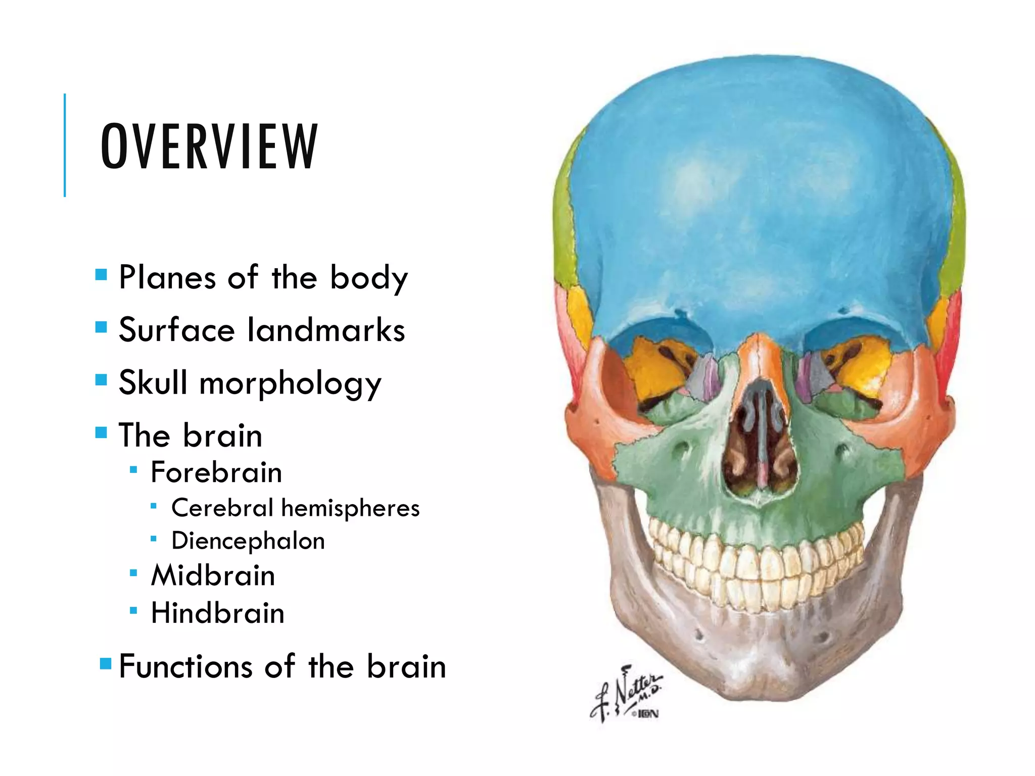
OVERVIEW
Planes ofthe body
Surface landmarks
Skull morphology
The brain
Forebrain
Cerebral hemispheres
Diencephalon
Midbrain
Hindbrain
Functions of the brain
3.
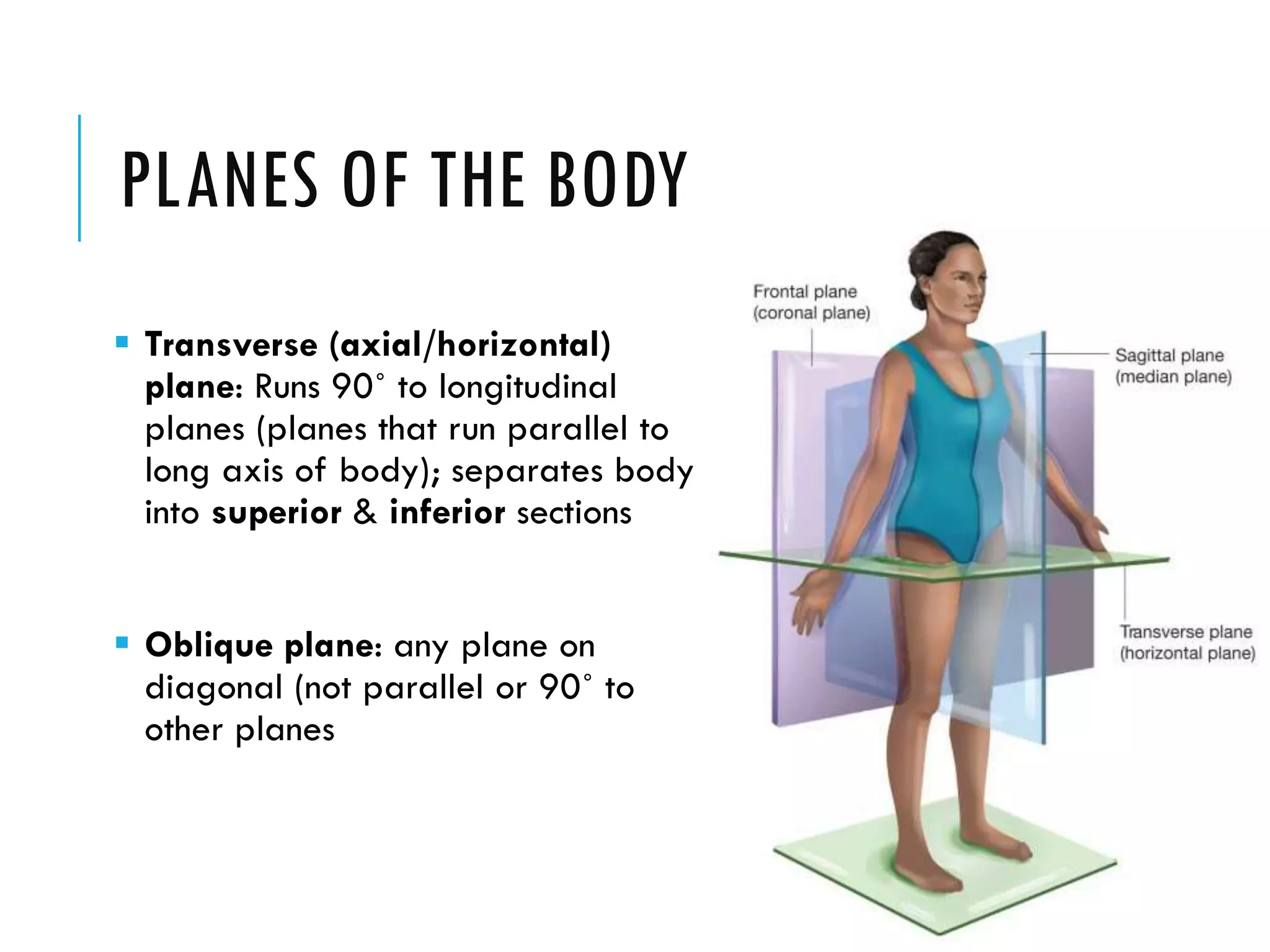
PLANES OF THEBODY
Transverse (axial/horizontal)
plane: Runs 90˚ to longitudinal
planes (planes that run parallel to
long axis of body); separates body
into superior & inferior sections
Oblique plane: any plane on
diagonal (not parallel or 90˚ to
other planes
4.
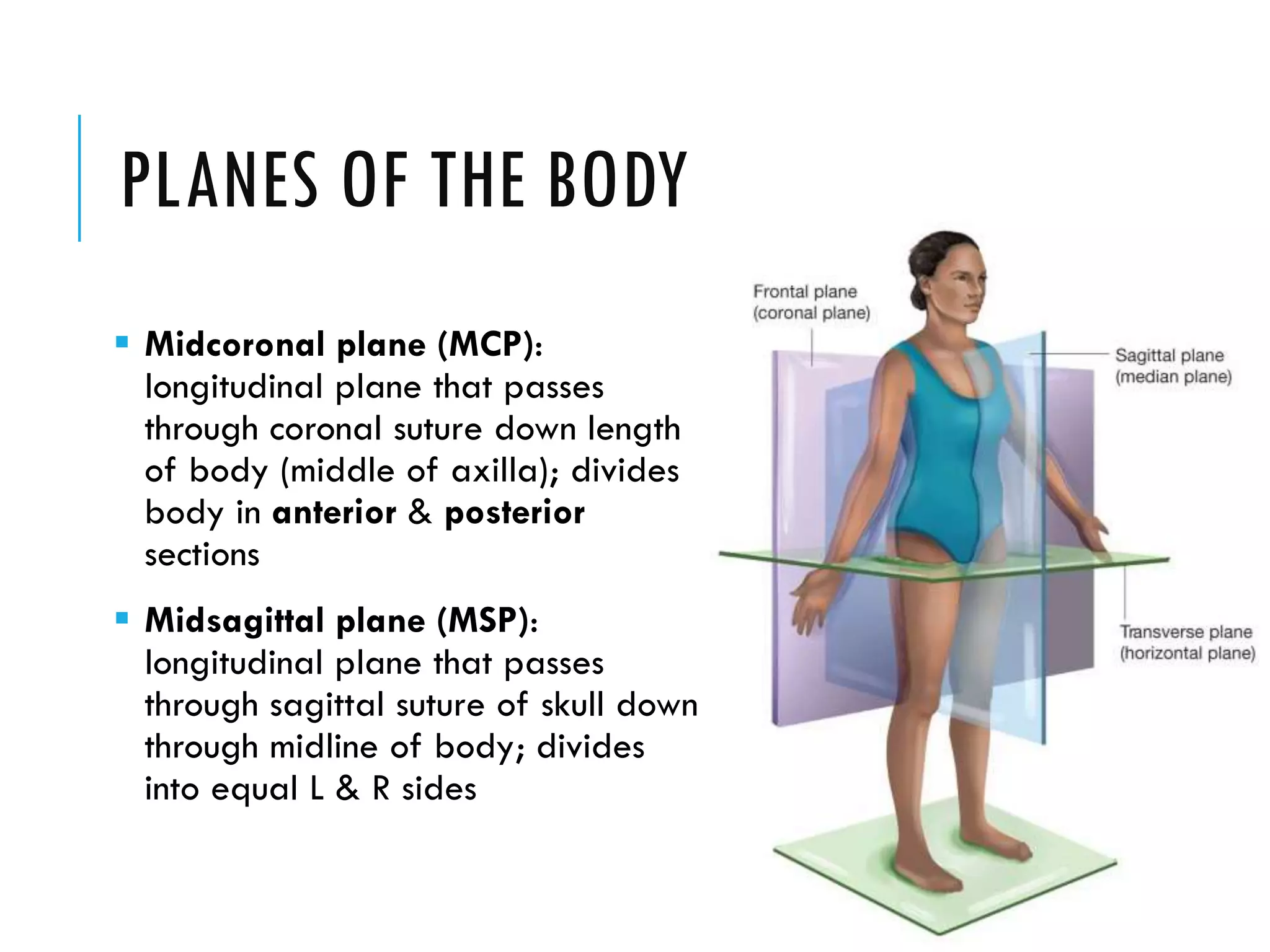
PLANES OF THEBODY
Midcoronal plane (MCP):
longitudinal plane that passes
through coronal suture down length
of body (middle of axilla); divides
body in anterior & posterior
sections
Midsagittal plane (MSP):
longitudinal plane that passes
through sagittal suture of skull down
through midline of body; divides
into equal L & R sides
5.
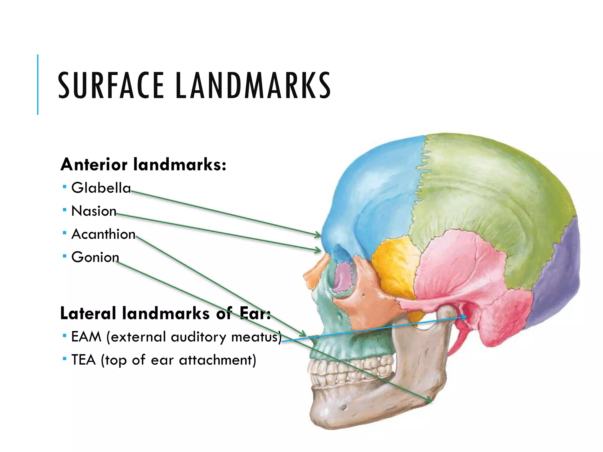
SURFACE LANDMARKS
Anterior landmarks:
Glabella
Nasion
Acanthion
Gonion
Lateral landmarks of Ear:
EAM (external auditory meatus)
TEA (top of ear attachment)
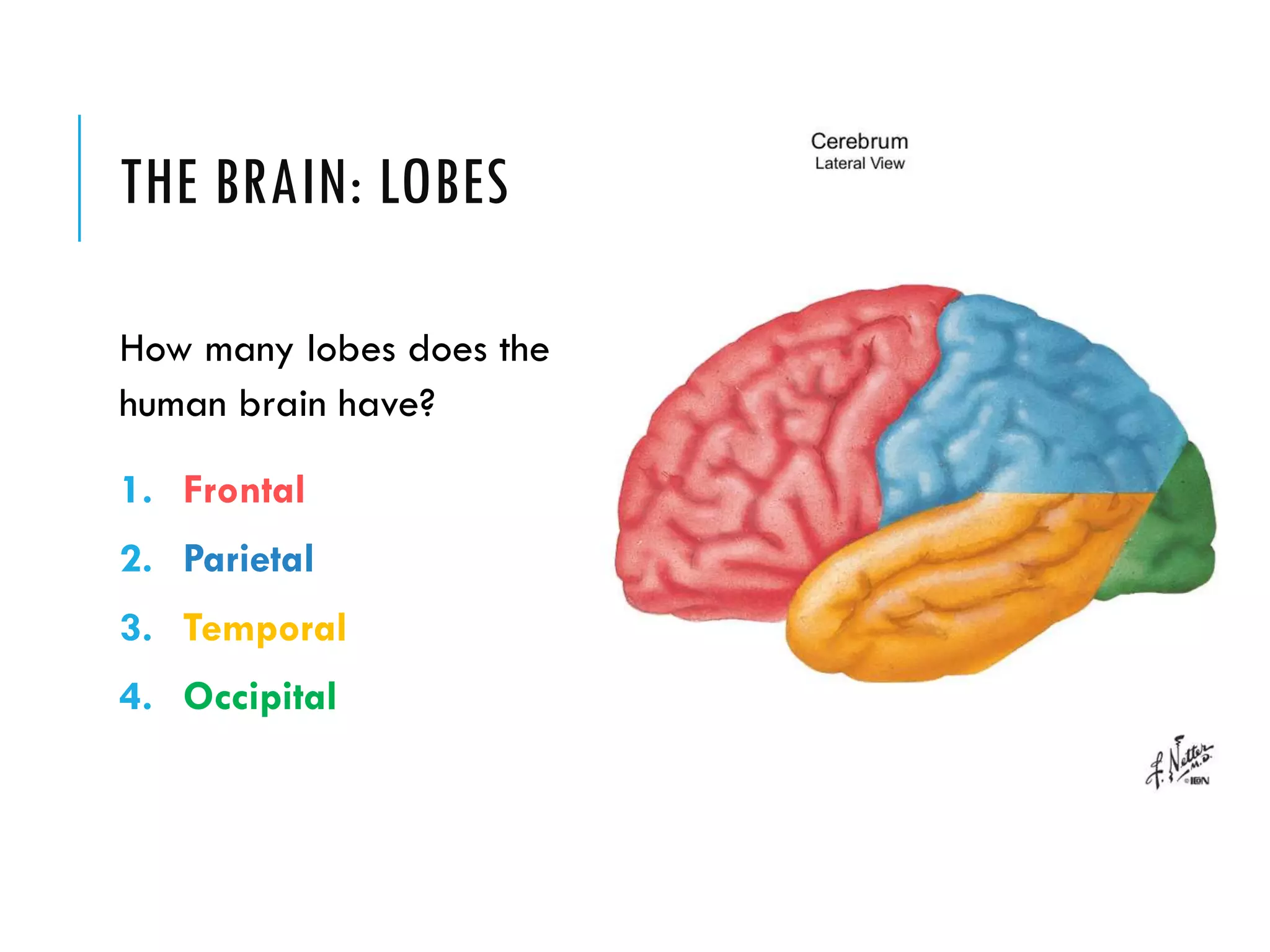
THE BRAIN: LOBES
Howmany lobes does the
human brain have?
1. Frontal
2. Parietal
3. Temporal
4. Occipital
9.
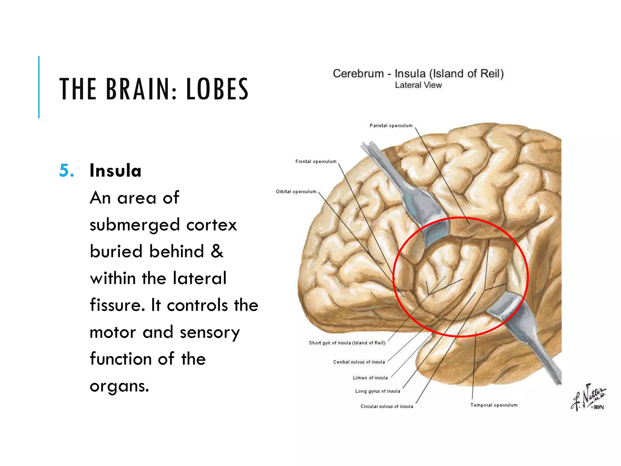
THE BRAIN: LOBES
5.Insula
An area of
submerged cortex
buried behind &
within the lateral
fissure. It controls the
motor and sensory
function of the
organs.
10.
FOREBRAIN: CEREBRUM (TELENCEPHALON)
Largest part of the brain
The outer surface, the cerebral cortex, is composed of gray
matter, which consists of neuron cell bodies and unmyelinated
fibers
Beneath the cerebral cortex is white matter, which consists of
nerve fibers covered in myelin
Consists of two cerebral hemispheres
The two hemispheres are connected by a mass of white matter
called the corpus callosum
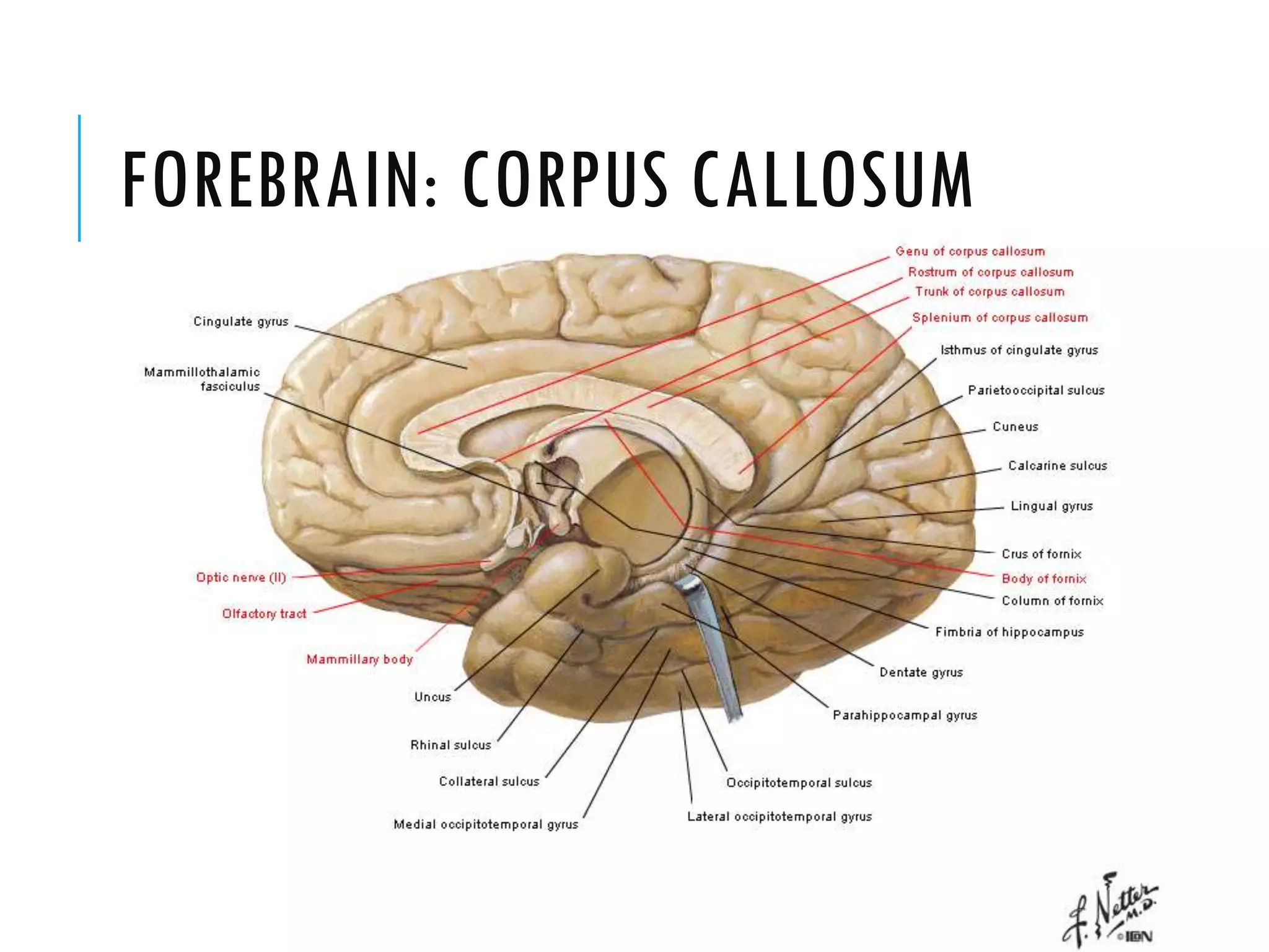
FOREBRAIN: CORPUS CALLOSUM
Splenium: large rounded posterior portion that
overhangs the posterior aspect of the thalamus
Body: large arched central portion superior to the
septum pellucidum
Genu: rounded anterior end which forms the anterior
wall of the lateral ventricle on each side of the midline
Rostrum: this portion which attaches to the anterior
commissure in anterior wall of 3rd ventricle
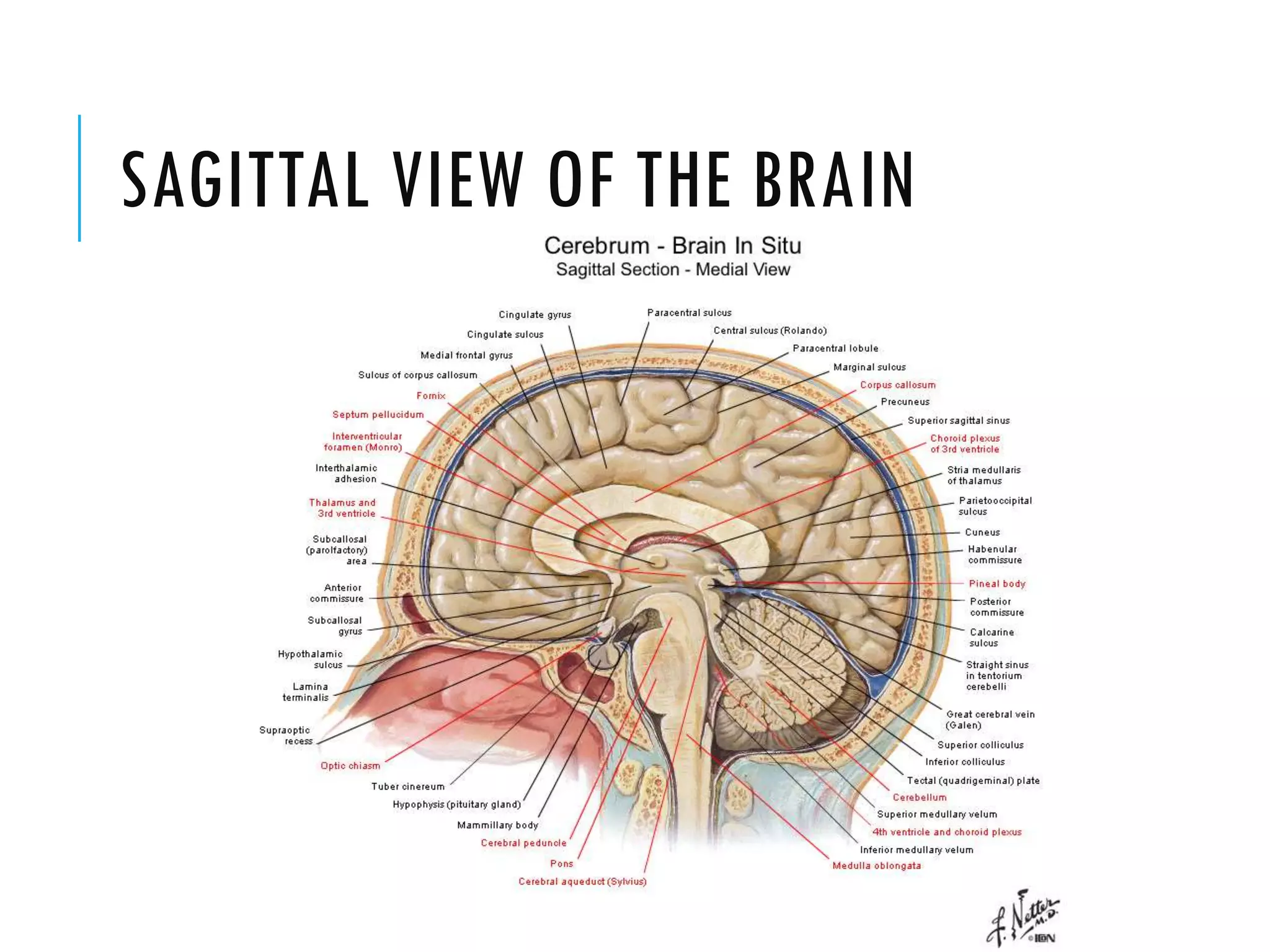
FOREBRAIN: DIENCEPHALON
Centrallylocated, surrounded by cerebral hemispheres, &
consists of:
Epithalamus: forms roof of 3rd ventricle
Thalamus: egg-shaped, largest portion, mass of gray matter
that forms superolateral walls of 3rd ventricle
Hypothalamus: forms floor of 3rd ventricle; on its inferior
aspect can be found:
Infundibulum (or pituitary stalk that attaches to pituitary gland)
Optic chiasm (where optic nerves cross over and then emerge as optic
tract)
Mammillary bodies: two pea-shaped masses of gray matter surrounded
by a layer of white matter
15.
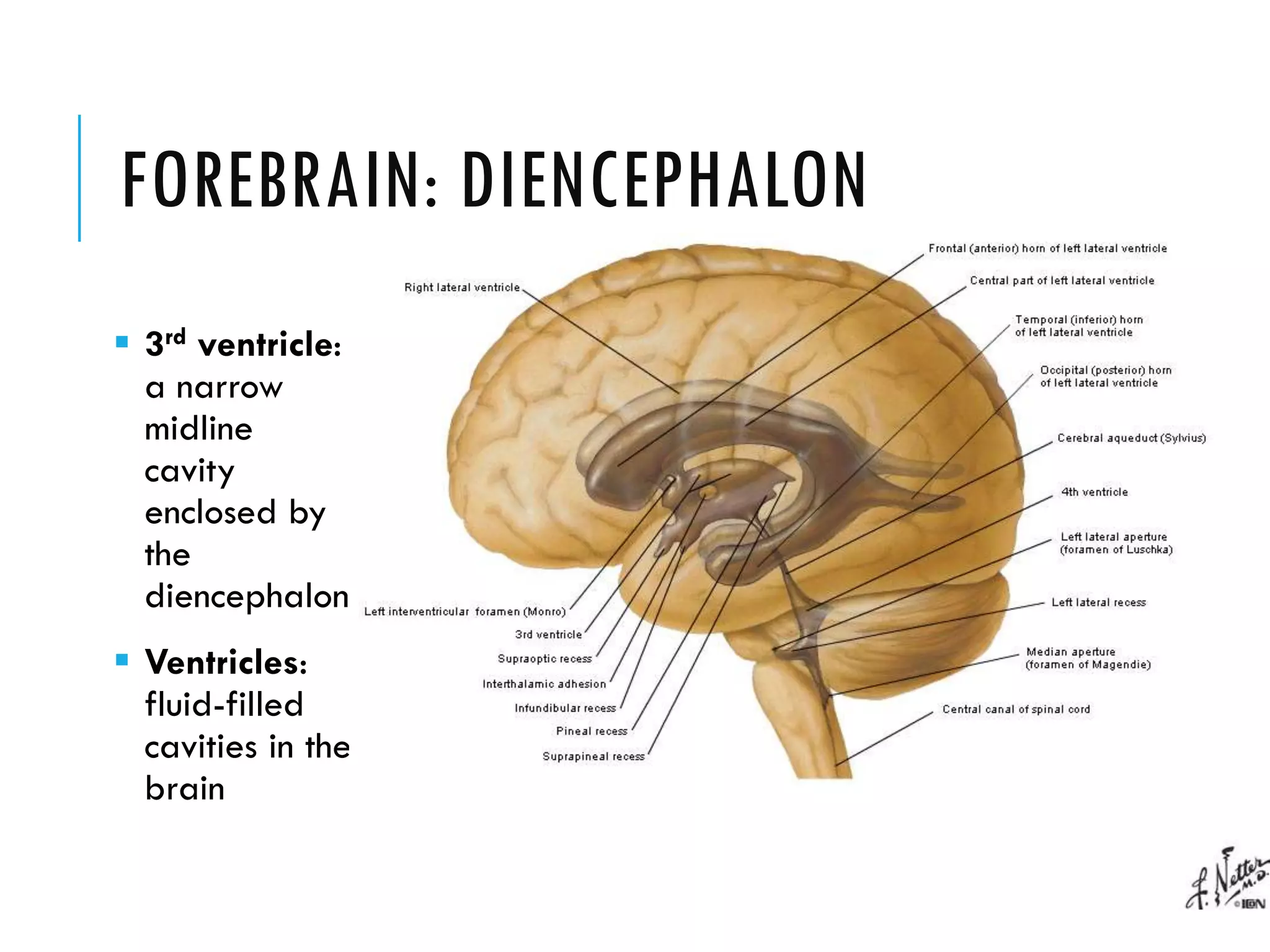
FOREBRAIN: DIENCEPHALON
3rdventricle:
a narrow
midline
cavity
enclosed by
the
diencephalon
Ventricles:
fluid-filled
cavities in the
brain
16.
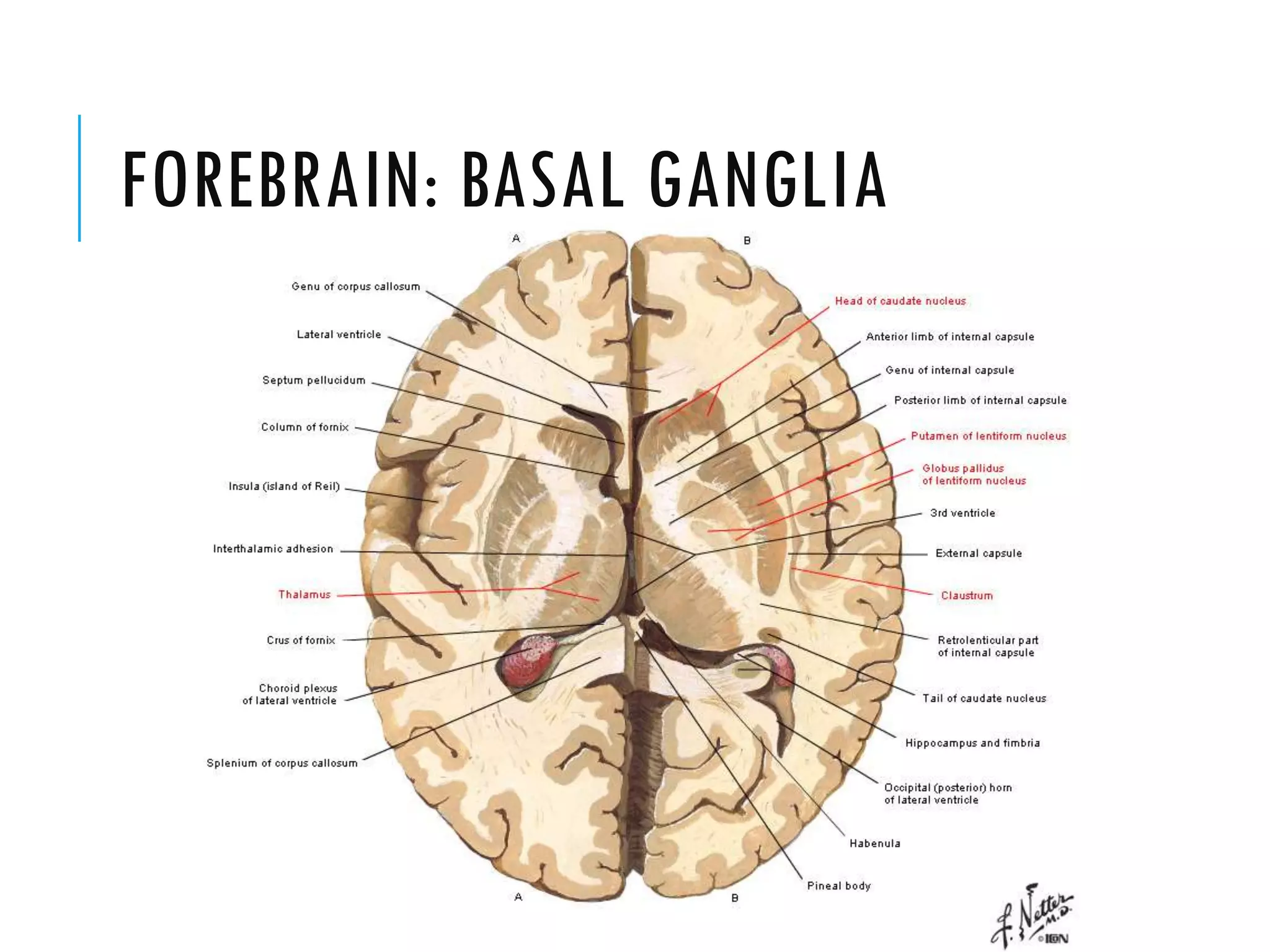
FOREBRAIN: BASAL GANGLIA
Located bilaterally between thalamus and insular
cortex
Consists of 4 main structures:
Caudate nucleus: adjacent to lateral ventricle
Claustrum: thin layer of gray matter located just lateral to
lentiform nucleus & medially to insula
Amygdaloid body
Lentiform nucleus: found centrally in each cerebral
hemisphere
Putamen: located lateral to globus pallidus
Globus pallidus
MIDBRAIN (MESENCEPHALON)
Cerebralaqueduct: passageway through the midbrain that
connects the 3rd ventricle with the 4th ventricle; also known as
aqueduct of Sylvius; divides the midbrain into an anterior
portion called the cerebral peduncles and a posterior portion
known as the tectum
Cerebral peduncles: on the ventral aspect of the midbrain;
composed of motor fibers that extend from the cerebral
cortex to the spinal cord; a narrow layer of deeply pigmented
gray matter called the substantia nigra crosses each cerebral
peduncle
Corpora quadrigemina: 4 rounded protuberances on
posterior aspect of midbrain; also referred to as superior and
inferior colliculi
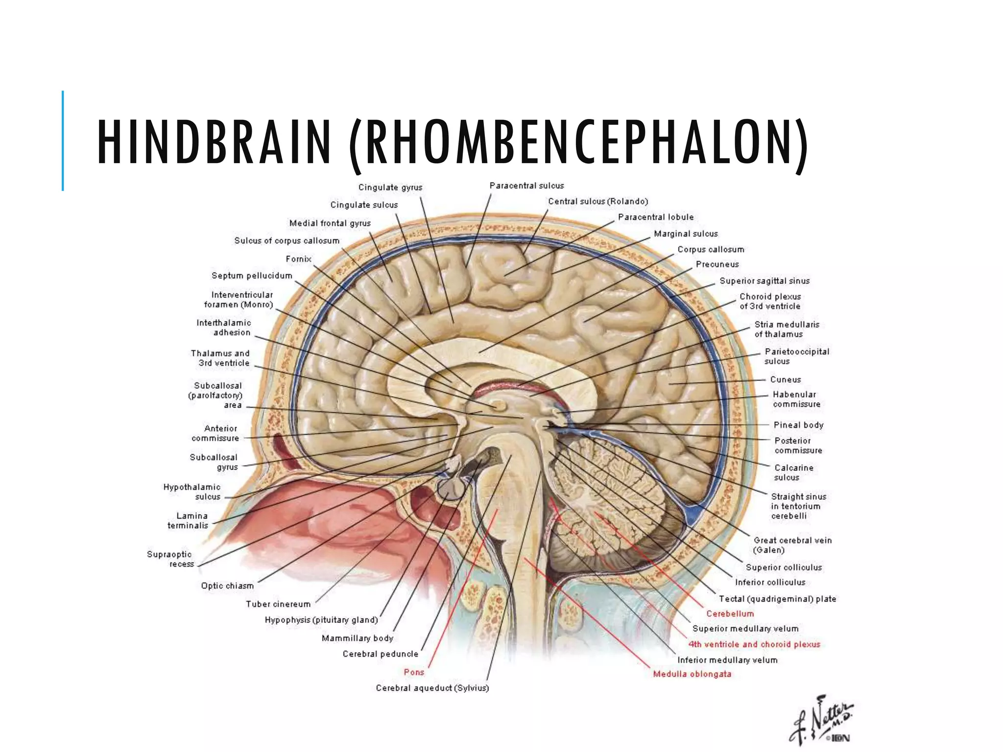
HINDBRAIN (RHOMBENCEPHALON)
Cerebellum:
Largest portion of hindbrain
Separated superiorly from occipital lobes by tentorium
cerebelli
Cerebellar cortex consists of gray matter arranged in narrow
folds called folia
Superior cerebellar peduncles connect cerebellum to midbrain
Middle cerebellar peduncles connect cerebellum to pons
Inferior cerebella peduncles connect cerebellum to medulla
oblongata
23.
HINDBRAIN (RHOMBENCEPHALON)
Pons:located between midbrain and medulla
oblongata; where fibers in cerebellum join those
from cerebrum and spinal cord
Medulla oblongata:
Forms the lower brainstem
Resembles cone that extends from pons to foramen
magnum, where it is continuous with the spinal cord
4th ventricle: closed by the cerebellum
posteriorly
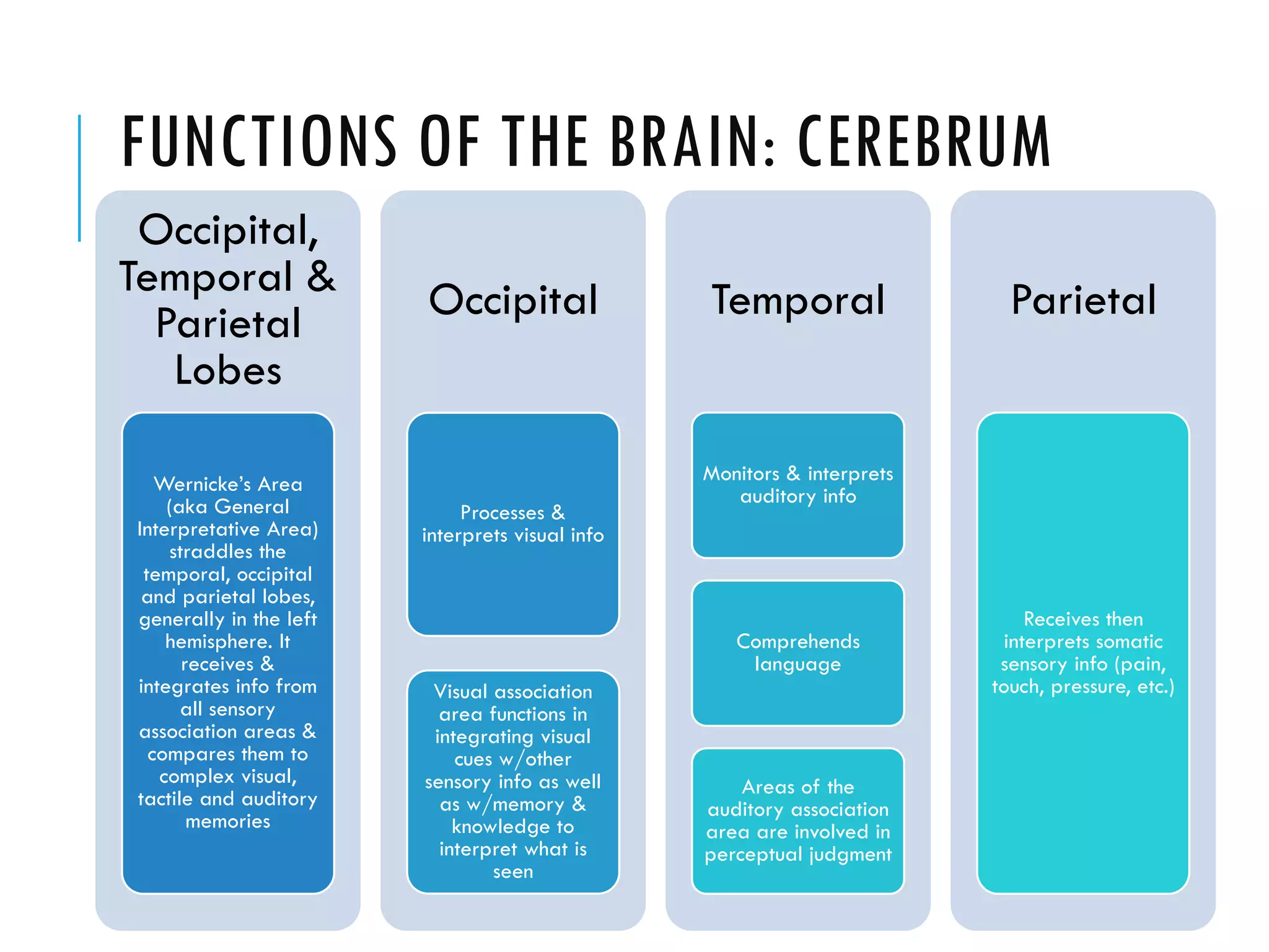
FUNCTIONS OF THEBRAIN: CEREBRUM
Occipital,
Temporal &
Parietal
Lobes
Wernicke’s Area
(aka General
Interpretative Area)
straddles the
temporal, occipital
and parietal lobes,
generally in the left
hemisphere. It
receives &
integrates info from
all sensory
association areas &
compares them to
complex visual,
tactile and auditory
memories
Occipital
Processes &
interprets visual info
Visual association
area functions in
integrating visual
cues w/other
sensory info as well
as w/memory &
knowledge to
interpret what is
seen
Temporal
Monitors & interprets
auditory info
Comprehends
language
Areas of the
auditory association
area are involved in
perceptual judgment
Parietal
Receives then
interprets somatic
sensory info (pain,
touch, pressure, etc.)
26.
FUNCTIONS OF THEBRAIN: CEREBRUM
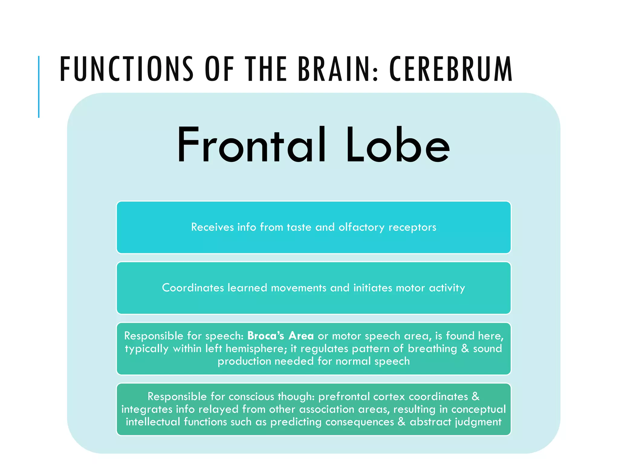
Frontal Lobe
Receives info from taste and olfactory receptors
Coordinates learned movements and initiates motor activity
Responsible for speech: Broca’s Area or motor speech area, is found here,
typically within left hemisphere; it regulates pattern of breathing & sound
production needed for normal speech
Responsible for conscious though: prefrontal cortex coordinates &
integrates info relayed from other association areas, resulting in conceptual
intellectual functions such as predicting consequences & abstract judgment
27.
FUNCTIONS OF THEBRAIN: BRAINSTEM
Medulla
oblongata
Relays sensory info to
other parts of brain
Relays motor info to other
parts of brain & spinal
cord
Regulates autonomic
functions (HR, BP, sneezing)
With other areas of brain,
functions in consciousness
& arousal
Pons
Connects one side of
cerebellum to other &
cerebellum to brainstem
Functions in somatic &
visceral motor control
Along with medulla,
controls breathing
Midbrain
Most complex &
integrative area of
brainstem
Relays motor impulses
from cerebral cortex to
pons
Relays sensory impulses
from spinal cord to
thalamus
Regulates auditory
reflexes
Regulates visual reflexes
28.
FUNCTIONS OF THEBRAIN
Thalamus
Provides data sorting
Relays sensory info to cerebral cortex after filtering
Responsible for crude perception of touch, pressure,
pain & temperature
Integrates some sensory info, influencing emotional
states
Functions in cognition and awareness
29.
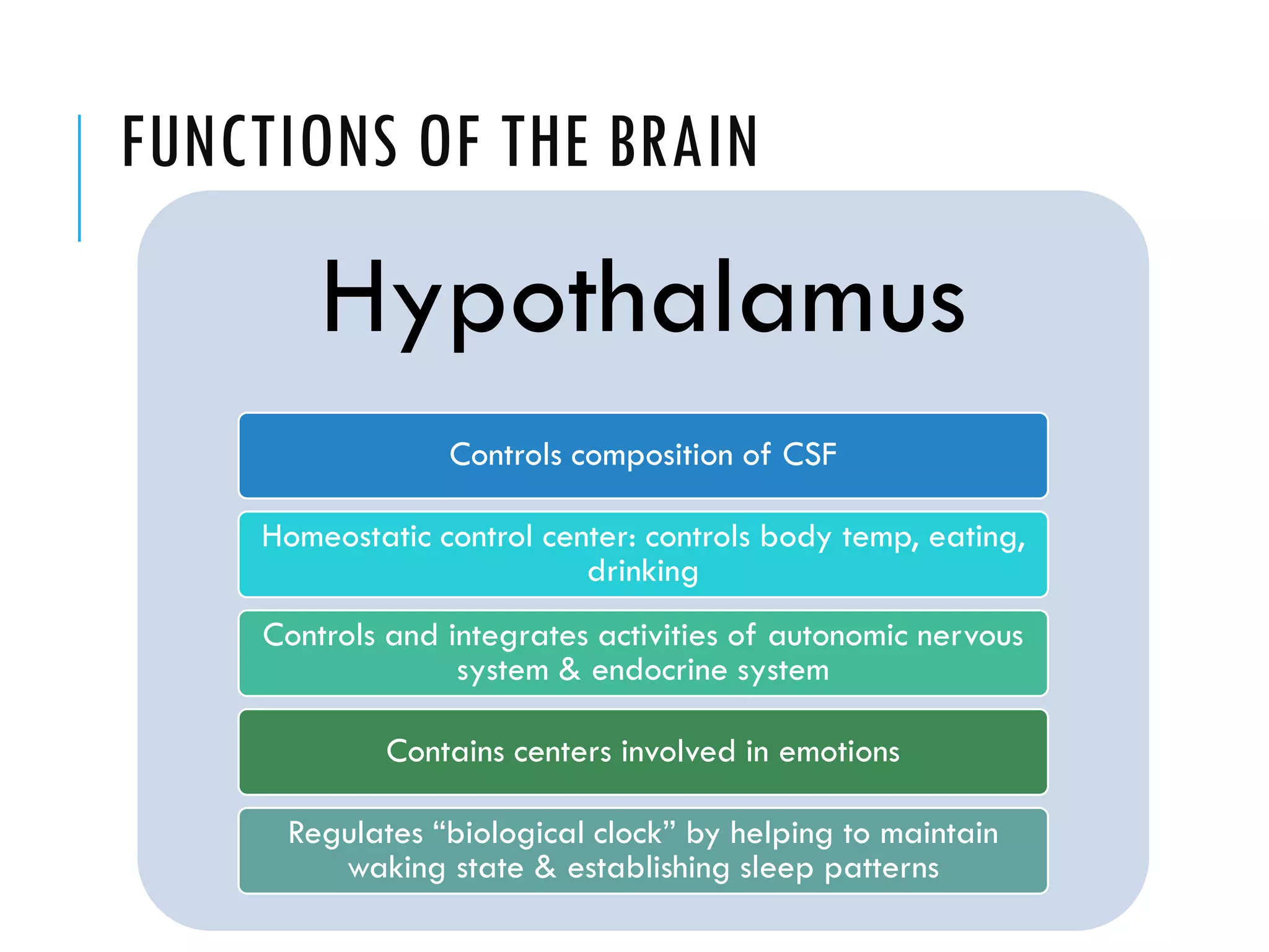
FUNCTIONS OF THEBRAIN
Hypothalamus
Controls composition of CSF
Homeostatic control center: controls body temp, eating,
drinking
Controls and integrates activities of autonomic nervous
system & endocrine system
Contains centers involved in emotions
Regulates “biological clock” by helping to maintain
waking state & establishing sleep patterns
30.
FUNCTIONS OF THEBRAIN
Cerebellum
Second-largest structure of brain
Coordinates & modulates in conscious & unconscious manner
motor commands coming from cerebral cortex & brainstem
Learns and remembers motor responses
Maintains balance & equilibrium of body