The document provides information on the functional anatomy and physiology of the nervous system. It discusses:
1. The three main functions of the nervous system: sensory input, integration, and motor output.
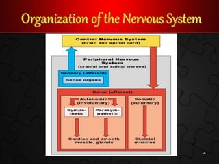
2. The structural classification of the central nervous system (CNS) which includes the brain and spinal cord, and the peripheral nervous system (PNS) which includes nerves outside the CNS.
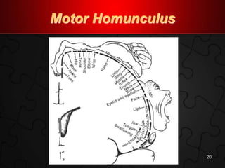
3. The organization and components of the nervous system including neurons, neuroglia, the CNS, brain regions and lobes, motor and sensory homunculi, and protection of the CNS by structures like the meninges and cerebrospinal fluid.


![14 [chapter 14 the brain and cranial nerves]](https://cdn.slidesharecdn.com/ss_thumbnails/14chapter14thebrainandcranialnerves-170828133437-thumbnail.jpg?width=640&height=640&fit=bounds)


