Downloaded 530 times


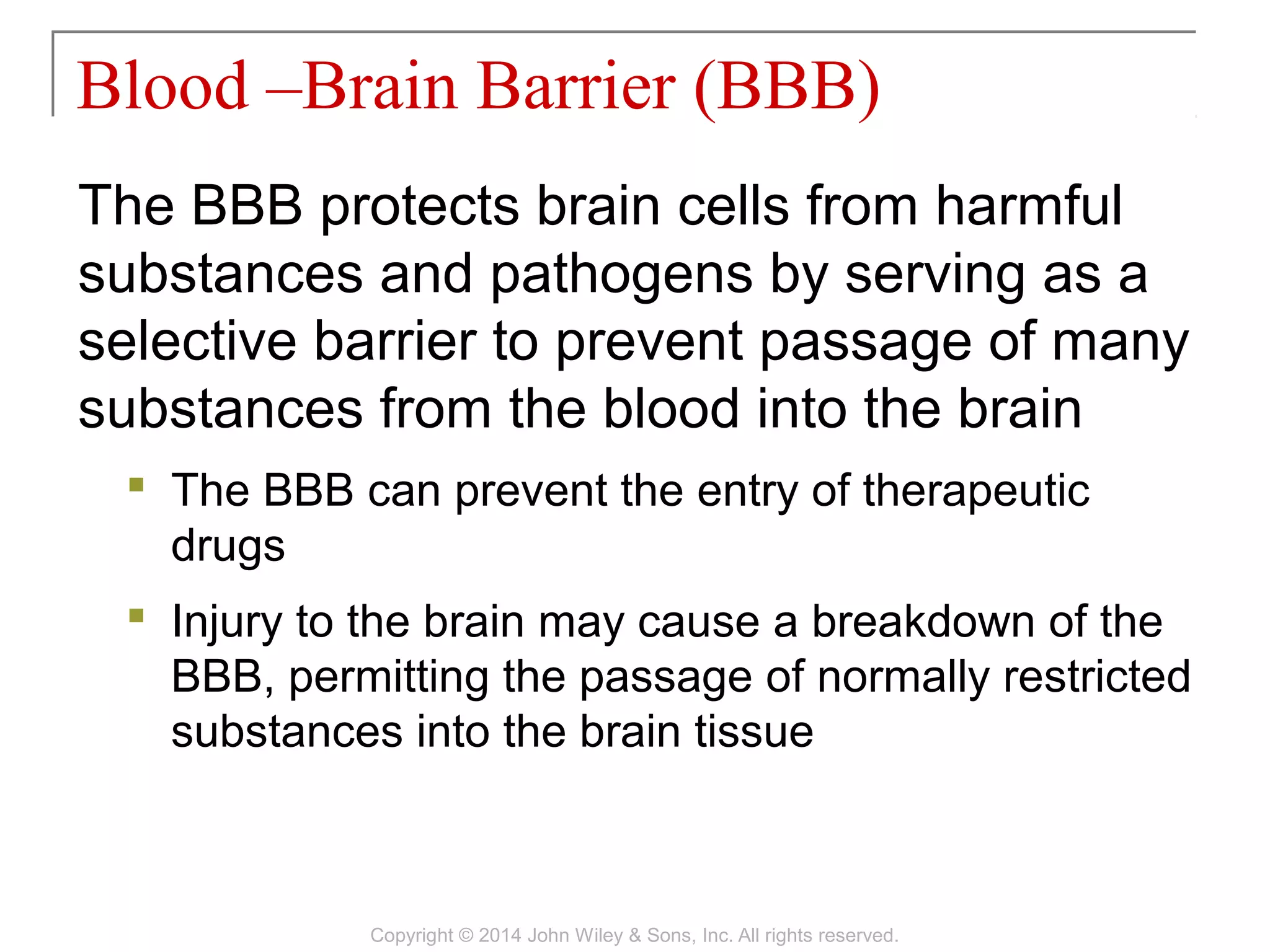
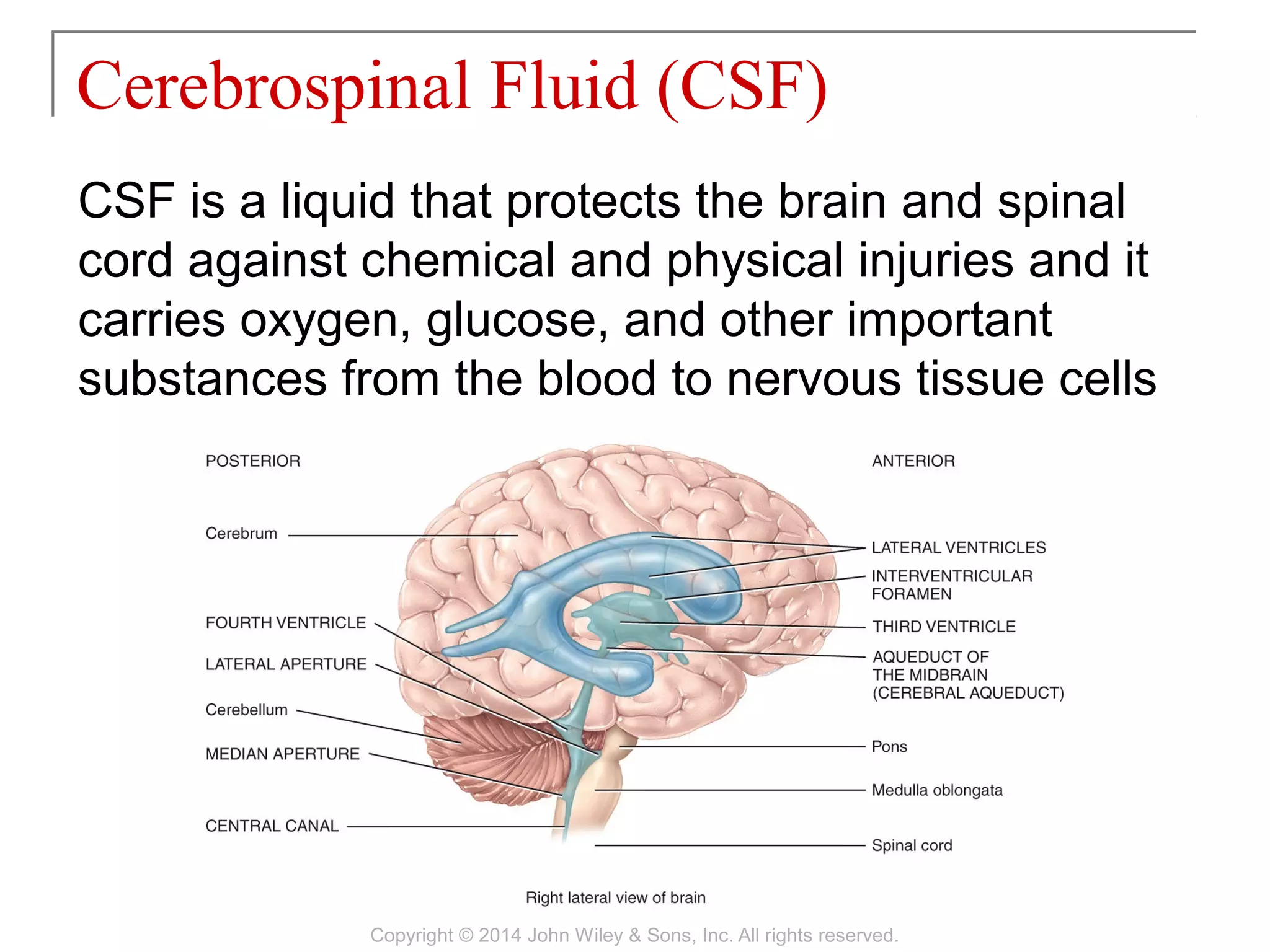
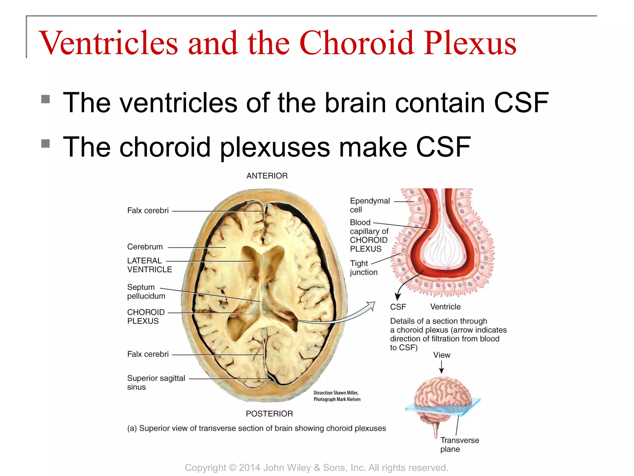


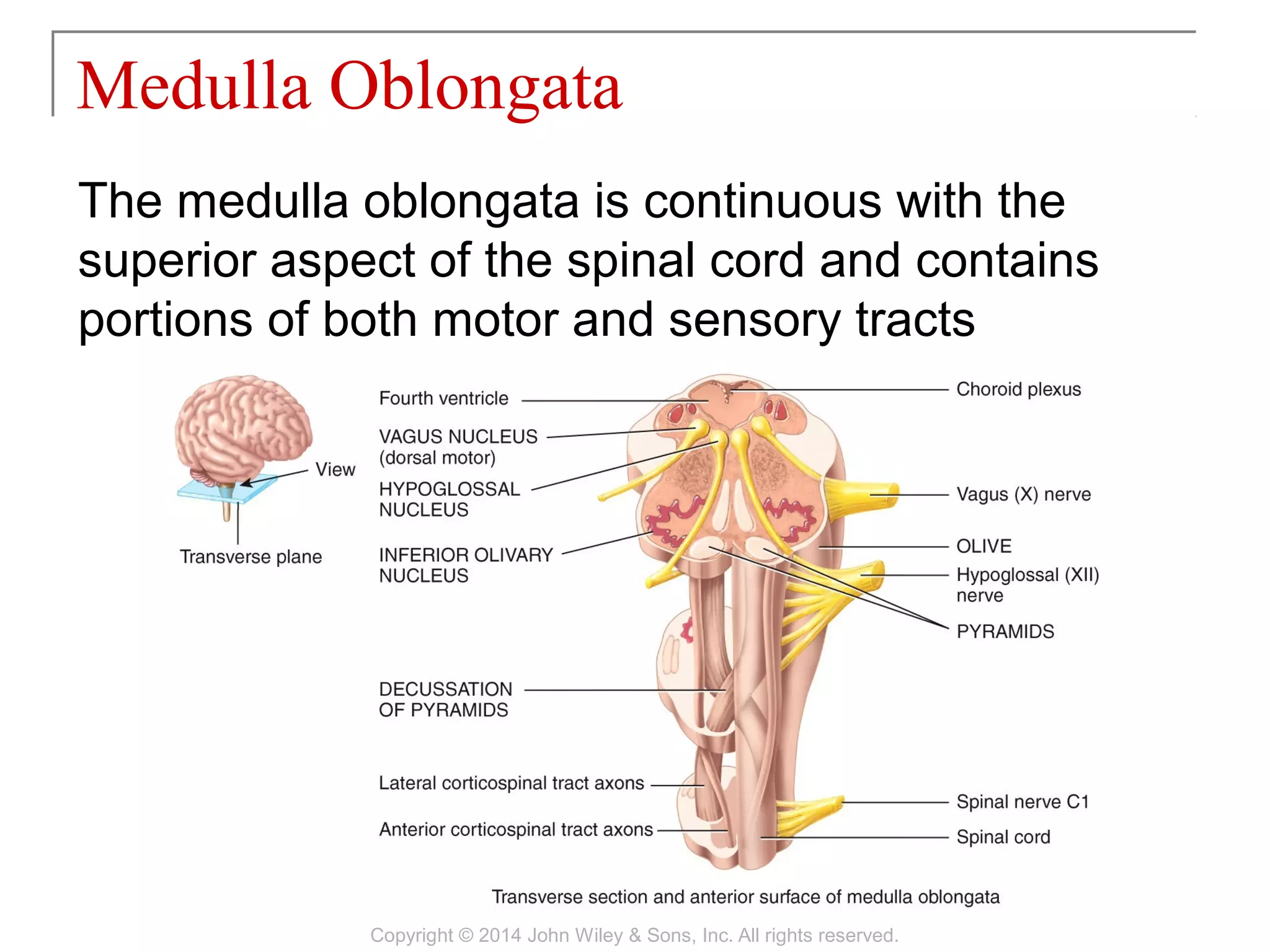


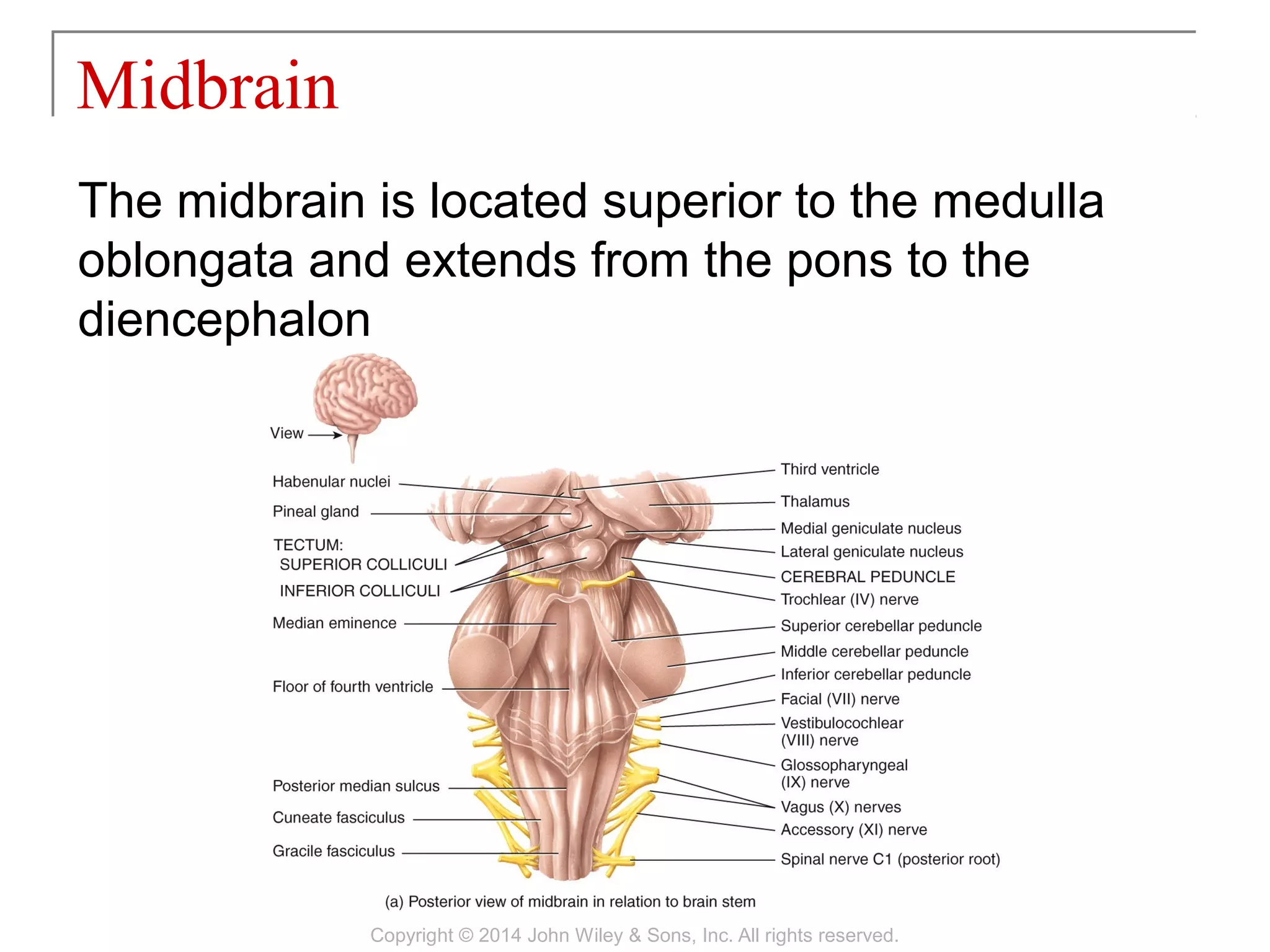

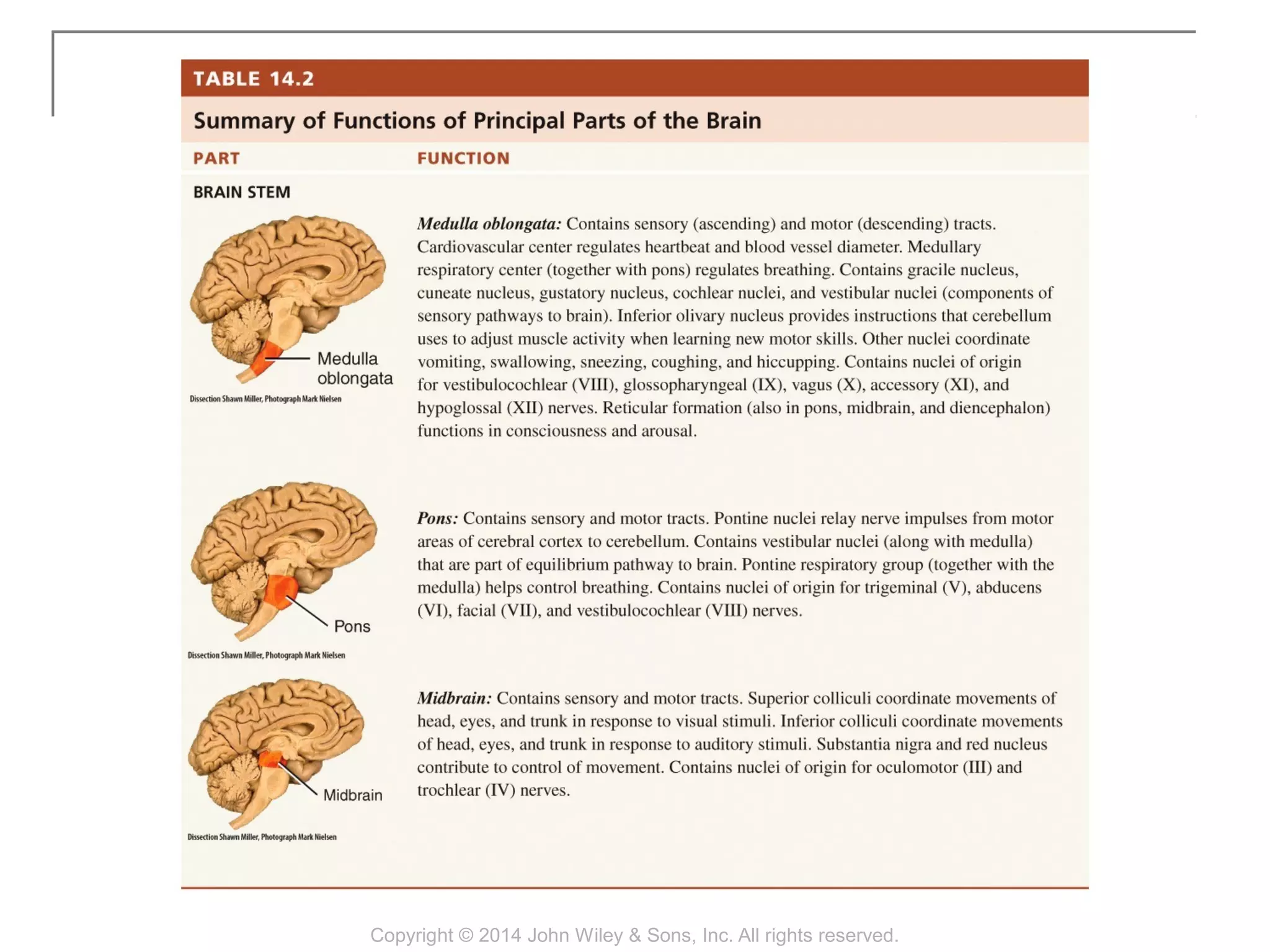

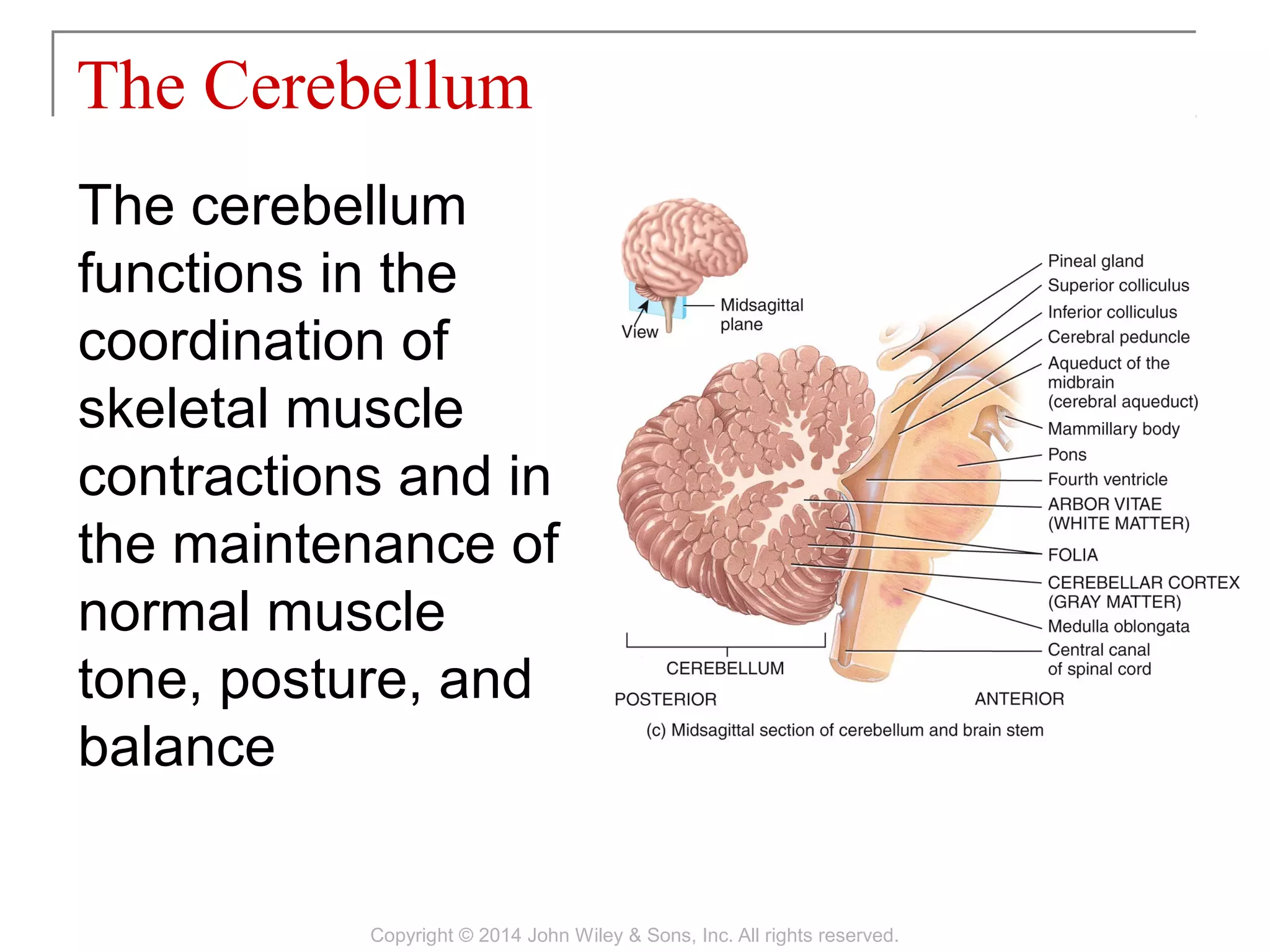
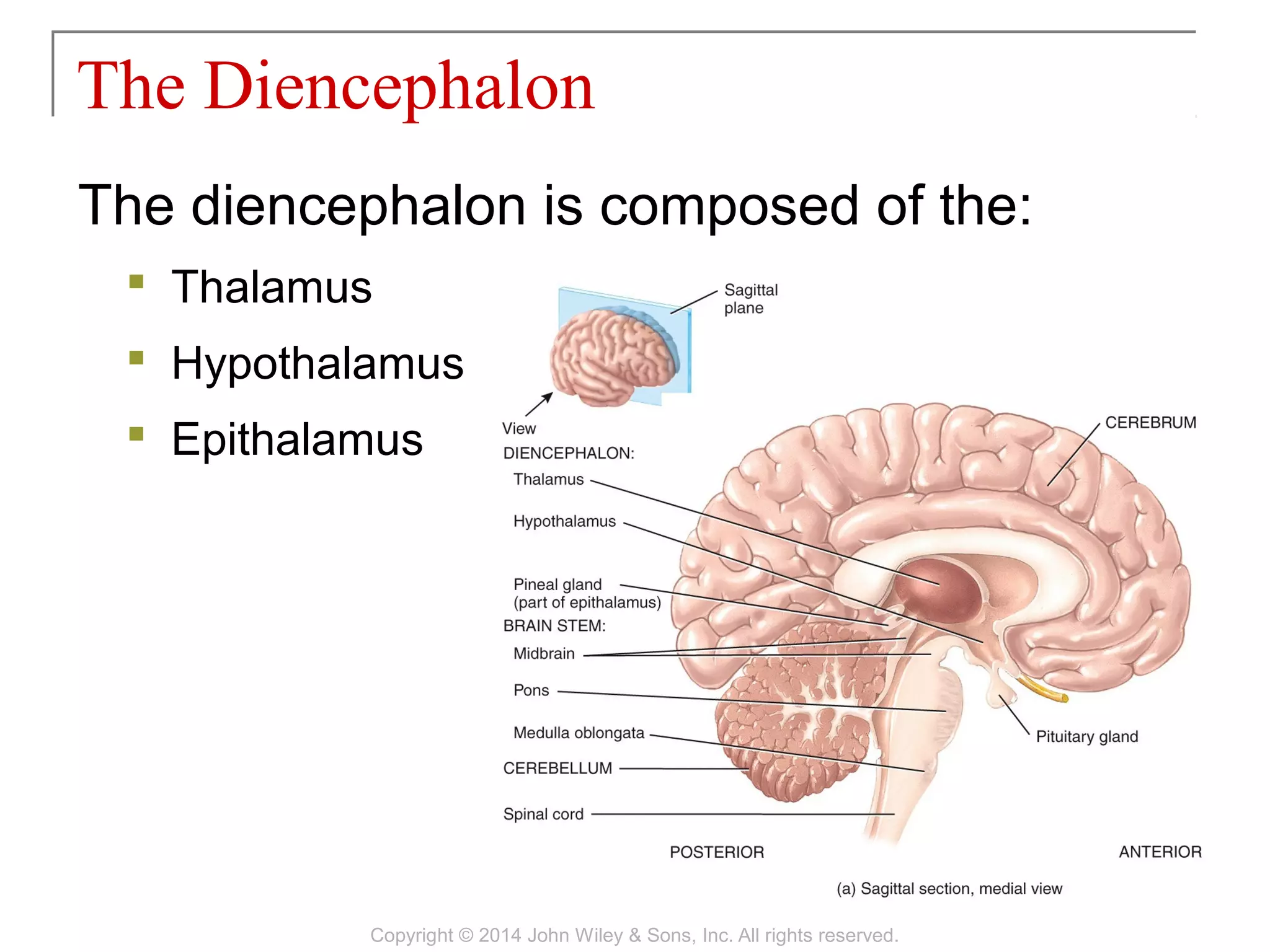

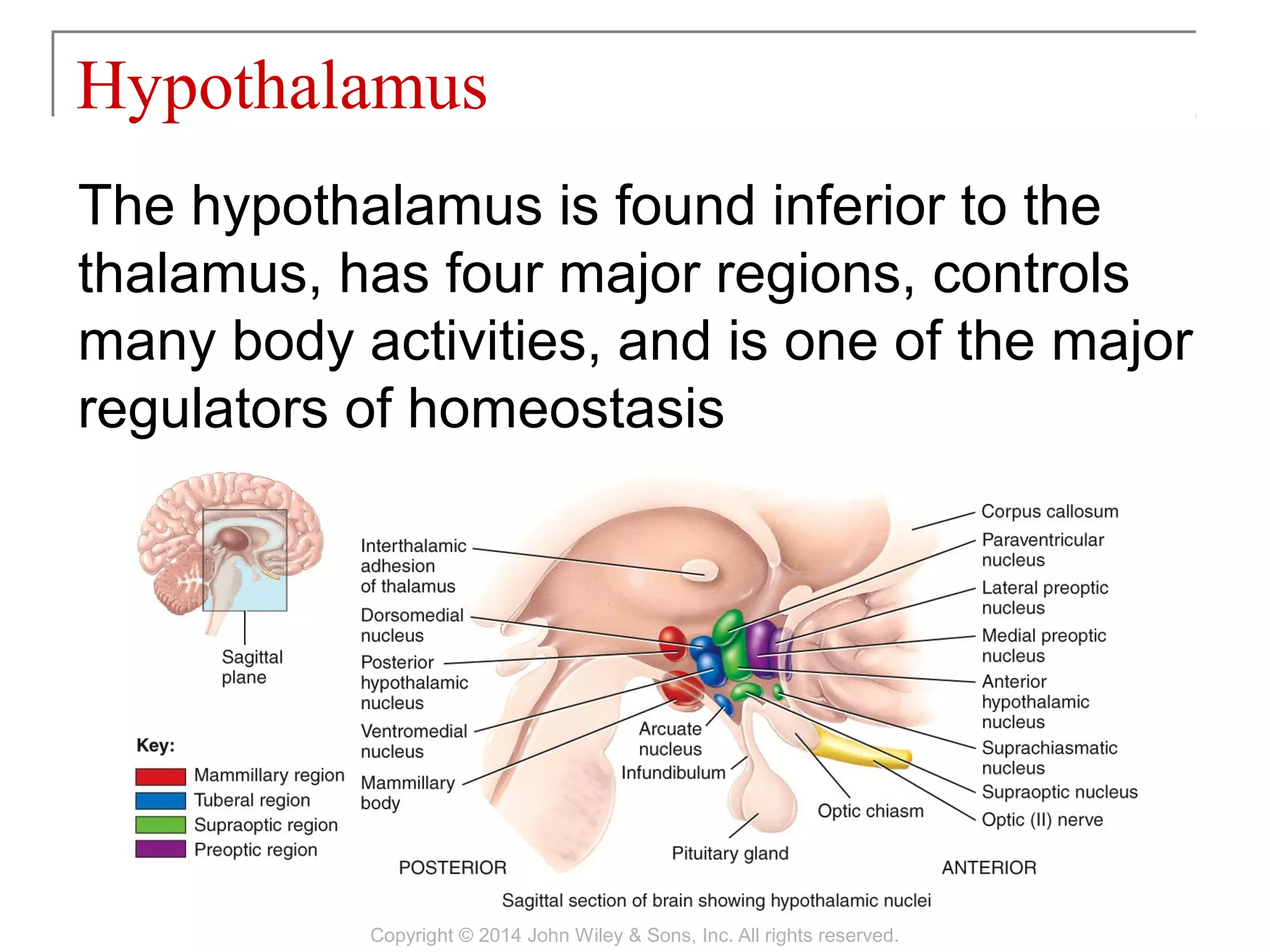
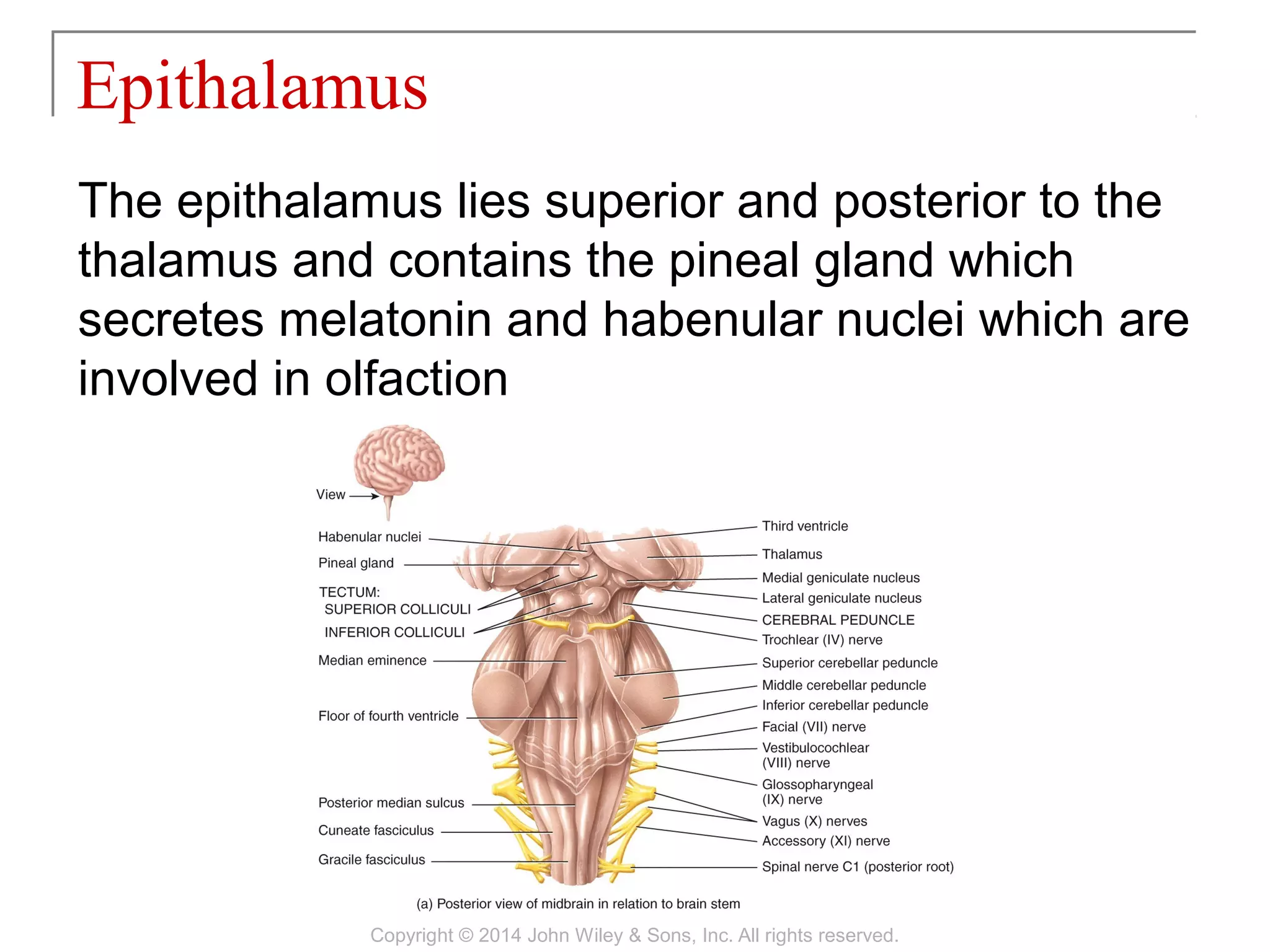


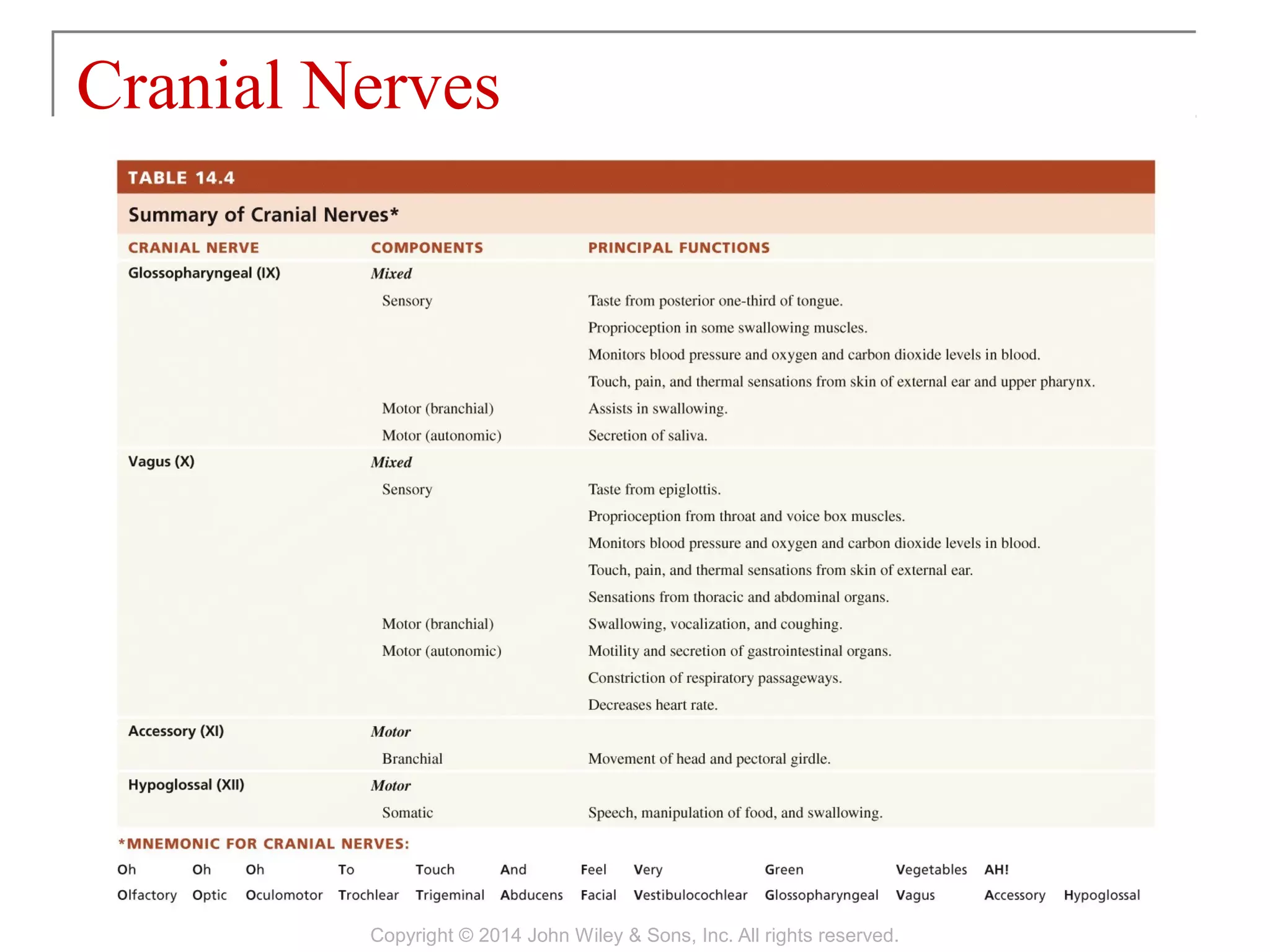


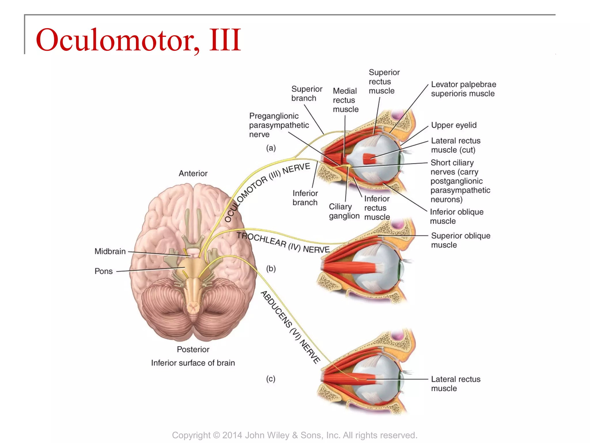


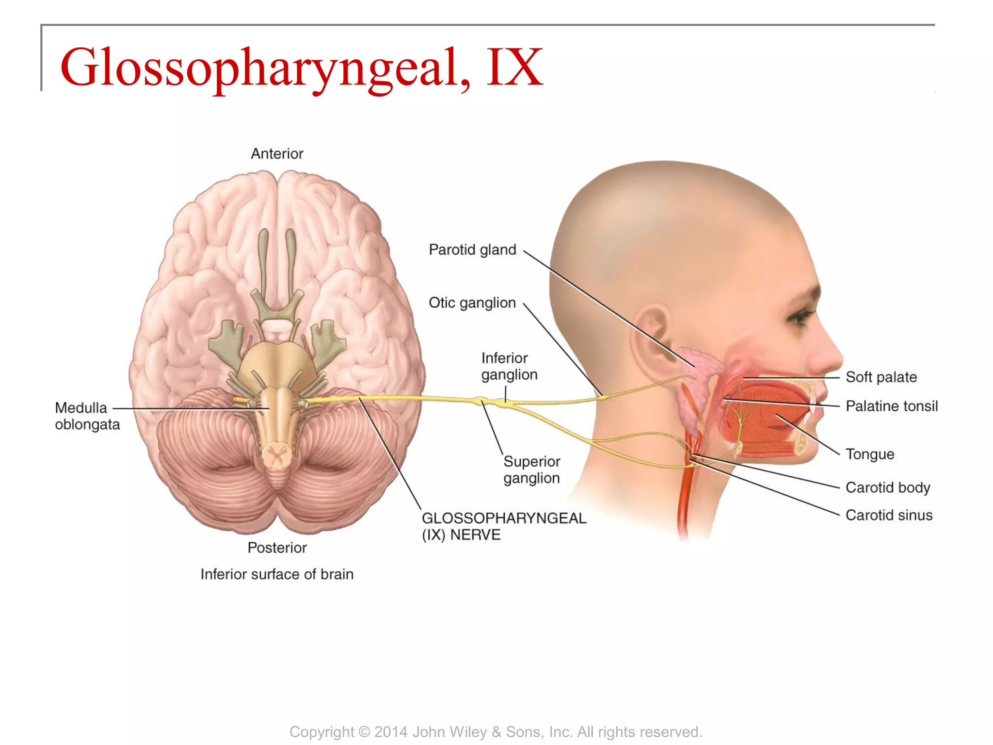
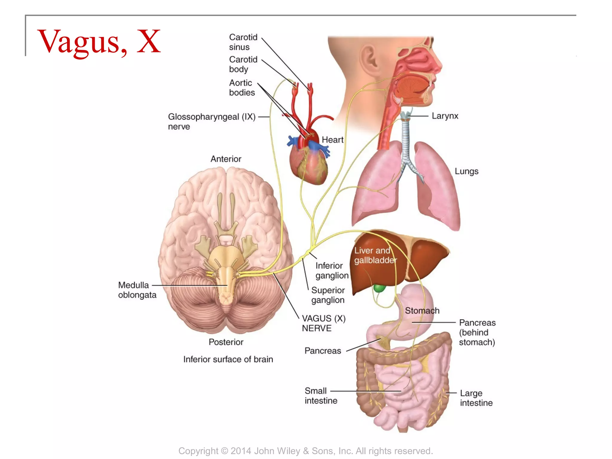
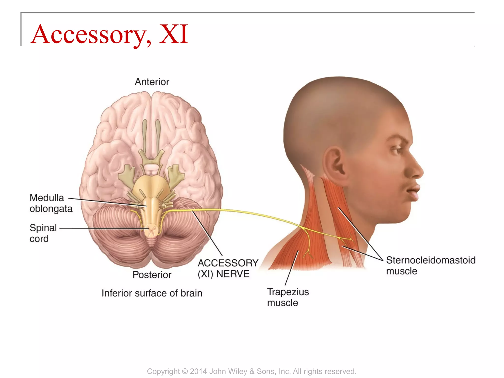
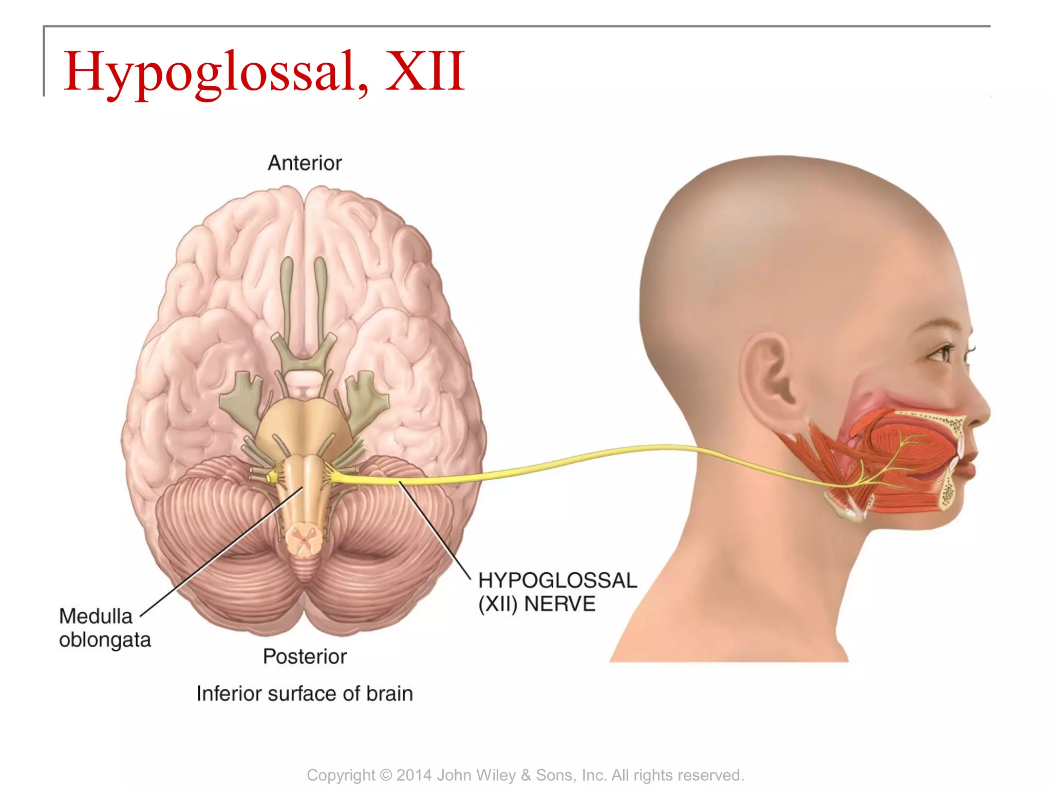


The chapter discusses the structure and function of the brain and cranial nerves. It describes how the brain is protected by cranial bones, meninges and cerebrospinal fluid, and receives blood flow through arteries and veins. The brain is organized into sections including the cerebrum, cerebellum, and brain stem. The brain stem contains the medulla, pons, and midbrain. The chapter also outlines the 12 pairs of cranial nerves and their functions.
![10 [chapter 10 muscular tissue]](https://cdn.slidesharecdn.com/ss_thumbnails/10chapter10musculartissue-170828040153-thumbnail.jpg?width=640&height=640&fit=bounds)
![13 [chapter 13 the spinal cord and spinal nerves]](https://cdn.slidesharecdn.com/ss_thumbnails/13chapter13thespinalcordandspinalnerves-170828040950-thumbnail.jpg?width=640&height=640&fit=bounds)
![15 [chapter 15 the autonomic nervous system]](https://cdn.slidesharecdn.com/ss_thumbnails/15chapter15theautonomicnervoussystem-170828041929-thumbnail.jpg?width=640&height=640&fit=bounds)
![12 [chapter 12 nervous tissue]](https://cdn.slidesharecdn.com/ss_thumbnails/12chapter12nervoustissue-170828041102-thumbnail.jpg?width=640&height=640&fit=bounds)
![05 [chapter 5 the integumentary system]](https://cdn.slidesharecdn.com/ss_thumbnails/05chapter5theintegumentarysystem-170828035624-thumbnail.jpg?width=640&height=640&fit=bounds)
![29 [chapter 29 development and inheritance]](https://cdn.slidesharecdn.com/ss_thumbnails/29chapter29developmentandinheritance-170828044352-thumbnail.jpg?width=640&height=640&fit=bounds)
![11 [chapter 11 the muscular system]](https://cdn.slidesharecdn.com/ss_thumbnails/11chapter11themuscularsystem-170828041038-thumbnail.jpg?width=640&height=640&fit=bounds)
![22 [chapter 22 the lymphatic system and immunity]](https://cdn.slidesharecdn.com/ss_thumbnails/22chapter22thelymphaticsystemandimmunity-170828153258-thumbnail.jpg?width=640&height=640&fit=bounds)
![17 [chapter 17 the special senses]](https://cdn.slidesharecdn.com/ss_thumbnails/17chapter17thespecialsenses-170828041636-thumbnail.jpg?width=640&height=640&fit=bounds)
![18 [chapter 18 the endocrine system]](https://cdn.slidesharecdn.com/ss_thumbnails/18chapter18theendocrinesystem-170828042016-thumbnail.jpg?width=640&height=640&fit=bounds)
![26 [chapter 26 the urinary system]](https://cdn.slidesharecdn.com/ss_thumbnails/26chapter26theurinarysystem-170828044011-thumbnail.jpg?width=640&height=640&fit=bounds)
![20 [chapter 20 the cardiovascular system the heart]](https://cdn.slidesharecdn.com/ss_thumbnails/20chapter20thecardiovascularsystem-theheart-170828133506-thumbnail.jpg?width=640&height=640&fit=bounds)
![23 [chapter 23 the respiratory system]](https://cdn.slidesharecdn.com/ss_thumbnails/23chapter23therespiratorysystem-170828043650-thumbnail.jpg?width=640&height=640&fit=bounds)
![07 [chapter 7 the skeletal system the axial skeleton]](https://cdn.slidesharecdn.com/ss_thumbnails/07chapter7theskeletalsystem-theaxialskeleton-170828035650-thumbnail.jpg?width=640&height=640&fit=bounds)
![19 [chapter 19 the cardiovascular system the blood]](https://cdn.slidesharecdn.com/ss_thumbnails/19chapter19thecardiovascularsystem-theblood-170828042033-thumbnail.jpg?width=640&height=640&fit=bounds)
![16 [chapter 16 sensory, motor, and integrative systems]](https://cdn.slidesharecdn.com/ss_thumbnails/16chapter16sensorymotorandintegrativesystems-170828041940-thumbnail.jpg?width=640&height=640&fit=bounds)
![01 [chapter 1 an introduction to the human body]](https://cdn.slidesharecdn.com/ss_thumbnails/01chapter1anintroductiontothehumanbody-170828035545-thumbnail.jpg?width=640&height=640&fit=bounds)
![21 [chapter 21 the cardiovascular system blood vessels and hemodynamics][11e]](https://cdn.slidesharecdn.com/ss_thumbnails/21chapter21thecardiovascularsystem-bloodvesselsandhemodynamics11e-170828043342-thumbnail.jpg?width=640&height=640&fit=bounds)

![24 [chapter 24 the digestive system][11e]](https://cdn.slidesharecdn.com/ss_thumbnails/24chapter24thedigestivesystem11e-170828043714-thumbnail.jpg?width=640&height=640&fit=bounds)
![06 [chapter 6 the skeletal system bone tissue]](https://cdn.slidesharecdn.com/ss_thumbnails/06chapter6theskeletalsystem-bonetissue-170828035633-thumbnail.jpg?width=640&height=640&fit=bounds)
![08 [chapter 8 the skeletal system appendicular skeleton]](https://cdn.slidesharecdn.com/ss_thumbnails/08chapter8theskeletalsystem-appendicularskeleton-170828041008-thumbnail.jpg?width=640&height=640&fit=bounds)
![09 [chapter 9 joints]](https://cdn.slidesharecdn.com/ss_thumbnails/09chapter9joints-170828041032-thumbnail.jpg?width=640&height=640&fit=bounds)
![04 [chapter 4 the tissue level of organization][11e]](https://cdn.slidesharecdn.com/ss_thumbnails/04chapter4thetissueleveloforganization11e-170828035609-thumbnail.jpg?width=640&height=640&fit=bounds)
![25 [chapter 25 metabolism and nutrition]](https://cdn.slidesharecdn.com/ss_thumbnails/25chapter25metabolismandnutrition-170828145139-thumbnail.jpg?width=640&height=640&fit=bounds)

![03 [chapter 3 the cellular level of organization]](https://cdn.slidesharecdn.com/ss_thumbnails/03chapter3thecellularleveloforganization-170828035521-thumbnail.jpg?width=640&height=640&fit=bounds)


![BrainCranialNerves [Autosaved].ppt](https://cdn.slidesharecdn.com/ss_thumbnails/braincranialnervesautosaved-230901083354-a5739a96-thumbnail.jpg?width=640&height=640&fit=bounds)
![BrainCranialNerves [Autosaved].ppt](https://cdn.slidesharecdn.com/ss_thumbnails/braincranialnervesautosaved-230916133851-323a0d58-thumbnail.jpg?width=640&height=640&fit=bounds)


