This document provides an overview of the muscles to be identified in Activities 5 & 6, which focus on appendicular and axial muscles. It lists the muscles of the pectoral girdle, rotator cuff, glenohumeral joint, arm, forearm, and thigh. For each muscle, it identifies the origin, insertion, action, and relevant textbook references and figures. The document is organized by muscle group and includes over 35 muscles total.

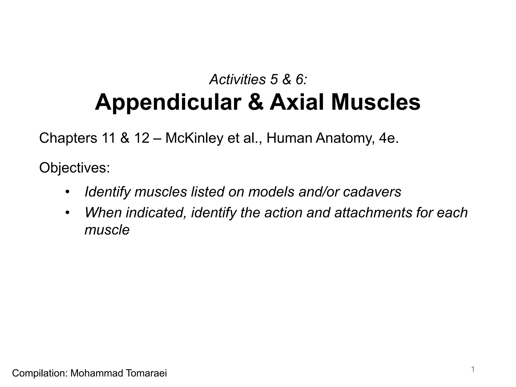
![Appendicular Muscles – Pectoral Girdle Muscles [6]
• Trapezius
• Action:
• Superior: elevates &
superiorly rotates scapula;
elevates clavicle
• Middle: retracts scapula
• Inferior: depresses scapula,
extends head
• Textbook Reference:
• Description: p. 354-355
• Figures: 11.1b, 12.2, 12.4
2](https://image.slidesharecdn.com/activity5-6-160526165849/85/Activity-5-6-2-320.jpg)
![Appendicular Muscles – Pectoral Girdle Muscles [6]
• Levator scapulae
• Action:
• Elevates scapula
• Textbook Reference:
• Description: p. 355
• Figures: 12.2, 12.3, 12.4b
3](https://image.slidesharecdn.com/activity5-6-160526165849/85/Activity-5-6-3-320.jpg)
![Appendicular Muscles – Pectoral Girdle Muscles [6]
• Serratus anterior
• Action:
• Protracts & stabilizes scapula
• Textbook Reference:
• Description: p. 354
• Figures: 11.1b, 11.14a, 12.1,
12.4a
4](https://image.slidesharecdn.com/activity5-6-160526165849/85/Activity-5-6-4-320.jpg)
![Appendicular Muscles – Pectoral Girdle Muscles [6]
• Pectoralis minor
• Action:
• Protracts & depresses scapula
• Textbook Reference:
• Description: p. 354
• Figures: 12.1, 12.4a
5](https://image.slidesharecdn.com/activity5-6-160526165849/85/Activity-5-6-5-320.jpg)
![Appendicular Muscles – Pectoral Girdle Muscles [6]
• Rhomboid major
• Action:
• Elevates & retracts (adducts)
scapula
• Rotates scapula inferiorly
• Textbook Reference:
• Description: p. 355
• Figures: 12.2, 12.4b
6](https://image.slidesharecdn.com/activity5-6-160526165849/85/Activity-5-6-6-320.jpg)
![Appendicular Muscles – Pectoral Girdle Muscles [6]
• Rhomboid minor
• Action:
• Elevates & retracts (adducts)
scapula
• Rotates scapula inferiorly
• Textbook Reference:
• Description: p. 355
• Figures: 12.2, 12.4b
7](https://image.slidesharecdn.com/activity5-6-160526165849/85/Activity-5-6-7-320.jpg)
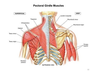
![Appendicular Muscles – Rotator Cuff Muscles [4]
• Supraspinatus
• Origin:
• Supraspinous fossa of scapula
• Insertion:
• Greater tubercle of humerus
• Action (rotator cuff muscles
together):
• Stabilize & rotate the
glenohumeral joint
• Textbook Reference:
• Description: p. 359
• Figures: 12.2, 12.4a & b
8](https://image.slidesharecdn.com/activity5-6-160526165849/85/Activity-5-6-8-320.jpg)
![Appendicular Muscles – Rotator Cuff Muscles [4]
• Infraspinatus
• Origin:
• Infraspinous fossa of scapula
• Insertion:
• Greater tubercle of humerus
• Action (rotator cuff muscles
together):
• Stabilize & rotate the
glenohumeral joint
• Textbook Reference:
• Description: p. 359
• Figures: 12.2, 12.4b
9](https://image.slidesharecdn.com/activity5-6-160526165849/85/Activity-5-6-9-320.jpg)
![Appendicular Muscles – Rotator Cuff Muscles [4]
• Teres minor
• Origin:
• Lateral border of scapula
• Insertion:
• Greater tubercle of humerus
• Action (rotator cuff muscles
together):
• Stabilize & rotate the
glenohumeral joint
• Textbook Reference:
• Description: p. 359
• Figures: 12.2, 12.4b
10](https://image.slidesharecdn.com/activity5-6-160526165849/85/Activity-5-6-10-320.jpg)
![Appendicular Muscles – Rotator Cuff Muscles [4]
• Subscapularis
• Origin:
• Subscapular fossa of scapula
• Insertion:
• Lesser tubercle of humerus
• Action (rotator cuff muscles
together):
• Stabilize & rotate the
glenohumeral joint
• Textbook Reference:
• Description: p. 359
• Figures: 12.4a, 12.5a
11](https://image.slidesharecdn.com/activity5-6-160526165849/85/Activity-5-6-11-320.jpg)
![Appendicular Muscles – Other Glenohumeral Joint Muscles [4]
• Teres major
• Origin:
• Lateral border & angle of
scapula
• Insertion:
• Lesser tubercle &
intertubercular sulcus of
humerus
• Action:
• Extends, adducts, & medially
rotates arm
• Textbook Reference:
• Description: p. 359
• Figures: 12.2, 12.4b
12](https://image.slidesharecdn.com/activity5-6-160526165849/85/Activity-5-6-12-320.jpg)
![Appendicular Muscles – Other Glenohumeral Joint Muscles [4]
• Latissimus dorsi
• Origin:
• Spinous processes of lower
thoracic vertebrae
• Lower ribs (8-12)
• Iliac crest
• Insertion:
• Intertubercular sulcus of
humerus
• Action:
• Extends, adducts, & medially
rotates arm
• Draws arm inferiorly &
posteriorly
• Textbook Reference:
• Description: p. 358
• Figures: 11.1, 12.1, 12.2,
12.4a & b 13](https://image.slidesharecdn.com/activity5-6-160526165849/85/Activity-5-6-13-320.jpg)
![Appendicular Muscles – Other Glenohumeral Joint Muscles [4]
• Deltoid
• Origin:
• Acromial end of clavicle
• Acromion & spine of scapula
• Insertion:
• Deltoid tuberosity of humerus
• Action:
• Abducts, flexes, extends, &
rotates arm
• Textbook Reference:
• Description: p. 358
• Figures: 11.1, 12.1, 12.2,
12.4a & b
14](https://image.slidesharecdn.com/activity5-6-160526165849/85/Activity-5-6-14-320.jpg)
![Appendicular Muscles – Other Glenohumeral Joint Muscles [4]
• Pectoralis major
• Origin:
• Clavicle
• Costal cartilages
• Insertion:
• Greater tubercle & lateral
intertubercular sulcus of
humerus
• Action:
• Flexes, adducts, & medially
rotates arm
• Textbook Reference:
• Description: p. 358
• Figures: 11.1, 12.1, 12.4a
15](https://image.slidesharecdn.com/activity5-6-160526165849/85/Activity-5-6-15-320.jpg)
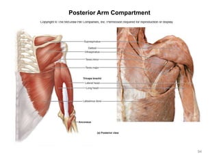
![Appendicular Muscles – Posterior Arm Compartment [1]
• Triceps brachii (3 heads)
• Origin:
• Long head: Infraglenoid
tubercle of scapula
• Lateral head: posterior
shaft of humerus
• Medial head: posterior
shaft of humerus, distal to
radial groove
• Insertion:
• Olecranon process of ulna
• Action:
• Extends forearm & assists
in arm adduction
• Textbook Reference:
• Description: p. 364
• Figures: 12.8a & b
16
Right arm, posterior view](https://image.slidesharecdn.com/activity5-6-160526165849/85/Activity-5-6-16-320.jpg)
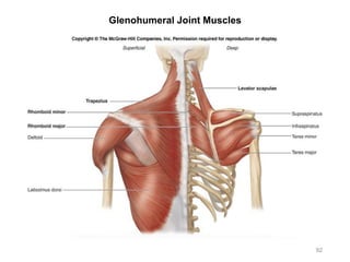
![Appendicular Muscles – Anterior Arm Compartment [5]
• Biceps brachii (2 heads)
• Origin:
• Long head: supraglenoid
tubercle of scapula
• Short head: coracoid process
of scapula
• Insertion:
• Radial tuberosity
• Action:
• Flexes arm (glenohumeral
joint)
• Flexes & supinates forearm
(elbow joint)
• Textbook Reference:
• Description: p. 363
• Figures: 12.7a & b, table 12.4
17](https://image.slidesharecdn.com/activity5-6-160526165849/85/Activity-5-6-17-320.jpg)
![Appendicular Muscles – Anterior Arm Compartment [5]
• Coracobrachialis
• Origin:
• Coracoid process of scapula
• Insertion:
• Middle medial shaft of
humerus
• Action:
• Adducts & flexes arm
(glenohumeral joint)
• Textbook Reference:
• Description: p. 363
• Figures: 12.7a & b
18](https://image.slidesharecdn.com/activity5-6-160526165849/85/Activity-5-6-18-320.jpg)
![Appendicular Muscles – Anterior Arm Compartment [5]
• Brachialis
• Origin:
• Distal, anterior surface of
humerus
• Insertion:
• Coronoid process of ulna
• Action:
• Flexes forearm (elbow joint)
• Textbook Reference:
• Description: p. 363
• Figures: 12.7a & b
19](https://image.slidesharecdn.com/activity5-6-160526165849/85/Activity-5-6-19-320.jpg)
![Appendicular Muscles – Anterior Arm Compartment [5]
• Brachioradialis
• Origin:
• Lateral humerus
• Insertion:
• Styloid process of radius
• Action:
• Flexes forearm (elbow joint)
• Textbook Reference:
• Description: p. 363
• Figures: 12.7a, 12.11a
• Landmark importance:
• Separates anterior forearm
flexors from posterior forearm
extensors
20](https://image.slidesharecdn.com/activity5-6-160526165849/85/Activity-5-6-20-320.jpg)
![Appendicular Muscles – Anterior Forearm Compartment [6+1]
• Pronator teres
• Action:
• Pronates forearm
• Textbook Reference:
• Description: p. 364
• Figures: 12.9, 12.11, 12.12
21](https://image.slidesharecdn.com/activity5-6-160526165849/85/Activity-5-6-21-320.jpg)
![Appendicular Muscles – Anterior Forearm Compartment [6+1]
• Flexor carpi radialis
• Action:
• Flexes wrist
• Abducts hand
• Textbook Reference:
• Description: p. 369
• Figures: 12.11, 12.12
22](https://image.slidesharecdn.com/activity5-6-160526165849/85/Activity-5-6-22-320.jpg)
![Appendicular Muscles – Anterior Forearm Compartment [6+1]
• Palmaris longus
• Action:
• Weakly flexes wrist
• Tenses fascia of palm
• Textbook Reference:
• Description: p. 369
• Figures: 12.11, 12.12
• Cadaver hint:
• Not all cadavers have this
muscle (for genetic reasons)
• Ends in a slender, flattened
tendon, passing over the
upper part of the flexor
retinaculum
23](https://image.slidesharecdn.com/activity5-6-160526165849/85/Activity-5-6-23-320.jpg)
![Appendicular Muscles – Anterior Forearm Compartment [6+1]
• Flexor carpi ulnaris
• Action:
• Flexes wrist
• Adducts hand
• Textbook Reference:
• Description: p. 369
• Figures: 12.11, 12.12, 12.13a
24](https://image.slidesharecdn.com/activity5-6-160526165849/85/Activity-5-6-24-320.jpg)
![Appendicular Muscles – Anterior Forearm Compartment [6+1]
• Flexor digitorum superficialis
• Action:
• Flexes wrist
• Flexes the 2nd to 5th
metacarpophalangeal (MP) &
proximal interphalangeal (PIP)
joints
• Textbook Reference:
• Description: p. 369
• Figures: 12.11b
25](https://image.slidesharecdn.com/activity5-6-160526165849/85/Activity-5-6-25-320.jpg)
![Appendicular Muscles – Anterior Forearm Compartment [6+1]
• Flexor digitorum profundus
• Action:
• Flexes wrist
• Flexes the 2nd to 5th
metacarpophalangeal (MP),
proximal interphalangeal
(PIP), & distal interphalangeal
(DIP) joints
• Textbook Reference:
• Description: p. 369
• Figures: 12.11c
26](https://image.slidesharecdn.com/activity5-6-160526165849/85/Activity-5-6-26-320.jpg)
![Appendicular Muscles – Anterior Forearm Compartment [6+1]
• Flexor retinaculum
(associated structure)
• Significance:
• Fibrous band of connective
tissue that covers the palmar
surface of the carpals
• Textbook Reference:
• Description: p. 366
• Figures: 12.11a, 12.14a
27](https://image.slidesharecdn.com/activity5-6-160526165849/85/Activity-5-6-27-320.jpg)
![Appendicular Muscles – Posterior Forearm Compartment [8+1]
• Extensor carpi radialis longus
• Action:
• Extends wrist
• Abducts hand
• Textbook Reference:
• Description: p. 369, 371
• Figures: 12.13a & b
28](https://image.slidesharecdn.com/activity5-6-160526165849/85/Activity-5-6-28-320.jpg)
![Appendicular Muscles – Posterior Forearm Compartment [8+1]
• Extensor carpi radialis brevis
• Action:
• Extends wrist
• Abducts hand
• Textbook Reference:
• Description: p. 369, 371
• Figures: 12.13a & b
29](https://image.slidesharecdn.com/activity5-6-160526165849/85/Activity-5-6-29-320.jpg)
![Appendicular Muscles – Posterior Forearm Compartment [8+1]
• Extensor carpi ulnaris
• Action:
• Extends wrist
• Adducts hand
• Textbook Reference:
• Description: p. 369, 371
• Figures: 12.13a & b
30](https://image.slidesharecdn.com/activity5-6-160526165849/85/Activity-5-6-30-320.jpg)
![Appendicular Muscles – Posterior Forearm Compartment [8+1]
• Extensor digitorum
• Action:
• Extends wrist
• Extends the 2nd to 5th
metacarpophalangeal (MP),
proximal interphalangeal
(PIP), & distal interphalangeal
(DIP) joints
• Textbook Reference:
• Description: p. 369, 371
• Figures: 12.13a & b
31](https://image.slidesharecdn.com/activity5-6-160526165849/85/Activity-5-6-31-320.jpg)
![Appendicular Muscles – Posterior Forearm Compartment [8+1]
• Abductor pollicis longus
• Action:
• Abducts thumb
• Weakly extends wrist
• Textbook Reference:
• Description: p. 369, 371
• Figures: 12.13a & b
32](https://image.slidesharecdn.com/activity5-6-160526165849/85/Activity-5-6-32-320.jpg)
![Appendicular Muscles – Posterior Forearm Compartment [8+1]
• Extensor pollicis longus
• Action:
• Extends metacarpophalangeal
(MP) & interphalangeal (IP)
joints of thumb
• Weakly extends wrist
• Textbook Reference:
• Description: p. 369, 371
• Figures: 12.13a & b
33](https://image.slidesharecdn.com/activity5-6-160526165849/85/Activity-5-6-33-320.jpg)
![Appendicular Muscles – Posterior Forearm Compartment [8+1]
• Extensor pollicis brevis
• Action:
• Extends metacarpophalangeal
(MP) joint of thumb
• Weakly extends wrist
• Textbook Reference:
• Description: p. 369, 371
• Figures: 12.13a & b
34](https://image.slidesharecdn.com/activity5-6-160526165849/85/Activity-5-6-34-320.jpg)
![Appendicular Muscles – Posterior Forearm Compartment [8+1]
• Supinator
• Action:
• Supinates forearm
• Textbook Reference:
• Description: p. 364
• Figures: 12.9, 12.13b
• Cadaver hint:
• Visible on a well-dissected
cadaver
35](https://image.slidesharecdn.com/activity5-6-160526165849/85/Activity-5-6-35-320.jpg)
![Appendicular Muscles – Posterior Forearm Compartment [8+1]
• Extensor retinaculum
• Significance:
• Fibrous band of connective
tissue that covers the dorsal
surface of the carpals
• Textbook Reference:
• Description: p. 369
• Figures: 12.13b, 12.14c
36](https://image.slidesharecdn.com/activity5-6-160526165849/85/Activity-5-6-36-320.jpg)
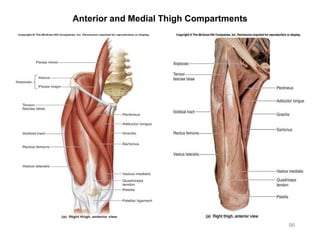
![Appendicular Muscles – Anterior Thigh Compartment [5]
Quadriceps Femoris Group [4]
• Rectus femoris
• Origin:
• Anterior inferior iliac spine
• Insertion:
• Patella via quadriceps tendon
and then tibial tuberosity via
patellar ligament
• Action:
• Extends leg
• Flexes thigh
• Textbook Reference:
• Description: p. 381
• Figures: 12.71a & b
37](https://image.slidesharecdn.com/activity5-6-160526165849/85/Activity-5-6-37-320.jpg)
![Appendicular Muscles – Anterior Thigh Compartment [5]
Quadriceps Femoris Group [4]
• Vastus lateralis
• Insertion:
• Patella via quadriceps tendon
and then tibial tuberosity via
patellar ligament
• Action:
• Extends leg
• Textbook Reference:
• Description: p. 381
• Figures: 12.17a & b, 12.15
38](https://image.slidesharecdn.com/activity5-6-160526165849/85/Activity-5-6-38-320.jpg)
![Appendicular Muscles – Anterior Thigh Compartment [5]
Quadriceps Femoris Group [4]
• Vastus medialis
• Insertion:
• Patella via quadriceps tendon
and then tibial tuberosity via
patellar ligament
• Action:
• Extends leg
• Textbook Reference:
• Description: p. 381
• Figures: 12.17a & b
39](https://image.slidesharecdn.com/activity5-6-160526165849/85/Activity-5-6-39-320.jpg)
![Appendicular Muscles – Anterior Thigh Compartment [5]
Quadriceps Femoris Group [4]
• Vastus intermedius
• Insertion:
• Patella via quadriceps tendon
and then tibial tuberosity via
patellar ligament
• Action:
• Extends leg
• Textbook Reference:
• Description: p. 381
• Figures: 12.17a & b
• Cadaver hint:
• This muscle is deep to rectus
femoris
40](https://image.slidesharecdn.com/activity5-6-160526165849/85/Activity-5-6-40-320.jpg)
![Appendicular Muscles – Anterior Thigh Compartment [5]
• Sartorius
• Origin:
• Anterior superior iliac spine
• Insertion:
• Tibial tuberosity, medial side
• Action:
• Flexes, abducts, & laterally
rotates thigh
• Flexes leg & rotates leg
medially (sitting cross-legged
on the floor)
• Textbook Reference:
• Description: p. 375, 381
• Figures: 12.17a & b
• Trivia:
• Longest muscle in the body
41](https://image.slidesharecdn.com/activity5-6-160526165849/85/Activity-5-6-41-320.jpg)
![Appendicular Muscles – Iliopsoas Group [2]
• Iliacus
• Origin:
• Iliac fossa
• Insertion:
• Lesser trochanter of femur
• Action:
• Flexes thigh
• Textbook Reference:
• Description: p. 375
• Figures: 12.15a, 12.17a
• Cadaver hint:
• Look inside the
abdominopelvic cavity
• Has a common insertion with
psoas major muscle
42](https://image.slidesharecdn.com/activity5-6-160526165849/85/Activity-5-6-42-320.jpg)
![Appendicular Muscles – Iliopsoas Group [2]
• Psoas major
• Origin:
• T12-L5 vertebrae
• Insertion:
• Lesser trochanter of femur
• Action:
• Flexes thigh
• Textbook Reference:
• Description: p. 375
• Figures: 12.15a, 12.17a
• Cadaver hint:
• Look inside the
abdominopelvic cavity
• Has a common insertion with
iliacus muscle
43](https://image.slidesharecdn.com/activity5-6-160526165849/85/Activity-5-6-43-320.jpg)
![Appendicular Muscles – Medial Thigh Compartment [5]
• Pectineus
• Action:
• Adducts thigh
• Weakly flexes thigh
• Textbook Reference:
• Description: p. 376
• Figures: 12.15, 12.17
44
Mnemonic:
Great Major League Baseball Players](https://image.slidesharecdn.com/activity5-6-160526165849/85/Activity-5-6-44-320.jpg)
![Appendicular Muscles – Medial Thigh Compartment [5]
• Adductor longus
• Action:
• Adducts thigh
• Weakly flexes thigh
• Textbook Reference:
• Description: p. 376
• Figures: 12.15, 12.17
45
Mnemonic:
Great Major League Baseball Players](https://image.slidesharecdn.com/activity5-6-160526165849/85/Activity-5-6-45-320.jpg)
![Appendicular Muscles – Medial Thigh Compartment [5]
• Adductor brevis
• Action:
• Adducts thigh
• Weakly flexes thigh
• Textbook Reference:
• Description: p. 376
• Figures: 12.15, 12.17
46
Mnemonic:
Great Major League Baseball Players](https://image.slidesharecdn.com/activity5-6-160526165849/85/Activity-5-6-46-320.jpg)
![Appendicular Muscles – Medial Thigh Compartment [5]
• Adductor magnus
• Action:
• Adducts thigh
• Flexes or extends, and
laterally rotates thigh
(depending on starting
position)
• Textbook Reference:
• Description: p. 376
• Figures: 12.15, 12.18
47
Mnemonic:
Great Major League Baseball Players](https://image.slidesharecdn.com/activity5-6-160526165849/85/Activity-5-6-47-320.jpg)
![Appendicular Muscles – Medial Thigh Compartment [5]
• Gracilis
• Origin:
• Inferior ramus & body of pubis
• Insertion:
• Upper medial surface of tibia
• Action:
• Weakly adducts & weakly
flexes thigh
• Flexes leg
• Textbook Reference:
• Description: p. 376, 381
• Figures: 12.15a, 12.17a
• Cadaver hint:
• NOT to be confused with
sartorius (compare origins)
48
Mnemonic:
Great Major League Baseball Players](https://image.slidesharecdn.com/activity5-6-160526165849/85/Activity-5-6-48-320.jpg)
![Appendicular Muscles – Posterior Thigh Compartment [3]
Hamstrings Group [3]
• Semitendinosus
• Origin:
• Ischial tuberosity
• Insertion:
• Proximal medial surface of
tibia
• Action:
• Extends thigh
• Flexes leg
• Medially rotates leg
• Textbook Reference:
• Description: p. 381
• Figures: 12.18a & b
• Identification hint:
• Semi (half) tendinosus
(tendon), means half of this
muscle consists of a tendon 49](https://image.slidesharecdn.com/activity5-6-160526165849/85/Activity-5-6-49-320.jpg)
![Appendicular Muscles – Posterior Thigh Compartment [3]
Hamstrings Group [3]
• Semimembranosus
• Origin:
• Ischial tuberosity
• Insertion:
• Medial condyle of tibia
• Action:
• Extends thigh
• Flexes leg
• Medially rotates leg
• Textbook Reference:
• Description: p. 381
• Figures: 12.18a & b, 12.15b
• Identification hint:
• Semi (half) membranosus
(membrane), means that half
of this muscle has a broad
membranous tendon 50](https://image.slidesharecdn.com/activity5-6-160526165849/85/Activity-5-6-50-320.jpg)
![Appendicular Muscles – Posterior Thigh Compartment [3]
Hamstrings Group [3]
• Biceps femoris (2 heads)
• Origin:
• Long head: ischial tuberosity
• Short head: linea aspera of
femur
• Insertion:
• Head of fibula
• Action:
• Extends thigh
• Flexes leg
• Laterally rotates leg
• Textbook Reference:
• Description: p. 381
• Figures: 12.18a & b, 12.15b
• Cadaver hint:
• By touching the origin of the short
head of biceps femoris, you can
feel the linea aspera of femur 51](https://image.slidesharecdn.com/activity5-6-160526165849/85/Activity-5-6-51-320.jpg)
![Appendicular Muscles – Gluteal Muscles [5+1]
• Tensor fasciae latae
• Origin:
• Iliac crest
• Anterior superior iliac spine
• Insertion:
• Iliotibial tract
• Action:
• Abducts thigh
• Medially rotates thigh
• Textbook Reference:
• Description: p. 396
• Figures: 12.15b, 12.17
52](https://image.slidesharecdn.com/activity5-6-160526165849/85/Activity-5-6-52-320.jpg)
![Appendicular Muscles – Gluteal Muscles [5+1]
• Gluteus maximus
• Origin:
• Iliac crest
• Sacrum
• Coccyx
• Insertion:
• Iliotibial tract
• Linea aspera
• Action:
• Extends thigh
• Laterally rotates thigh
• Textbook Reference:
• Description: p. 377
• Figures: 12.15b, 12.18
53](https://image.slidesharecdn.com/activity5-6-160526165849/85/Activity-5-6-53-320.jpg)
![Appendicular Muscles – Gluteal Muscles [5+1]
• Gluteus medius
• Origin:
• Iliac crest
• Insertion:
• Greater trochanter of
femur
• Action:
• Abducts thigh
• Medially rotates thigh
• Textbook Reference:
• Description: p. 377
• Figures: 12.15b, 12.18a
54](https://image.slidesharecdn.com/activity5-6-160526165849/85/Activity-5-6-54-320.jpg)
![Appendicular Muscles – Gluteal Muscles [5+1]
• Gluteus minimus
• Origin:
• Lateral surface of ilium
• Insertion:
• Greater trochanter of femur
• Action:
• Abducts thigh
• Medially rotates thigh
• Textbook Reference:
• Description: p. 377
• Figures: 12.15c
55](https://image.slidesharecdn.com/activity5-6-160526165849/85/Activity-5-6-55-320.jpg)
![Appendicular Muscles – Gluteal Muscles [5+1]
• Piriformis
• Origin:
• Anterolateral surface of
sacrum
• Insertion:
• Greater trochanter of femur
• Action:
• Laterally rotates thigh
• Textbook Reference:
• Description: p. 377
• Figures: 12.15c
56](https://image.slidesharecdn.com/activity5-6-160526165849/85/Activity-5-6-56-320.jpg)
![Appendicular Muscles – Gluteal Muscles [5+1]
• Iliotibial tract / band
(associated structure)
• Significance:
• Thick band of fascia
continuous with the tensor
fasciae latae muscle
• Textbook Reference:
• Description: p. 376
• Figures: 12.15, 12.17, 12.18
57
Mnemonic:
Follow the Iliotibial Tract to reach Starbucks &
get a Tensor Fasciae Latae](https://image.slidesharecdn.com/activity5-6-160526165849/85/Activity-5-6-57-320.jpg)
![Appendicular Muscles – Anterior Leg Compartment [3]
• Tibialis anterior
• Origin:
• Lateral condyle & proximal
shaft of tibia
• Insertion:
• Metatarsal I
• Medial cuneiform
• Action:
• Dorsiflexes foot
• Inverts foot
• Textbook Reference:
• Description: p. 383
• Figures: 12.20a & b, 12.21
58
Mnemonic (anterior, medial to lateral):
Tom – Harry – Dick](https://image.slidesharecdn.com/activity5-6-160526165849/85/Activity-5-6-58-320.jpg)
![Appendicular Muscles – Anterior Leg Compartment [3]
• Extensor digitorum longus
• Action:
• Extends toes 2-5
• Dorsiflexes foot
• Textbook Reference:
• Description: p. 383
• Figures: 12.20a & b, 12.21
• Identification hint:
• Think of how the tendons of
this muscle insert over the
digits to extend them
59
Mnemonic (anterior, medial to lateral):
Tom – Harry – Dick](https://image.slidesharecdn.com/activity5-6-160526165849/85/Activity-5-6-59-320.jpg)
![Appendicular Muscles – Anterior Leg Compartment [3]
• Extensor hallucis longus
• Action:
• Extends hallux
• Dorsiflexes foot
• Textbook Reference:
• Description: p. 383
• Figures: 12.20a & b, 12.21
60
Mnemonic (anterior, medial to lateral):
Tom – Harry – Dick
Harry likes to hide!](https://image.slidesharecdn.com/activity5-6-160526165849/85/Activity-5-6-60-320.jpg)
![Appendicular Muscles – Lateral Leg Compartment [2]
• Fibularis longus
• Action:
• Everts foot
• Weakly plantar flexes foot
• Textbook Reference:
• Description: p. 383
• Figures: 12.21a & b, 12.22b
• Identification hint:
• Thinking logically, the name of
fibularis longus suggests that
this muscle sits on top of
fibula, and has a longer
tendon relative to another
muscle of similar name and
location
61](https://image.slidesharecdn.com/activity5-6-160526165849/85/Activity-5-6-61-320.jpg)
![Appendicular Muscles – Lateral Leg Compartment [2]
• Fibularis brevis
• Action:
• Everts foot
• Weakly plantar flexes foot
• Textbook Reference:
• Description: p. 383
• Figures: 12.21a & b, 12.22b
• Identification hint:
• This muscle is deep to
fibularis longus and has a
relatively shorter tendon
62](https://image.slidesharecdn.com/activity5-6-160526165849/85/Activity-5-6-62-320.jpg)
![Appendicular Muscles – Posterior Leg Compartment [5]
• Gastrocnemius
• Origin:
• Lateral & medial condyles of
femur
• Insertion:
• Calcaneus via calcaneal
(Achilles) tendon
• Action:
• Plantar flexes foot
• Flexes leg
• Textbook Reference:
• Description: p. 383
• Figures: 12.20a, 12.21a, 12.22a
63](https://image.slidesharecdn.com/activity5-6-160526165849/85/Activity-5-6-63-320.jpg)
![Appendicular Muscles – Posterior Leg Compartment [5]
• Soleus
• Origin:
• Head & proximal shaft of
fibula
• Medial border of tibia
• Insertion:
• Calcaneus via calcaneal
(Achilles) tendon
• Action:
• Plantar flexes foot
• Textbook Reference:
• Description: p. 383
• Figures: 12.21a, 12.22a
64](https://image.slidesharecdn.com/activity5-6-160526165849/85/Activity-5-6-64-320.jpg)
![Appendicular Muscles – Posterior Leg Compartment [5]
• Tibialis posterior
• Action:
• Plantar flexes foot
• Inverts foot
• Textbook Reference:
• Description: p. 383
• Figures: 12.22b & c
• Identification hint:
• Since the posterior leg muscles
have relatively deep bodies, it’s
easier to look at their tendons
near the medial malleolus of tibia
65
Mnemonic (posterior, medial to lateral):
Tom – Dick – Harry](https://image.slidesharecdn.com/activity5-6-160526165849/85/Activity-5-6-65-320.jpg)
![Appendicular Muscles – Posterior Leg Compartment [5]
• Flexor digitorum longus
• Action:
• Plantar flexes foot
• Flexes toes 2-5,
metacarpophalangeal (MP),
proximal interphalangeal
(PIP), & distal interphalangeal
(DIP) joints
• Textbook Reference:
• Description: p. 383
• Figures: 12.22b & c
• Identification hint:
• Since the posterior leg muscles
have relatively deep bodies, it’s
easier to look at their tendons
near the medial malleolus of tibia
66
Mnemonic (posterior, medial to lateral):
Tom – Dick – Harry](https://image.slidesharecdn.com/activity5-6-160526165849/85/Activity-5-6-66-320.jpg)
![Appendicular Muscles – Posterior Leg Compartment [5]
• Flexor hallucis longus
• Action:
• Plantar flexes foot
• Flexes metacarpophalangeal
(MP) and interphalangeal (IP)
joints of hallux
• Textbook Reference:
• Description: p. 383
• Figures: 12.22b & c
• Identification hint:
• Since the posterior leg muscles
have relatively deep bodies, it’s
easier to look at their tendons
near the medial malleolus of tibia
67
Mnemonic (posterior, medial to lateral):
Tom – Dick – Harry
Harry likes to hide!](https://image.slidesharecdn.com/activity5-6-160526165849/85/Activity-5-6-67-320.jpg)
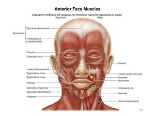
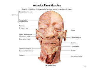
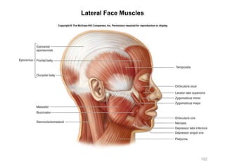
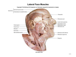
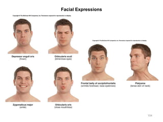
![Axial Muscles – Facial Expression Muscles [8]
• Frontalis
(fontal belly of occipitofrontalis)
• Action:
• Draws scalp forward
• Raises eyebrows
• Wrinkles forehead
• Textbook Reference:
• Description: p. 322
• Figures: 11.2a & b
68](https://image.slidesharecdn.com/activity5-6-160526165849/85/Activity-5-6-68-320.jpg)
![Axial Muscles – Facial Expression Muscles [8]
• Occipitalis
(occipital belly of
occipitofrontalis)
• Action:
• Draws scalp backward
• Textbook Reference:
• Description: p. 322
• Figures: 11.1b, 11.2b
69](https://image.slidesharecdn.com/activity5-6-160526165849/85/Activity-5-6-69-320.jpg)
![Axial Muscles – Facial Expression Muscles [8]
• Orbicularis oris
• Action:
• Compresses & purses lips
(kiss muscle)
• Textbook Reference:
• Description: p. 322
• Figures: 11.2a & b
70](https://image.slidesharecdn.com/activity5-6-160526165849/85/Activity-5-6-70-320.jpg)
![Axial Muscles – Facial Expression Muscles [8]
• Orbicularis oculi
• Action:
• Closes eye (blink muscle)
• Textbook Reference:
• Description: p. 322
• Figures: 11.2a & b
71](https://image.slidesharecdn.com/activity5-6-160526165849/85/Activity-5-6-71-320.jpg)
![Axial Muscles – Facial Expression Muscles [8]
• Platysma
• Action:
• Pulls lower lip inferiorly
• Tenses skin of neck
• Textbook Reference:
• Description: p. 322
• Figures: 11.2a & b
72](https://image.slidesharecdn.com/activity5-6-160526165849/85/Activity-5-6-72-320.jpg)
![Axial Muscles – Facial Expression Muscles [8]
• Zygomaticus major
• Action:
• Pulls corners of mouth
superiorly (smiling muscle)
• Textbook Reference:
• Description: p. 322
• Figures: 11.2a & b
• Identification hint:
• Typically, muscles ending with
major are below those ending with
minor (be careful of exceptions)
73](https://image.slidesharecdn.com/activity5-6-160526165849/85/Activity-5-6-73-320.jpg)
![Axial Muscles – Facial Expression Muscles [8]
• Zygomaticus minor
• Action:
• Pulls corners of mouth
superiorly (smiling muscle)
• Textbook Reference:
• Description: p. 322
• Figures: 11.2a & b
• Identification hint:
• Typically, muscles ending with
major are below those ending with
minor (be careful of exceptions)
74](https://image.slidesharecdn.com/activity5-6-160526165849/85/Activity-5-6-74-320.jpg)
![Axial Muscles – Facial Expression Muscles [8]
• Buccinator
• Action:
• Presses cheeks against molar
teeth, as in chewing, whistling,
playing a wind instrument, and
suckling in infants
• Textbook Reference:
• Description: p. 327
• Figures: 11.2a & b
75](https://image.slidesharecdn.com/activity5-6-160526165849/85/Activity-5-6-75-320.jpg)
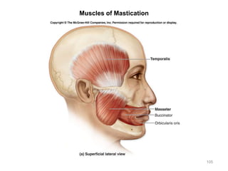
![Axial Muscles – Mastication (Chewing) Muscles [2]
• Temporalis
• Origin:
• Parietal bone
• Frontal bone
• Insertion:
• Coronoid process of mandible
• Action:
• Elevates & retracts mandible
• Textbook Reference:
• Description: p. 331
• Figures: 11.2b, 11.5
76](https://image.slidesharecdn.com/activity5-6-160526165849/85/Activity-5-6-76-320.jpg)
![Axial Muscles – Mastication (Chewing) Muscles [2]
• Masseter
• Origin:
• Zygomatic arch
• Insertion:
• Coronoid process, angle, &
ramus of mandible
• Action:
• Elevates & protracts mandible
• Jaw closure
• Textbook Reference:
• Description: p. 331
• Figures: 11.2a & b, 11.5
77](https://image.slidesharecdn.com/activity5-6-160526165849/85/Activity-5-6-77-320.jpg)
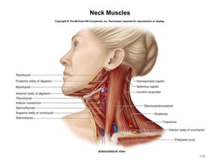
![Axial Muscles – Neck Muscles [3]
• Sternocleidomastoid
• Origin:
• Manubrium of sternum
• Sternal end of clavicle
• Insertion:
• Mastoid process of temporal
bone
• Action:
• One side: lateral flexion with
rotation of head to opposite
side
• Both sides: flexes head &
neck
• Textbook Reference:
• Description: p. 336
• Figures: 11.8, 11.9
78](https://image.slidesharecdn.com/activity5-6-160526165849/85/Activity-5-6-78-320.jpg)
![Axial Muscles – Neck Muscles [3]
• Splenius capitis
• Origin:
• Ligamentum nuchae
(connective tissue covering
the spinal processes of the
cervical vertebrae)
• Insertion:
• Occipital bone
• Mastoid process of temporal
bone
• Action:
• One side: turns head to same
side
• Both sides: extends head &
neck
• Textbook Reference:
• Description: p. 336
• Figures: 11.10, 11.11 79
Mnemonic:
PUT THE CAP ON TOP OF
CERVICIS](https://image.slidesharecdn.com/activity5-6-160526165849/85/Activity-5-6-79-320.jpg)
![Axial Muscles – Neck Muscles [3]
• Splenius cervicis
• Origin:
• Spinous processes of T3-T6
vertebrae
• Insertion:
• Transverse processes of
cervical vertebrae
• Action:
• One side: turns head to same
side
• Both sides: extends head &
neck
• Textbook Reference:
• Description: p. 336
• Figures: 11.10, 11.11
80
Mnemonic:
PUT THE CAP ON TOP OF
CERVICIS](https://image.slidesharecdn.com/activity5-6-160526165849/85/Activity-5-6-80-320.jpg)
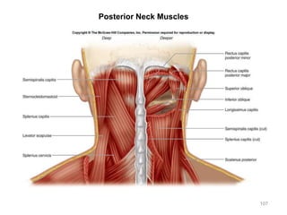
![Axial Muscles – Vertebral Column Muscles [3+1]
• Erector spinae groups
• Muscles
• Iliocostalis group (lateral)
• Longissimus group (middle)
• Spinalis group (medial)
• Action
• One side: laterally flexes the
vertebral column
• Both sides: extends vertebral
column
• Textbook Reference:
• Description: p. 339
• Figures: 11.11
81](https://image.slidesharecdn.com/activity5-6-160526165849/85/Activity-5-6-81-320.jpg)
![Axial Muscles – Vertebral Column Muscles [3+1]
• Quadratus lumborum
• Action:
• One side: laterally flexes the
vertebral column
• Both sides: extends vertebral
column
• Textbook Reference:
• Description: p. 339
• Figures: 11.11
82](https://image.slidesharecdn.com/activity5-6-160526165849/85/Activity-5-6-82-320.jpg)
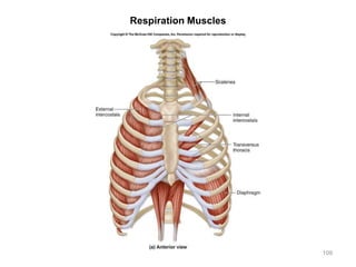
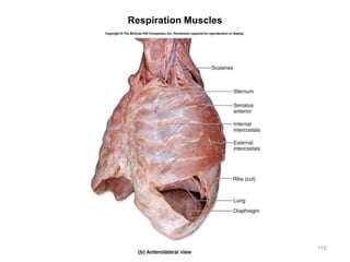
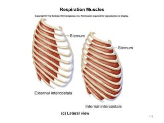
![Axial Muscles – Respiration Muscles [3]
• External intercostals
• Action:
• Elevates ribs during normal
inspiration (inhalation)
• Textbook Reference:
• Description: p. 342
• Figures: 11.11, 11.13
• Cadaver hint:
• Looking from bottom to top,
notice that external
intercostals point in a direction
away from the body (to the
shoulders)
83](https://image.slidesharecdn.com/activity5-6-160526165849/85/Activity-5-6-83-320.jpg)
![Axial Muscles – Respiration Muscles [3]
• Internal intercostals
• Action:
• Depresses ribs during forced
exhalation
• Textbook Reference:
• Description: p. 342
• Figures: 11.13
• Cadaver hint:
• Looking from bottom to top,
notice that internal intercostals
point in a direction into the
body (to the chin)
84](https://image.slidesharecdn.com/activity5-6-160526165849/85/Activity-5-6-84-320.jpg)
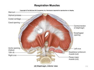
![Axial Muscles – Respiration Muscles [3]
• Diaphragm
• Action:
• Expands the thoracic cavity
during normal inspiration
• Textbook Reference:
• Description: p. 342
• Figures: 11.13
85](https://image.slidesharecdn.com/activity5-6-160526165849/85/Activity-5-6-85-320.jpg)
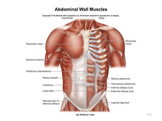
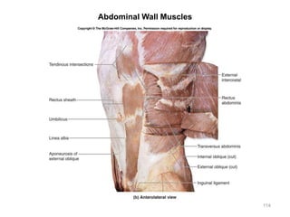
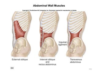
![Axial Muscles – Abdominal Wall Muscles [4+1]
• External oblique
• Action:
• Both sides: flexes vertebral
column & compresses
abdominal wall
• One side: laterally flexes
vertebral column
• Textbook Reference:
• Description: p. 344
• Figures: 11.14a & b
• Cadaver hint:
• Looking from bottom to top,
notice that external oblique
points in a direction away from
the body (to the shoulders)
86](https://image.slidesharecdn.com/activity5-6-160526165849/85/Activity-5-6-86-320.jpg)
![Axial Muscles – Abdominal Wall Muscles [4+1]
• Internal oblique
• Action:
• Both sides: flexes vertebral
column & compresses
abdominal wall
• One side: laterally flexes
vertebral column
• Textbook Reference:
• Description: p. 344
• Figures: 11.14a & b
• Cadaver hint:
• Looking from bottom to top,
notice that internal oblique
points in a direction into the
body (to the chin)
87](https://image.slidesharecdn.com/activity5-6-160526165849/85/Activity-5-6-87-320.jpg)
![Axial Muscles – Abdominal Wall Muscles [4+1]
• Transversus abdominis
• Action:
• Both sides: flexes vertebral
column & compresses
abdominal wall
• One side: laterally flexes
vertebral column
• Textbook Reference:
• Description: p. 344
• Figures: 11.14a & b
• Cadaver hint:
• Notice the horizontal direction
of this muscle’s fibers
88](https://image.slidesharecdn.com/activity5-6-160526165849/85/Activity-5-6-88-320.jpg)
![Axial Muscles – Abdominal Wall Muscles [4+1]
• Rectus abdominis
• Action:
• Flexes vertebral column &
compresses abdominal wall
• Textbook Reference:
• Description: p. 344
• Figures: 11.14a & b
• Trivia:
• This is the six-pack abs
muscle
89](https://image.slidesharecdn.com/activity5-6-160526165849/85/Activity-5-6-89-320.jpg)
![Axial Muscles – Abdominal Wall Muscles [4+1]
• Inguinal ligament
(associated structure)
• Significance:
• Formed by the aponeurosis of
the external oblique muscle
• Contains tissues coursing
from the trunk to the lower
limb
• Textbook Reference:
• Description: p. 344
• Figures: 11.14a & b
90](https://image.slidesharecdn.com/activity5-6-160526165849/85/Activity-5-6-90-320.jpg)


![Triangles of the_neck[1]](https://cdn.slidesharecdn.com/ss_thumbnails/trianglesoftheneck1-180125150132-thumbnail.jpg?width=640&height=640&fit=bounds)


![L9 muscles of upper limb [Autosaved].pptx](https://cdn.slidesharecdn.com/ss_thumbnails/l9musclesofupperlimbautosaved-230601011342-d18f2c9a-thumbnail.jpg?width=640&height=640&fit=bounds)


