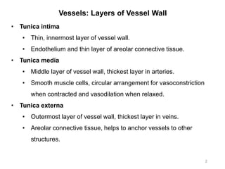
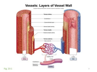
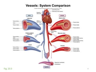
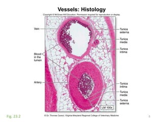
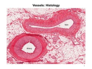
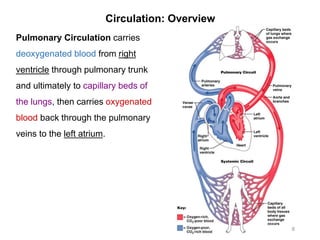
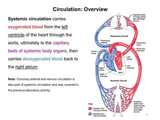
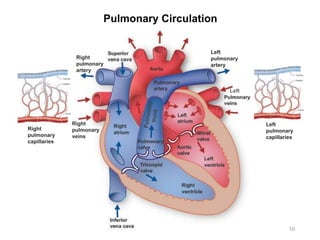
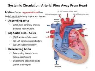
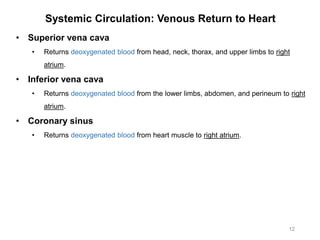
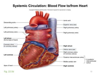
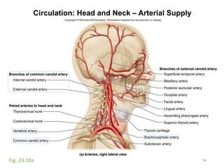
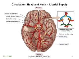
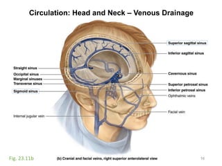
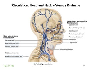
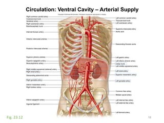
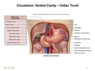
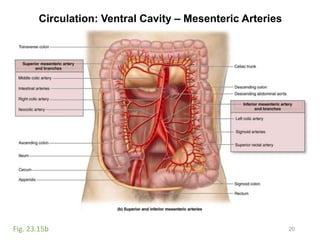
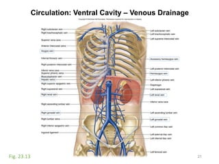
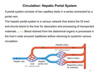
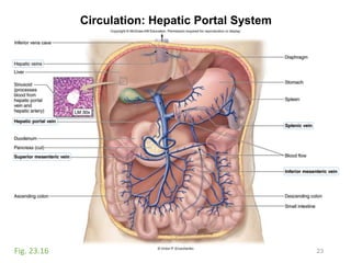
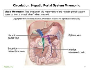
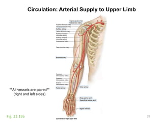
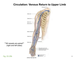
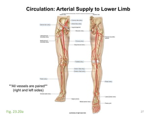
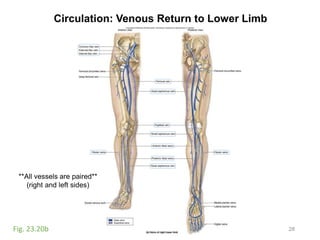
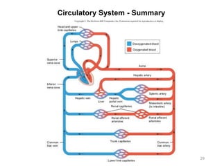
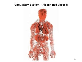
Activity #10:
Vessels and Circulation
Chapter 23 – McKinley et al., Human Anatomy, 4e.
Objectives:
• Observe and sketch histology slide of an artery and a vein
and identify structures on each.
• Identify structures and vessels involved in pulmonary
circulation on cadavers and classroom models.
• Identify structures and vessels involved in systemic circulation
(by region) on cadavers and classroom models.
1Compilation: Lisa Radmall
Vessels: Layers of Vessel Wall
2
• Tunica intima
• Thin, innermost layer of vessel wall.
• Endothelium and thin layer of areolar connective tissue.
• Tunica media
• Middle layer of vessel wall, thickest layer in arteries.
• Smooth muscle cells, circular arrangement for vasoconstriction
when contracted and vasodilation when relaxed.
• Tunica externa
• Outermost layer of vessel wall, thickest layer in veins.
• Areolar connective tissue, helps to anchor vessels to other
structures.
Vessels: Layers of Vessel Wall
3Fig. 23.1
Vessels: System Comparison
4Fig. 23.3
Vessels: Histology
5Fig. 23.2
Vessels: Histology
6
Vessels: Identification on Cadavers
7
• Arteries
• Round shape
• Collapse when ‘pinched’ (hollow lumen)
• Rebounds to round shape
• Veins
• Flat shape
• Usually collapsed
• Often dark in color
• Nerves
• Round shape
• Does not collapse when ‘pinched’ (contains nerve fibers)
Circulation: Overview
8
Pulmonary Circulation carries
deoxygenated blood from right
ventricle through pulmonary trunk
and ultimately to capillary beds of
the lungs, then carries oxygenated
blood back through the pulmonary
veins to the left atrium.
Circulation: Overview
9
Systemic circulation carries
oxygenated blood from the left
ventricle of the heart through the
aorta, ultimately to the capillary
beds of systemic body organs, then
carries deoxygenated blood back to
the right atrium
Note: Coronary arterial and venous circulation is
also part of systemic circulation and was covered in
the previous laboratory activity.
Pulmonary Circulation
10
Left
Right
pulmonary
veins
Right
pulmonary
artery
Left
pulmonary
artery
Left
pulmonary
capillaries
Right
pulmonary
capillaries
Systemic Circulation: Arterial Flow Away From Heart
11
Aorta – Carries oxygenated blood from
the left ventricle to body organs and tissues.
• Ascending aorta
• Left & right coronary arteries.
Supplies heart muscle.
• (A) Aortic arch - ABCs
• (B) Brachiocephalic trunk
• (C) Left common carotid artery
• (S) Left subclavian artery
• Descending Aorta
• Descending thoracic aorta
(above diaphragm)
• Descending abdominal aorta
(below diaphragm)
Aortic Arch
(B) Brachiocephalic trunk
(C) Left Common Carotid Artery
(S) Left Subclavian Artery
Systemic Circulation: Venous Return to Heart
12
• Superior vena cava
• Returns deoxygenated blood from head, neck, thorax, and upper limbs to right
atrium.
• Inferior vena cava
• Returns deoxygenated blood from the lower limbs, abdomen, and perineum to right
atrium.
• Coronary sinus
• Returns deoxygenated blood from heart muscle to right atrium.
Systemic Circulation: Blood Flow to/from Heart
13Fig. 22.5b
Circulation: Head and Neck – Arterial Supply
14Fig. 23.10a
Circulation: Head and Neck – Arterial Supply
15Fig. 23.11a
Circulation: Head and Neck – Venous Drainage
16Fig. 23.11b
Circulation: Head and Neck – Venous Drainage
17Fig. 23.10b
Circulation: Ventral Cavity – Arterial Supply
18Fig. 23.12
Circulation: Ventral Cavity – Celiac Trunk
19Fig. 23.15a
Circulation: Ventral Cavity – Mesenteric Arteries
20Fig. 23.15b
Circulation: Ventral Cavity – Venous Drainage
21Fig. 23.13
Circulation: Hepatic Portal System
22
A portal system consists of two capillary beds in a series connected by a
portal vein.
The hepatic portal system is a venous network that drains the GI tract
and shunts blood to the liver for absorption and processing of transported
materials. (p.703) Blood drained from the abdominal organs is processed in
the liver’s wide sinusoid capillaries before returning to systemic venous
circulation.
(1st capillary beds)
(2nd capillary bed)
Circulation: Hepatic Portal System
23Fig. 23.16
Circulation: Hepatic Portal System Mnemonic
24Table 23.3
Visual Mnemonic: The location of the main veins of the hepatic portal system
seem to form a visual “chair” when isolated.
Circulation: Arterial Supply to Upper Limb
25Fig. 23.19a
**All vessels are paired**
(right and left sides)
Circulation: Venous Return to Upper Limb
26Fig. 23.19b
**All vessels are paired**
(right and left sides)
Circulation: Arterial Supply to Lower Limb
27Fig. 23.20a
**All vessels are paired**
(right and left sides)
Circulation: Venous Return to Lower Limb
28Fig. 23.20b
**All vessels are paired**
(right and left sides)
Circulatory System - Summary
29
Circulatory System – Plastinated Vessels
30
Image References
31
Pulmonary Circulation: https://healthconspiracyblog.files.wordpress.com/2015/01/heart.jpg, www.ck12.org

Activity 10-vessels-circulation

  • 1.
    Activity #10: Vessels andCirculation Chapter 23 – McKinley et al., Human Anatomy, 4e. Objectives: • Observe and sketch histology slide of an artery and a vein and identify structures on each. • Identify structures and vessels involved in pulmonary circulation on cadavers and classroom models. • Identify structures and vessels involved in systemic circulation (by region) on cadavers and classroom models. 1Compilation: Lisa Radmall
  • 2.
    Vessels: Layers ofVessel Wall 2 • Tunica intima • Thin, innermost layer of vessel wall. • Endothelium and thin layer of areolar connective tissue. • Tunica media • Middle layer of vessel wall, thickest layer in arteries. • Smooth muscle cells, circular arrangement for vasoconstriction when contracted and vasodilation when relaxed. • Tunica externa • Outermost layer of vessel wall, thickest layer in veins. • Areolar connective tissue, helps to anchor vessels to other structures.
  • 3.
    Vessels: Layers ofVessel Wall 3Fig. 23.1
  • 4.
  • 5.
  • 6.
  • 7.
    Vessels: Identification onCadavers 7 • Arteries • Round shape • Collapse when ‘pinched’ (hollow lumen) • Rebounds to round shape • Veins • Flat shape • Usually collapsed • Often dark in color • Nerves • Round shape • Does not collapse when ‘pinched’ (contains nerve fibers)
  • 8.
    Circulation: Overview 8 Pulmonary Circulationcarries deoxygenated blood from right ventricle through pulmonary trunk and ultimately to capillary beds of the lungs, then carries oxygenated blood back through the pulmonary veins to the left atrium.
  • 9.
    Circulation: Overview 9 Systemic circulationcarries oxygenated blood from the left ventricle of the heart through the aorta, ultimately to the capillary beds of systemic body organs, then carries deoxygenated blood back to the right atrium Note: Coronary arterial and venous circulation is also part of systemic circulation and was covered in the previous laboratory activity.
  • 10.
  • 11.
    Systemic Circulation: ArterialFlow Away From Heart 11 Aorta – Carries oxygenated blood from the left ventricle to body organs and tissues. • Ascending aorta • Left & right coronary arteries. Supplies heart muscle. • (A) Aortic arch - ABCs • (B) Brachiocephalic trunk • (C) Left common carotid artery • (S) Left subclavian artery • Descending Aorta • Descending thoracic aorta (above diaphragm) • Descending abdominal aorta (below diaphragm) Aortic Arch (B) Brachiocephalic trunk (C) Left Common Carotid Artery (S) Left Subclavian Artery
  • 12.
    Systemic Circulation: VenousReturn to Heart 12 • Superior vena cava • Returns deoxygenated blood from head, neck, thorax, and upper limbs to right atrium. • Inferior vena cava • Returns deoxygenated blood from the lower limbs, abdomen, and perineum to right atrium. • Coronary sinus • Returns deoxygenated blood from heart muscle to right atrium.
  • 13.
    Systemic Circulation: BloodFlow to/from Heart 13Fig. 22.5b
  • 14.
    Circulation: Head andNeck – Arterial Supply 14Fig. 23.10a
  • 15.
    Circulation: Head andNeck – Arterial Supply 15Fig. 23.11a
  • 16.
    Circulation: Head andNeck – Venous Drainage 16Fig. 23.11b
  • 17.
    Circulation: Head andNeck – Venous Drainage 17Fig. 23.10b
  • 18.
    Circulation: Ventral Cavity– Arterial Supply 18Fig. 23.12
  • 19.
    Circulation: Ventral Cavity– Celiac Trunk 19Fig. 23.15a
  • 20.
    Circulation: Ventral Cavity– Mesenteric Arteries 20Fig. 23.15b
  • 21.
    Circulation: Ventral Cavity– Venous Drainage 21Fig. 23.13
  • 22.
    Circulation: Hepatic PortalSystem 22 A portal system consists of two capillary beds in a series connected by a portal vein. The hepatic portal system is a venous network that drains the GI tract and shunts blood to the liver for absorption and processing of transported materials. (p.703) Blood drained from the abdominal organs is processed in the liver’s wide sinusoid capillaries before returning to systemic venous circulation. (1st capillary beds) (2nd capillary bed)
  • 23.
    Circulation: Hepatic PortalSystem 23Fig. 23.16
  • 24.
    Circulation: Hepatic PortalSystem Mnemonic 24Table 23.3 Visual Mnemonic: The location of the main veins of the hepatic portal system seem to form a visual “chair” when isolated.
  • 25.
    Circulation: Arterial Supplyto Upper Limb 25Fig. 23.19a **All vessels are paired** (right and left sides)
  • 26.
    Circulation: Venous Returnto Upper Limb 26Fig. 23.19b **All vessels are paired** (right and left sides)
  • 27.
    Circulation: Arterial Supplyto Lower Limb 27Fig. 23.20a **All vessels are paired** (right and left sides)
  • 28.
    Circulation: Venous Returnto Lower Limb 28Fig. 23.20b **All vessels are paired** (right and left sides)
  • 29.
  • 30.
    Circulatory System –Plastinated Vessels 30
  • 31.
    Image References 31 Pulmonary Circulation:https://healthconspiracyblog.files.wordpress.com/2015/01/heart.jpg, www.ck12.org