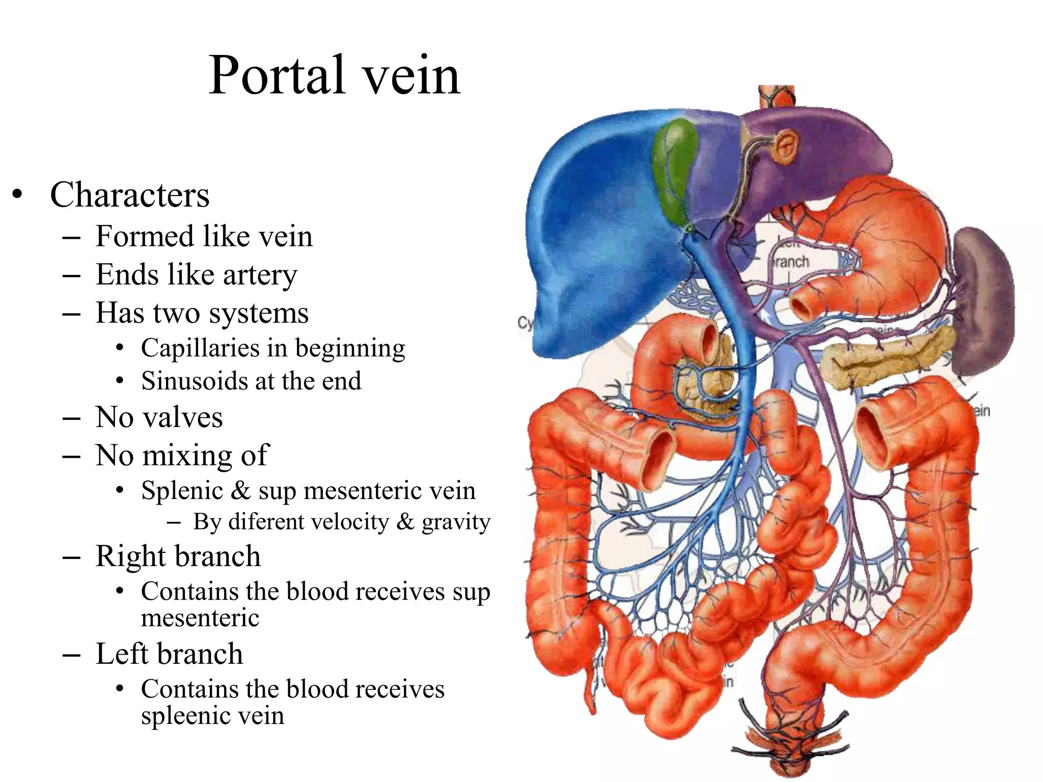
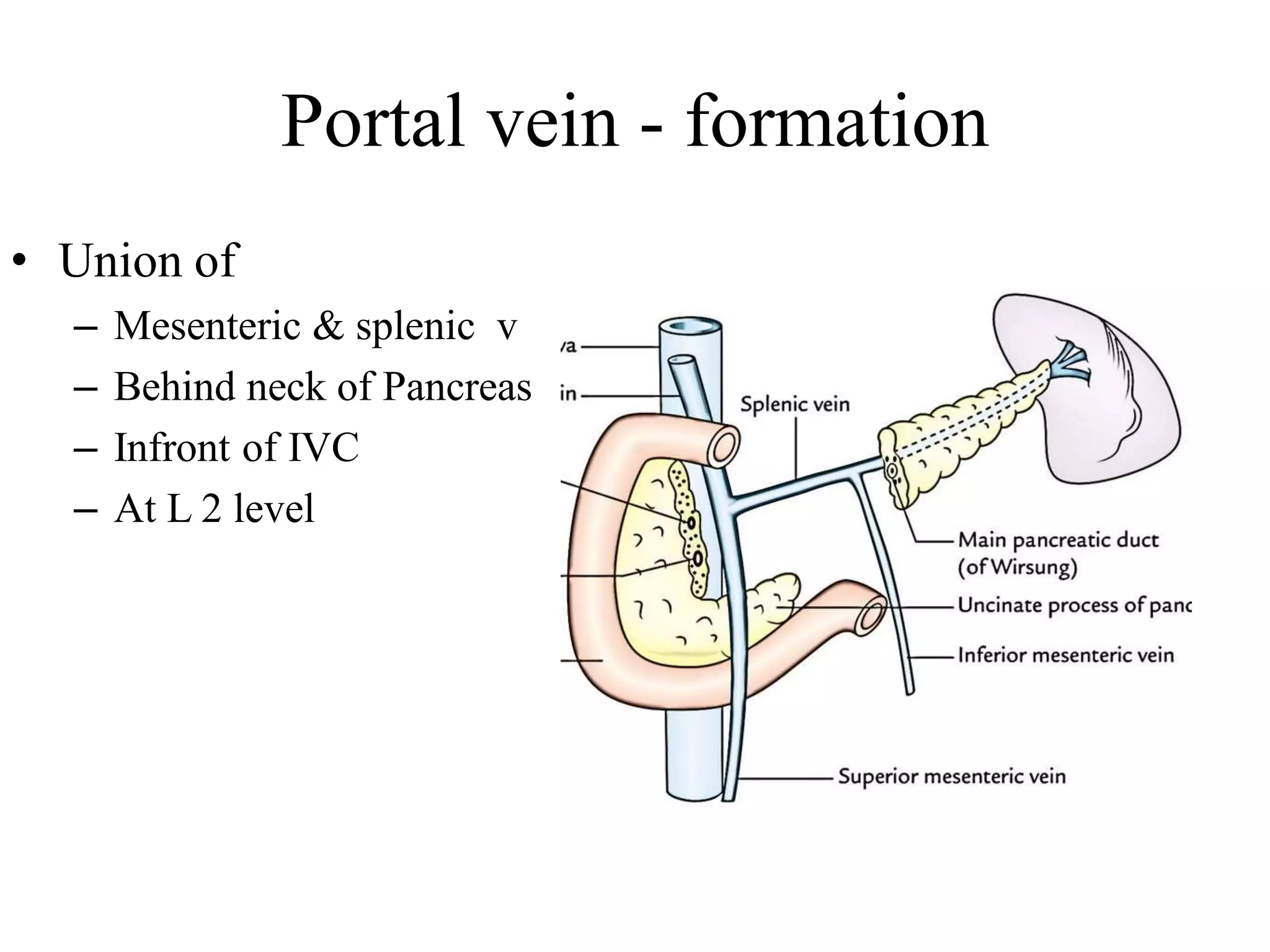
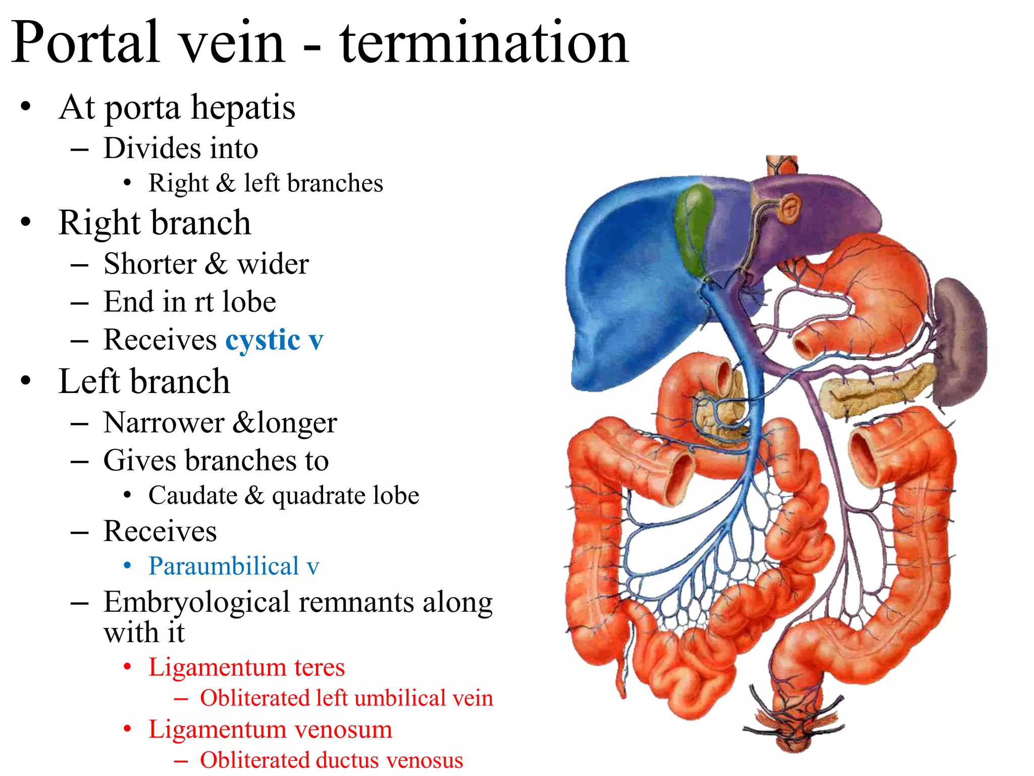
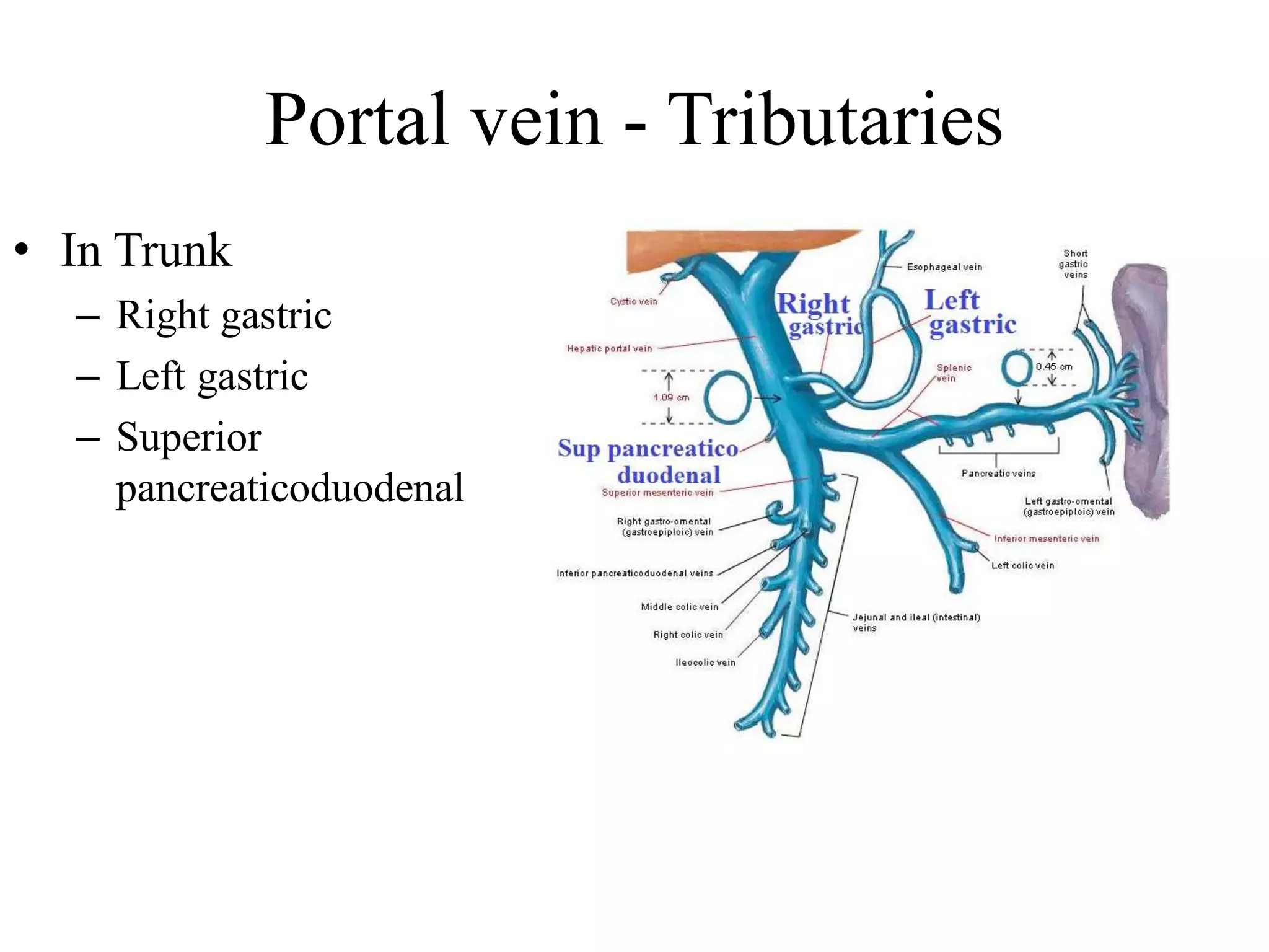
Spleen & Portal vein
Transpyloric plane (L1 vertebra)
passes through
Spleen
• Haemolymph organ
• Womb & Tomb of RBC
– Formation of RBC
• During intra-uterine life
(till the 5th month)
– Destruction of old RBC
• Made up of
– Lymphoid tissue
• Reservoir of blood and
iron
Spleen - Location
• Lies between
– Fundus of stomach
and diaphragm
• Lies in
– Left hypochondrium
• Extends in between
– 9th to 11th ribs
Measurements
• 1- inch Thickness
• 3 - inch Breadth
• 5 – inch Length
• 7 oz – Weight (150 gm)
• 9-11 ribs related – (left
side)
Harris dictum of odd numbers on Spleen :1,3,5,7,9 -11
External features
• Two ends
• Three borders
• Two surfaces
• Hilum
Ends
• Anterior or lateral end
– Expanded or more like a
border
– Directed downwards,
forwards and reaches mid
axillary line
• Posterior or medial end
– Rounded
– Directed upwards
backwards, medially
– Rest on upper pole of left
kidney
Borders
• Superior border
– Presents one or two notches
near anterior end
– Notches indicate spleen is
lobulated in development
• (fusion of different masses of
lymphoid tissue)
• Inferior border
– Rounded
• Intermediate border
– Thick, incomplete
– Extends from medial end till
hilum
Surfaces
• Diaphragmatic surface
– Convex and smooth
– Related to
• Diaphragm
• Lt lung & lts pleura
• Costodiaphragmatic recess
• 9th, 10th & 11th ribs of left
side
VISCERAL SURFACE
• Concave, irregular, directed
to abdominal cavity
• Contains
– Hilum and impressions for 4
abdominal organs
• Gastric impression
– Related to posterior wall of
fundus of stomach
• Renal impression
• Colic impression (left colic
flexure)
• Pancreatic impression (tail
of pancreas)
Peritoneum
• Covers entire spleen
– Except the hilum
• Ligaments related
– Gastro-splenic lig.
– Lieno-renal lig.
– Phrenico-colic
Peritoneal connections
& contents
• Gastrosplenic ligament
– Connections
• From fundus and greater curvature of
stomach
• To hilum of spleen
– Contents
• Short gastric vessels
• Left gastroepiploic vessels
• Lymph nodes
• Lienorenal ligament
– Connections
• From lower border of hilum of
spleen
• To anterior surface of kidney
– Contents
• Splenic vessels
• Tail of pancreas
• Lymph nodes
Applied importance of Gastro-splenic and lieno-renal ligament
• Accessory spleens
– Common in 10-15% of
people
– Found at the hilum of spleen
– Lieno-renal or the gastro-
splenic ligaments
• Tail of pancreas is in close
relation to hilum of spleen
– During surgical removal of
the spleen care must be
taken
Peritoneal
connections &
contents
• Phrenico-colic
– Sustentaculum lienis
– Connections
• From left colic flexure to
diaphragm
• Lieno phrenic
– Suspensory ligament of
spleen
– Connections
• Apex of Spleen to
diaphragm
Arterial Supply
• Splenic artery
– Branch of Coeliac artery
• Very tortuous
• End artery
– Course
• Along the upper border of
pancreas
• Crosses lt. supra renal &
upper part of kidney
• Enters lienorenal ligament
• Reaches hilum by
dividing into 4-5
segmental arteries
Splenomegaly
• Enlargement of spleen
– Causes
• Cirrhosis
• Malaria
• Myeloid leukaemia
– Normally not palpable
– Enlarged spleen is
palpable
• Under left costal margin
– During inspiration
Clinical angle of spleen
• Anterior basal angle
– Junction of superior border
with lateral end
– Lies in 9th intercostal space
behind left mid-axillary line
– First palpable in
splenomegaly
Portal vein
• Collects blood
– Entire abdominal part of GI
tract
• Except lower rectum & anal
canal
– And also collects venous
blood from
• Gall bladder, pancreas &
spleen
Portal vein
• Characters
– Formed like vein
– Ends like artery
– Has two systems
• Capillaries in beginning
• Sinusoids at the end
– No valves
– No mixing of
• Splenic & sup mesenteric vein
– By diferent velocity & gravity
– Right branch
• Contains the blood receives sup
mesenteric
– Left branch
• Contains the blood receives
spleenic vein
Portal vein - formation
• Union of
– Mesenteric & splenic v
– Behind neck of Pancreas
– Infront of IVC
– At L 2 level
Portal vein - course
• Divided into 3 parts
– Infra duodenal
– Retroduodenal
– Supraduodenal
Portal vein - Infra duodenal part
• Lies below
– First part of duodenum
• Relations
– Anterior
• Neck of pancreas
– Posterior
• IVC
Portal vein - Retro duodenal part
• Lies behind
– First part of duodenum
• Relations
– Anterior
• First part of duodenum
• Bile duct
• Gastroduodenal artery
– Posterior
• IVC
Portal vein - Supra duodenal part
• Lies above
– The first part of duodenum
– Lies in free margin of
lesser omentum
• Relations
– Anterior
• Hepatic artery
• Bile duct
– Posterior
• IVC
Portal vein - termination
• At porta hepatis
– Divides into
• Right & left branches
• Right branch
– Shorter & wider
– End in rt lobe
– Receives cystic v
• Left branch
– Narrower &longer
– Gives branches to
• Caudate & quadrate lobe
– Receives
• Paraumbilical v
– Embryological remnants along
with it
• Ligamentum teres
– Obliterated left umbilical vein
• Ligamentum venosum
– Obliterated ductus venosus
Portal vein - Tributaries
• Formative
– Splenic
– Superior mesenteric
Portal vein - Tributaries
• In Trunk
– Right gastric
– Left gastric
– Superior
pancreaticoduodenal
Portal vein - Tributaries
• In branches
– In Right branch
• Cystic
– In left branch
• Paraumbilical
Portal hypertension
• Obstruction in portal vein
– Increase portal venous pressure
• Causes :
– Pre hepatic : Thrombosis of
portal vein
– Hepatic: Cirrhosis
– Post hepatic: Budd-Chiari
Syndrome
• Effects:
– Splenomegaly,
– Ascites,
– Collateral circulation through sites
of Portasystemic anastomosis
Budd-Chiari
Syndrome
- hepatic veins are
blocked by a clot,
usually left lobe is
affected.
Sites of porto-caval anastomosis
• Five important sites of
portal -systemic
circulation
– Lower third of the
Esophagus
– Paraumbilical Area
– Rectum & Anal canal
– Bare area of liver
– Retroperitonial
Caput medusae
• Veins around umbilicus is
dilated & distended
– Due to portal obstruction
• Veins involved
– From portal system
• Paraumbilical veins
– Systemic veins
• From above umbilicus
– Lateral thoracic & superior
epigastric
• From below umbilicus
– Superficial epigastric, &
inferior epigastric
Oesophageal varices
• Dilatation of veins at
lower end of oesophagus
– Due to portal obstruction
– May rupture & cause
vomiting of blood
• Veins involved
– From portal system
• Oesophageal vein to left
gastric vein
– Systemic vein
• Oesophageal vein to
hemiazygos vein
Piles or haemorrhoids
• Enlargement of veins of
anal canal
– Due to portal obstruction
• Veins involved
– From portal system
• Superior rectal
– Systemic vein
• Middle & inferior rectal
Bare area of liver
• Veins involved
– From portal system
• Hepatic venules
– Right division of the portal vein
– Systemic vein
• Phrenic & intercostal veins
•
Retroperitoneal
organs
• Systemic vein
– Lt renal V
• Portal vein
– Splenic V

Spleen & portal vein

  • 1.
  • 2.
    Transpyloric plane (L1vertebra) passes through
  • 3.
    Spleen • Haemolymph organ •Womb & Tomb of RBC – Formation of RBC • During intra-uterine life (till the 5th month) – Destruction of old RBC • Made up of – Lymphoid tissue • Reservoir of blood and iron
  • 4.
    Spleen - Location •Lies between – Fundus of stomach and diaphragm • Lies in – Left hypochondrium • Extends in between – 9th to 11th ribs
  • 5.
    Measurements • 1- inchThickness • 3 - inch Breadth • 5 – inch Length • 7 oz – Weight (150 gm) • 9-11 ribs related – (left side) Harris dictum of odd numbers on Spleen :1,3,5,7,9 -11
  • 6.
    External features • Twoends • Three borders • Two surfaces • Hilum
  • 7.
    Ends • Anterior orlateral end – Expanded or more like a border – Directed downwards, forwards and reaches mid axillary line • Posterior or medial end – Rounded – Directed upwards backwards, medially – Rest on upper pole of left kidney
  • 8.
    Borders • Superior border –Presents one or two notches near anterior end – Notches indicate spleen is lobulated in development • (fusion of different masses of lymphoid tissue) • Inferior border – Rounded • Intermediate border – Thick, incomplete – Extends from medial end till hilum
  • 9.
    Surfaces • Diaphragmatic surface –Convex and smooth – Related to • Diaphragm • Lt lung & lts pleura • Costodiaphragmatic recess • 9th, 10th & 11th ribs of left side
  • 10.
    VISCERAL SURFACE • Concave,irregular, directed to abdominal cavity • Contains – Hilum and impressions for 4 abdominal organs • Gastric impression – Related to posterior wall of fundus of stomach • Renal impression • Colic impression (left colic flexure) • Pancreatic impression (tail of pancreas)
  • 11.
    Peritoneum • Covers entirespleen – Except the hilum • Ligaments related – Gastro-splenic lig. – Lieno-renal lig. – Phrenico-colic
  • 12.
    Peritoneal connections & contents •Gastrosplenic ligament – Connections • From fundus and greater curvature of stomach • To hilum of spleen – Contents • Short gastric vessels • Left gastroepiploic vessels • Lymph nodes • Lienorenal ligament – Connections • From lower border of hilum of spleen • To anterior surface of kidney – Contents • Splenic vessels • Tail of pancreas • Lymph nodes
  • 13.
    Applied importance ofGastro-splenic and lieno-renal ligament • Accessory spleens – Common in 10-15% of people – Found at the hilum of spleen – Lieno-renal or the gastro- splenic ligaments • Tail of pancreas is in close relation to hilum of spleen – During surgical removal of the spleen care must be taken
  • 14.
    Peritoneal connections & contents • Phrenico-colic –Sustentaculum lienis – Connections • From left colic flexure to diaphragm • Lieno phrenic – Suspensory ligament of spleen – Connections • Apex of Spleen to diaphragm
  • 15.
    Arterial Supply • Splenicartery – Branch of Coeliac artery • Very tortuous • End artery – Course • Along the upper border of pancreas • Crosses lt. supra renal & upper part of kidney • Enters lienorenal ligament • Reaches hilum by dividing into 4-5 segmental arteries
  • 16.
    Splenomegaly • Enlargement ofspleen – Causes • Cirrhosis • Malaria • Myeloid leukaemia – Normally not palpable – Enlarged spleen is palpable • Under left costal margin – During inspiration
  • 17.
    Clinical angle ofspleen • Anterior basal angle – Junction of superior border with lateral end – Lies in 9th intercostal space behind left mid-axillary line – First palpable in splenomegaly
  • 18.
    Portal vein • Collectsblood – Entire abdominal part of GI tract • Except lower rectum & anal canal – And also collects venous blood from • Gall bladder, pancreas & spleen
  • 19.
    Portal vein • Characters –Formed like vein – Ends like artery – Has two systems • Capillaries in beginning • Sinusoids at the end – No valves – No mixing of • Splenic & sup mesenteric vein – By diferent velocity & gravity – Right branch • Contains the blood receives sup mesenteric – Left branch • Contains the blood receives spleenic vein
  • 20.
    Portal vein -formation • Union of – Mesenteric & splenic v – Behind neck of Pancreas – Infront of IVC – At L 2 level
  • 21.
    Portal vein -course • Divided into 3 parts – Infra duodenal – Retroduodenal – Supraduodenal
  • 22.
    Portal vein -Infra duodenal part • Lies below – First part of duodenum • Relations – Anterior • Neck of pancreas – Posterior • IVC
  • 23.
    Portal vein -Retro duodenal part • Lies behind – First part of duodenum • Relations – Anterior • First part of duodenum • Bile duct • Gastroduodenal artery – Posterior • IVC
  • 24.
    Portal vein -Supra duodenal part • Lies above – The first part of duodenum – Lies in free margin of lesser omentum • Relations – Anterior • Hepatic artery • Bile duct – Posterior • IVC
  • 25.
    Portal vein -termination • At porta hepatis – Divides into • Right & left branches • Right branch – Shorter & wider – End in rt lobe – Receives cystic v • Left branch – Narrower &longer – Gives branches to • Caudate & quadrate lobe – Receives • Paraumbilical v – Embryological remnants along with it • Ligamentum teres – Obliterated left umbilical vein • Ligamentum venosum – Obliterated ductus venosus
  • 26.
    Portal vein -Tributaries • Formative – Splenic – Superior mesenteric
  • 27.
    Portal vein -Tributaries • In Trunk – Right gastric – Left gastric – Superior pancreaticoduodenal
  • 28.
    Portal vein -Tributaries • In branches – In Right branch • Cystic – In left branch • Paraumbilical
  • 29.
    Portal hypertension • Obstructionin portal vein – Increase portal venous pressure • Causes : – Pre hepatic : Thrombosis of portal vein – Hepatic: Cirrhosis – Post hepatic: Budd-Chiari Syndrome • Effects: – Splenomegaly, – Ascites, – Collateral circulation through sites of Portasystemic anastomosis Budd-Chiari Syndrome - hepatic veins are blocked by a clot, usually left lobe is affected.
  • 30.
    Sites of porto-cavalanastomosis • Five important sites of portal -systemic circulation – Lower third of the Esophagus – Paraumbilical Area – Rectum & Anal canal – Bare area of liver – Retroperitonial
  • 31.
    Caput medusae • Veinsaround umbilicus is dilated & distended – Due to portal obstruction • Veins involved – From portal system • Paraumbilical veins – Systemic veins • From above umbilicus – Lateral thoracic & superior epigastric • From below umbilicus – Superficial epigastric, & inferior epigastric
  • 32.
    Oesophageal varices • Dilatationof veins at lower end of oesophagus – Due to portal obstruction – May rupture & cause vomiting of blood • Veins involved – From portal system • Oesophageal vein to left gastric vein – Systemic vein • Oesophageal vein to hemiazygos vein
  • 33.
    Piles or haemorrhoids •Enlargement of veins of anal canal – Due to portal obstruction • Veins involved – From portal system • Superior rectal – Systemic vein • Middle & inferior rectal
  • 34.
    Bare area ofliver • Veins involved – From portal system • Hepatic venules – Right division of the portal vein – Systemic vein • Phrenic & intercostal veins •
  • 35.
    Retroperitoneal organs • Systemic vein –Lt renal V • Portal vein – Splenic V