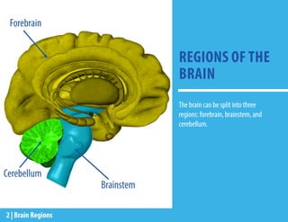
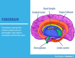
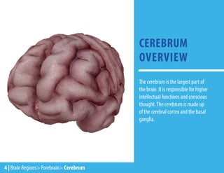
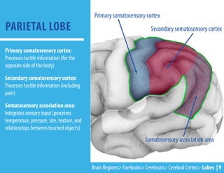
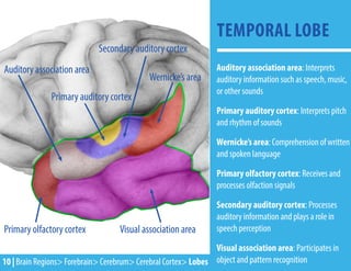
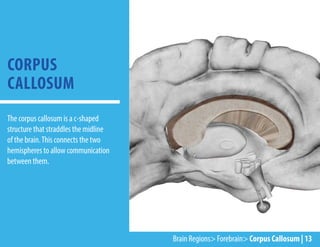
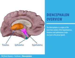
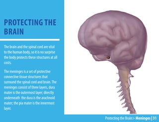
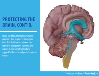
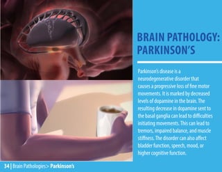
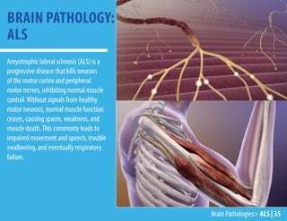
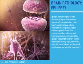
ByTheVisibleBodyTeam&ProfessorBlytheNilson
Located inside the skull, the brain is an organ of the
central nervous system that receives sensory input
and information to form perception and thought. It
controls activities such as speech, movement, and
maintaining homeostasis.
THE BIG
BRAIN
EBOOK
REGIONS OF THE
BRAIN
The brain can be split into three
regions: forebrain, brainstem, and
cerebellum.
Forebrain
Brainstem
Cerebellum
2 | Brain Regions
Cerebral Cortex
Diencephalon Limbic system
Corpus Callosum
Basal Ganglia
FOREBRAIN
Brain Regions>Forebrain | 3
The forebrain is made up of the
cerebrum (cerebral cortex and
basal ganglia), corpus callosum,
diencephalon, and the limbic system.
CEREBRUM
OVERVIEW
4 | Brain Regions> Forebrain> Cerebrum
The cerebrum is the largest part of
the brain. It is responsible for higher
intellectual functions and conscious
thought.The cerebrum is made up
of the cerebral cortex and the basal
ganglia.
The cerebral cortex is the surface of the
cerebrum.The cortex contains around 16
billion neurons (cells that relay sensory
information and motor commands as
electric signals throughout the body). In
order for the cerebral cortex to fit inside
the skull, the cerebral cortex has folds
called gyri and grooves called sulci.
CEREBRAL
CORTEX OVERVIEW
sulcus
gyrus
Neurondiagram
Brain Regions> Forebrain> Cerebrum> Cerebral Cortex | 5
The cerebral cortex consists of a left
and a right hemisphere.The left is
typically responsible for language,
calculation, comprehension, and
writing.The right is generally
responsible for creativity, visual,
emotional, and artistic awareness.
Each hemisphere controls the opposite
side of the body.
CEREBRAL CORTEX
HEMISPHERES
Left hemisphere Right hemisphere
6 | Brain Regions> Forebrain> Cerebrum> Cerebral Cortex
Each hemisphere has four lobes:
frontal, parietal, temporal,
and occipital lobe. Each lobe is
responsible for different brain
functions.
LOBES OF THE
CEREBRAL CORTEX
Frontal Lobe Parietal Lobe
Temporal Lobe Occipital Lobe
Brain Regions> Forebrain> Cerebrum> Cerebral Cortex> Lobes | 7
FRONTAL LOBE
Prefrontal association area: Executive
function (thought, cognition, planning,
and personality)
Premotor cortex: Planning movement
Supplementary motor area: Planning
limb and eye movement
Broca’s area: Speech production and
language processing
Gustatory cortex: Receives and
processes taste signals
Primary motor area:Voluntary
movement control
Prefrontal association area
Supplementary motor area (SMA)
Gustatory cortex
Primary motor area
Broca’s area
Premotor cortex
8 | Brain Regions> Forebrain> Cerebrum> Cerebral Cortex> Lobes
Primary somatosensory cortex:
Processes tactile information (for the
opposite side of the body)
Secondary somatosensory cortex:
Processes tactile information (including
pain)
Somatosensory association area:
Integrates sensory input (perceives
temperature, pressure, size, texture, and
relationships between touched objects)
PARIETAL LOBE
Primary somatosensory cortex
Secondary somatosensory cortex
Somatosensory association area
Brain Regions> Forebrain> Cerebrum> Cerebral Cortex> Lobes | 9
Auditory association area: Interprets
auditory information such as speech, music,
or other sounds
Primary auditory cortex: Interprets pitch
and rhythm of sounds
Wernicke’s area: Comprehension of written
and spoken language
Primary olfactory cortex: Receives and
processes olfaction signals
Secondary auditory cortex: Processes
auditory information and plays a role in
speech perception
Visual association area: Participates in
object and pattern recognition
TEMPORAL LOBE
Wernicke’s area
Visual association area
Primary olfactory cortex
Auditory association area
Secondary auditory cortex
Primary auditory cortex
10 | Brain Regions> Forebrain> Cerebrum> Cerebral Cortex> Lobes
Primary visual cortex: Receives
and processes visual signals from the
opposite visual field via the thalamus
Secondary visual cortex: Interprets
visual signals and contributes to visual
mapping
Tertiary visual cortex: Participates in
object recognition and motion sensing
OCCIPITAL LOBE
Tertiary
visual
cortex
Secondary visual cortex
Primary
visual
cortex
Brain Regions>Forebrain>Cerebrum>Cerebral Cortex> Lobes | 11
12 | Brain Regions> Forebrain> Cerebrum> Basal Ganglia
The basal ganglia contain nuclei inside
the cerebrum that influence body
movement and muscle tone.
BASAL GANGLIA
Brain Regions> Forebrain> Corpus Callosum | 13
The corpus callosum is a c-shaped
structure that straddles the midline
of the brain.This connects the two
hemispheres to allow communication
between them.
CORPUS
CALLOSUM
Hypothalamus
Thalamus Epithalamus
The diencephalon is a region of the
brain that consists of the hypothalamus,
thalamus and epithalamus (main
structure is the pineal gland).
DIENCEPHALON
OVERVIEW
14 | Brain Regions> Forebrain> Diencephalon
The hypothalamus regulates autonomic
nervous functions, emotions, behavior,
food consumption, circadian rhythms,
and body temperature. It also produces
hormones that influence the pituitary
gland and regulate endocrine functions.
At the base of the hypothalamus, the
optic chiasm is the crossing point of the
optic nerves.
HYPOTHALAMUS
Brain Regions> Forebrain> Diencephalon> Hypothalamus | 15
The thalamus plays a role in learning
and memory. It consists of several
nuclei that pass sensory information to
different regions of the cerebral cortex.
The thalamus also determines what
sensory information is important enough
to send to the cerebral cortex to allow
for conscious awareness of the sensory
input.
THALAMUS
16 | Brain Regions> Forebrain> Diencephalon> Thalamus
PITUITARY
Hormones secreted by the
hypothalamus activate hormone
secretion by the pituitary gland.
These hormones drive endocrine
functions (like sexual development
and promotion of bone and muscle
growth) by targeting glands and
tissues.
Brain Regions> Forebrain> Diencephalon> Pituitary | 17
The limbic system is located on the
inner border of the cerebrum, above the
diencephalon, forming a pair of rings
between the two cerebral hemispheres.
It manages a range of emotions and
contributes to the processing of smell
memory.
Amygdala = memory and emotion
Hippocampus = long term memory
Mammillary body = smell memory
Septal nucleus = feeling of pleasure
Cingulate gyrus = expressing emotions
through gestures
LIMBIC SYSTEM
18 | Brain Regions> Forebrain> Limbic System
Amygdala
Hippocampus
Mammillary body
Septal nucleus
Cingulate gyrus
Midbrain
Medulla oblongata
Pons
The brainstem (consisting of the
midbrain, pons, and medulla oblongata)
connects the brain to the spinal cord.
The brainstem controls the involuntary
functions of the respiratory, digestive,
and circulatory systems, while also
contributing to hearing, balance, and
gustation.
BRAINSTEM
Brain Regions> Brainstem | 19
The midbrain contains nuclei, called
colliculi, which control visual and auditory
reflexes. It also contains nuclei for the
oculomotor (CN03) and trochlear (CN04)
nerves. Both help control eye movement.
MIDBRAIN
20 | Brain Regions> Brain Stem> Midbrain
The pons connects the brain to the brain
stem through tracts of white fibers that
carry signals between them. It houses
pontine nuclei, which affect breathing
rhythms.
PONS
Brain Regions> Brain Stem> Pons | 21
The medulla oblongata controls
involuntary functions of the respiratory,
digestive, and circulatory system
and contributes to hearing, balance,
and taste.The medulla plays a role in
homeostatis, acting as the autonomic
reflex center for heart contraction,
vasomotor control, respiratory rate and
depth, as well as other reflexes such as
swallowing, coughing, vomiting, and
sneezing.
MEDULLA
OBLONGATA
22 | Brain Regions> Brain Stem> Medulla Oblongata
CEREBELLUM
The cerebellum is mostly considered to
be a motor structure. Even though motor
commands do not directly come from the
cerebellum, it modifies the commands
to make the movements more accurate.
This is why cerebellar damage leads
to impairment in motor control. Other
functions of the cerebellum include:
maintaining balance and posture,
coordination of voluntary movements,
and motor learning, as well as some
cognitive functions related to word
association and puzzle solving.
Brain Regions> Cerebellum | 23
24 | Nervous System> Overview
The brain is connected with the rest of
the body via the nervous system.The
nervous system is responsible for the
correlation and integration of various
bodily processes, producing reactions
and adjustments based on the current
environment.The nervous system can
be split into two different regions: the
central nervous system (CNS) and the
peripheral nervous system (PNS).
PARTS OF THE
NERVOUS SYSTEM
Peripheral nervous
system
Central nervous
system
Brain
Spinal cord
The central nervous system (CNS)
consists of the brain and the spinal cord.
The average length of a male spinal cord
is 45 cm and for females, it is around 43
cm.The spinal cord carries incoming and
outgoing messages between the brain
and the rest of the body.
CENTRAL NERVOUS
SYSTEM
Nervous System> Central Nervous System | 25
26 | Nervous System> Peripheral Nervous System
The peripheral nervous system (PNS)
consists of all the nerves and ganglia
outside of the central nervous system
connecting it to all the regions of the body.
The PNS can be split into two different
divisions: the somatic nervous system
(SNS) and the autonomic nervous system
(ANS).
The somatic nervous system - controls all
voluntary movement plus somatic reflexes
The autonomic nervous system - regulates
involuntary movement such as heartbeat
and smooth muscle contraction.
PERIPHERAL
NERVOUS SYSTEM
CRANIAL NERVES
Nervous System> PNS> Cranial Nerves | 27
The cranial nerves are a group of 12
paired nerves in the peripheral nervous
system that connect the muscles and
organs of the head and some organs
outside the head directly to the brain.
CN01: olfactory = smell
CN02: optic = sight
CN03: oculomotor = eye and pupil
movement
CN04: trochlear = eye movement
CN06: abducens = eye movement
CN08: vestibulocochlear = hearing,
balance
CN01
CN02
CN03
CN08
CN06
CN04
28 | Nervous System> PNS> Cranial Nerves
CRANIAL NERVES,
CONT’D.
CN07
CN11
CN09
CN12
CN05
CN05: trigeminal = face sensation
CN07: facial = face movement, salivation
CN09: glossopharyngeal = taste,
swallowing
CN10: vagus = heart rate, digestion
CN11: accessory = head movement
CN12: hypoglossal = tongue movement
CN10
Blood in the Brain> Arteries | 29
The arteries deliver oxygenated blood
from the heart to all the regions of the
body.The left and right internal carotid
and vertebral arteries supply the brain.
The blood is distributed throughout the
brain via the numerous branches of the
circle ofWillis.
BLOOD SUPPLY TO
THE BRAIN
Circle ofWillis
Internal carotid
Vertebral arteries
30 | Blood in the Brain> Veins
BLOOD SUPPLY TO
THE BRAIN
Veins return deoxygenated blood to the
heart. The largest vein in the brain is
the venous sinus, located between the
two hemispheres of the brain. It receives
blood from the left and right superficial
cerebral veins of the cerebrum. The
blood is then drained via the left and
right internal jugular veins
Venous sinus Superficial cerebral veins
Jugular veins
The brain and the spinal cord are vital
to the human body, so it is no surprise
the body protects these structures at all
costs.
The meninges is a set of protective
connective tissue structures that
surround the spinal cord and brain.The
meninges consist of three layers, dura
mater is the outermost layer; directly
underneath the dura is the arachnoid
mater; the pia mater is the innermost
layer.
PROTECTING THE
BRAIN
Protecting the Brain> Meninges | 31
Falx cerebri
Tentorium cerebelli
32 | Protecting the Brain> Meninges> Cranial Dural Septa
PROTECTING THE
BRAIN, CONT’D.
Within the brain there are double layers
of dura mater, called cranial dural septa,
that separate and support the brain.
Two of these, the falx cerebri and the
tentorium cerebelli, are highlighted in
the picture.
Protecting the Brain> Ventricles | 33
Inside the brain, there are networked
ventricles that produce cerebrospinal
fluid.This fluid moves between the
ventricles transporting nutrients and
wastes. It also provides structural
support to the brain and protects against
trauma.
PROTECTING THE
BRAIN, CONT’D.
Parkinson’s disease is a
neurodegenerative disorder that
causes a progressive loss of fine motor
movements. It is marked by decreased
levels of dopamine in the brain.The
resulting decrease in dopamine sent to
the basal ganglia can lead to difficulties
initiating movements.This can lead to
tremors, impaired balance, and muscle
stiffness.The disorder can also affect
bladder function, speech, mood, or
higher cognitive function.
BRAIN PATHOLOGY:
PARKINSON’S
34 | Brain Pathologies> Parkinson’s
Brain Pathologies> ALS | 35
Amyotrophic lateral sclerosis (ALS) is a
progressive disease that kills neurons
of the motor cortex and peripheral
motor nerves, inhibiting normal muscle
control.Without signals from healthy
motor neurons, normal muscle function
ceases, causing spasm, weakness, and
muscle death.This commonly leads to
impaired movement and speech, trouble
swallowing, and eventually respiratory
failure.
BRAIN PATHOLOGY:
ALS
36 | Brain Pathologies> Epilepsy
Epilepsy is a neurological disorder
characterized by recurrent seizures,
which result from disruptions to the
brain’s normal electrical activity.
Different forms of epilepsy produce
different types of seizures.The
most common forms of stroke and
hypertension can cause temporal lobe
epilepsy producing focal aware seizures
with altered hearing, sight, and speech,
or focal impaired seizures with impaired
consciousness and repetitive movements.
BRAIN PATHOLOGY:
EPILEPSY
Brain Pathologies> Alzheimer’s | 37
Alzheimer’s disease (AD) is the most
common form of dementia among older
people. Dementia is a brain disorder that
seriously affects a person’s ability to carry
out daily activities. AD begins slowly. It
first involves the parts of the brain that
control thought, memory, and language.
Over time, symptoms get worse. People
may not recognize family members
or have trouble speaking, reading, or
writing. Eventually, they need total care.
BRAIN PATHOLOGY:
ALZHEIMER’S
Healthy brain
Alzheimer’s disease
  ­€_‚ƒ
„ †_‡‚ˆ†€‡ ‡‚ƒ‰‚‡‰‰ƒ„‰_†‰ˆ€ Š

The brain E-book by Visible Body - excellent

  • 1.
    ByTheVisibleBodyTeam&ProfessorBlytheNilson Located inside theskull, the brain is an organ of the central nervous system that receives sensory input and information to form perception and thought. It controls activities such as speech, movement, and maintaining homeostasis. THE BIG BRAIN EBOOK
  • 2.
    REGIONS OF THE BRAIN Thebrain can be split into three regions: forebrain, brainstem, and cerebellum. Forebrain Brainstem Cerebellum 2 | Brain Regions
  • 3.
    Cerebral Cortex Diencephalon Limbicsystem Corpus Callosum Basal Ganglia FOREBRAIN Brain Regions>Forebrain | 3 The forebrain is made up of the cerebrum (cerebral cortex and basal ganglia), corpus callosum, diencephalon, and the limbic system.
  • 4.
    CEREBRUM OVERVIEW 4 | BrainRegions> Forebrain> Cerebrum The cerebrum is the largest part of the brain. It is responsible for higher intellectual functions and conscious thought.The cerebrum is made up of the cerebral cortex and the basal ganglia.
  • 5.
    The cerebral cortexis the surface of the cerebrum.The cortex contains around 16 billion neurons (cells that relay sensory information and motor commands as electric signals throughout the body). In order for the cerebral cortex to fit inside the skull, the cerebral cortex has folds called gyri and grooves called sulci. CEREBRAL CORTEX OVERVIEW sulcus gyrus Neurondiagram Brain Regions> Forebrain> Cerebrum> Cerebral Cortex | 5
  • 6.
    The cerebral cortexconsists of a left and a right hemisphere.The left is typically responsible for language, calculation, comprehension, and writing.The right is generally responsible for creativity, visual, emotional, and artistic awareness. Each hemisphere controls the opposite side of the body. CEREBRAL CORTEX HEMISPHERES Left hemisphere Right hemisphere 6 | Brain Regions> Forebrain> Cerebrum> Cerebral Cortex
  • 7.
    Each hemisphere hasfour lobes: frontal, parietal, temporal, and occipital lobe. Each lobe is responsible for different brain functions. LOBES OF THE CEREBRAL CORTEX Frontal Lobe Parietal Lobe Temporal Lobe Occipital Lobe Brain Regions> Forebrain> Cerebrum> Cerebral Cortex> Lobes | 7
  • 8.
    FRONTAL LOBE Prefrontal associationarea: Executive function (thought, cognition, planning, and personality) Premotor cortex: Planning movement Supplementary motor area: Planning limb and eye movement Broca’s area: Speech production and language processing Gustatory cortex: Receives and processes taste signals Primary motor area:Voluntary movement control Prefrontal association area Supplementary motor area (SMA) Gustatory cortex Primary motor area Broca’s area Premotor cortex 8 | Brain Regions> Forebrain> Cerebrum> Cerebral Cortex> Lobes
  • 9.
    Primary somatosensory cortex: Processestactile information (for the opposite side of the body) Secondary somatosensory cortex: Processes tactile information (including pain) Somatosensory association area: Integrates sensory input (perceives temperature, pressure, size, texture, and relationships between touched objects) PARIETAL LOBE Primary somatosensory cortex Secondary somatosensory cortex Somatosensory association area Brain Regions> Forebrain> Cerebrum> Cerebral Cortex> Lobes | 9
  • 10.
    Auditory association area:Interprets auditory information such as speech, music, or other sounds Primary auditory cortex: Interprets pitch and rhythm of sounds Wernicke’s area: Comprehension of written and spoken language Primary olfactory cortex: Receives and processes olfaction signals Secondary auditory cortex: Processes auditory information and plays a role in speech perception Visual association area: Participates in object and pattern recognition TEMPORAL LOBE Wernicke’s area Visual association area Primary olfactory cortex Auditory association area Secondary auditory cortex Primary auditory cortex 10 | Brain Regions> Forebrain> Cerebrum> Cerebral Cortex> Lobes
  • 11.
    Primary visual cortex:Receives and processes visual signals from the opposite visual field via the thalamus Secondary visual cortex: Interprets visual signals and contributes to visual mapping Tertiary visual cortex: Participates in object recognition and motion sensing OCCIPITAL LOBE Tertiary visual cortex Secondary visual cortex Primary visual cortex Brain Regions>Forebrain>Cerebrum>Cerebral Cortex> Lobes | 11
  • 12.
    12 | BrainRegions> Forebrain> Cerebrum> Basal Ganglia The basal ganglia contain nuclei inside the cerebrum that influence body movement and muscle tone. BASAL GANGLIA
  • 13.
    Brain Regions> Forebrain>Corpus Callosum | 13 The corpus callosum is a c-shaped structure that straddles the midline of the brain.This connects the two hemispheres to allow communication between them. CORPUS CALLOSUM
  • 14.
    Hypothalamus Thalamus Epithalamus The diencephalonis a region of the brain that consists of the hypothalamus, thalamus and epithalamus (main structure is the pineal gland). DIENCEPHALON OVERVIEW 14 | Brain Regions> Forebrain> Diencephalon
  • 15.
    The hypothalamus regulatesautonomic nervous functions, emotions, behavior, food consumption, circadian rhythms, and body temperature. It also produces hormones that influence the pituitary gland and regulate endocrine functions. At the base of the hypothalamus, the optic chiasm is the crossing point of the optic nerves. HYPOTHALAMUS Brain Regions> Forebrain> Diencephalon> Hypothalamus | 15
  • 16.
    The thalamus playsa role in learning and memory. It consists of several nuclei that pass sensory information to different regions of the cerebral cortex. The thalamus also determines what sensory information is important enough to send to the cerebral cortex to allow for conscious awareness of the sensory input. THALAMUS 16 | Brain Regions> Forebrain> Diencephalon> Thalamus
  • 17.
    PITUITARY Hormones secreted bythe hypothalamus activate hormone secretion by the pituitary gland. These hormones drive endocrine functions (like sexual development and promotion of bone and muscle growth) by targeting glands and tissues. Brain Regions> Forebrain> Diencephalon> Pituitary | 17
  • 18.
    The limbic systemis located on the inner border of the cerebrum, above the diencephalon, forming a pair of rings between the two cerebral hemispheres. It manages a range of emotions and contributes to the processing of smell memory. Amygdala = memory and emotion Hippocampus = long term memory Mammillary body = smell memory Septal nucleus = feeling of pleasure Cingulate gyrus = expressing emotions through gestures LIMBIC SYSTEM 18 | Brain Regions> Forebrain> Limbic System Amygdala Hippocampus Mammillary body Septal nucleus Cingulate gyrus
  • 19.
    Midbrain Medulla oblongata Pons The brainstem(consisting of the midbrain, pons, and medulla oblongata) connects the brain to the spinal cord. The brainstem controls the involuntary functions of the respiratory, digestive, and circulatory systems, while also contributing to hearing, balance, and gustation. BRAINSTEM Brain Regions> Brainstem | 19
  • 20.
    The midbrain containsnuclei, called colliculi, which control visual and auditory reflexes. It also contains nuclei for the oculomotor (CN03) and trochlear (CN04) nerves. Both help control eye movement. MIDBRAIN 20 | Brain Regions> Brain Stem> Midbrain
  • 21.
    The pons connectsthe brain to the brain stem through tracts of white fibers that carry signals between them. It houses pontine nuclei, which affect breathing rhythms. PONS Brain Regions> Brain Stem> Pons | 21
  • 22.
    The medulla oblongatacontrols involuntary functions of the respiratory, digestive, and circulatory system and contributes to hearing, balance, and taste.The medulla plays a role in homeostatis, acting as the autonomic reflex center for heart contraction, vasomotor control, respiratory rate and depth, as well as other reflexes such as swallowing, coughing, vomiting, and sneezing. MEDULLA OBLONGATA 22 | Brain Regions> Brain Stem> Medulla Oblongata
  • 23.
    CEREBELLUM The cerebellum ismostly considered to be a motor structure. Even though motor commands do not directly come from the cerebellum, it modifies the commands to make the movements more accurate. This is why cerebellar damage leads to impairment in motor control. Other functions of the cerebellum include: maintaining balance and posture, coordination of voluntary movements, and motor learning, as well as some cognitive functions related to word association and puzzle solving. Brain Regions> Cerebellum | 23
  • 24.
    24 | NervousSystem> Overview The brain is connected with the rest of the body via the nervous system.The nervous system is responsible for the correlation and integration of various bodily processes, producing reactions and adjustments based on the current environment.The nervous system can be split into two different regions: the central nervous system (CNS) and the peripheral nervous system (PNS). PARTS OF THE NERVOUS SYSTEM Peripheral nervous system Central nervous system
  • 25.
    Brain Spinal cord The centralnervous system (CNS) consists of the brain and the spinal cord. The average length of a male spinal cord is 45 cm and for females, it is around 43 cm.The spinal cord carries incoming and outgoing messages between the brain and the rest of the body. CENTRAL NERVOUS SYSTEM Nervous System> Central Nervous System | 25
  • 26.
    26 | NervousSystem> Peripheral Nervous System The peripheral nervous system (PNS) consists of all the nerves and ganglia outside of the central nervous system connecting it to all the regions of the body. The PNS can be split into two different divisions: the somatic nervous system (SNS) and the autonomic nervous system (ANS). The somatic nervous system - controls all voluntary movement plus somatic reflexes The autonomic nervous system - regulates involuntary movement such as heartbeat and smooth muscle contraction. PERIPHERAL NERVOUS SYSTEM
  • 27.
    CRANIAL NERVES Nervous System>PNS> Cranial Nerves | 27 The cranial nerves are a group of 12 paired nerves in the peripheral nervous system that connect the muscles and organs of the head and some organs outside the head directly to the brain. CN01: olfactory = smell CN02: optic = sight CN03: oculomotor = eye and pupil movement CN04: trochlear = eye movement CN06: abducens = eye movement CN08: vestibulocochlear = hearing, balance CN01 CN02 CN03 CN08 CN06 CN04
  • 28.
    28 | NervousSystem> PNS> Cranial Nerves CRANIAL NERVES, CONT’D. CN07 CN11 CN09 CN12 CN05 CN05: trigeminal = face sensation CN07: facial = face movement, salivation CN09: glossopharyngeal = taste, swallowing CN10: vagus = heart rate, digestion CN11: accessory = head movement CN12: hypoglossal = tongue movement CN10
  • 29.
    Blood in theBrain> Arteries | 29 The arteries deliver oxygenated blood from the heart to all the regions of the body.The left and right internal carotid and vertebral arteries supply the brain. The blood is distributed throughout the brain via the numerous branches of the circle ofWillis. BLOOD SUPPLY TO THE BRAIN Circle ofWillis Internal carotid Vertebral arteries
  • 30.
    30 | Bloodin the Brain> Veins BLOOD SUPPLY TO THE BRAIN Veins return deoxygenated blood to the heart. The largest vein in the brain is the venous sinus, located between the two hemispheres of the brain. It receives blood from the left and right superficial cerebral veins of the cerebrum. The blood is then drained via the left and right internal jugular veins Venous sinus Superficial cerebral veins Jugular veins
  • 31.
    The brain andthe spinal cord are vital to the human body, so it is no surprise the body protects these structures at all costs. The meninges is a set of protective connective tissue structures that surround the spinal cord and brain.The meninges consist of three layers, dura mater is the outermost layer; directly underneath the dura is the arachnoid mater; the pia mater is the innermost layer. PROTECTING THE BRAIN Protecting the Brain> Meninges | 31
  • 32.
    Falx cerebri Tentorium cerebelli 32| Protecting the Brain> Meninges> Cranial Dural Septa PROTECTING THE BRAIN, CONT’D. Within the brain there are double layers of dura mater, called cranial dural septa, that separate and support the brain. Two of these, the falx cerebri and the tentorium cerebelli, are highlighted in the picture.
  • 33.
    Protecting the Brain>Ventricles | 33 Inside the brain, there are networked ventricles that produce cerebrospinal fluid.This fluid moves between the ventricles transporting nutrients and wastes. It also provides structural support to the brain and protects against trauma. PROTECTING THE BRAIN, CONT’D.
  • 34.
    Parkinson’s disease isa neurodegenerative disorder that causes a progressive loss of fine motor movements. It is marked by decreased levels of dopamine in the brain.The resulting decrease in dopamine sent to the basal ganglia can lead to difficulties initiating movements.This can lead to tremors, impaired balance, and muscle stiffness.The disorder can also affect bladder function, speech, mood, or higher cognitive function. BRAIN PATHOLOGY: PARKINSON’S 34 | Brain Pathologies> Parkinson’s
  • 35.
    Brain Pathologies> ALS| 35 Amyotrophic lateral sclerosis (ALS) is a progressive disease that kills neurons of the motor cortex and peripheral motor nerves, inhibiting normal muscle control.Without signals from healthy motor neurons, normal muscle function ceases, causing spasm, weakness, and muscle death.This commonly leads to impaired movement and speech, trouble swallowing, and eventually respiratory failure. BRAIN PATHOLOGY: ALS
  • 36.
    36 | BrainPathologies> Epilepsy Epilepsy is a neurological disorder characterized by recurrent seizures, which result from disruptions to the brain’s normal electrical activity. Different forms of epilepsy produce different types of seizures.The most common forms of stroke and hypertension can cause temporal lobe epilepsy producing focal aware seizures with altered hearing, sight, and speech, or focal impaired seizures with impaired consciousness and repetitive movements. BRAIN PATHOLOGY: EPILEPSY
  • 37.
    Brain Pathologies> Alzheimer’s| 37 Alzheimer’s disease (AD) is the most common form of dementia among older people. Dementia is a brain disorder that seriously affects a person’s ability to carry out daily activities. AD begins slowly. It first involves the parts of the brain that control thought, memory, and language. Over time, symptoms get worse. People may not recognize family members or have trouble speaking, reading, or writing. Eventually, they need total care. BRAIN PATHOLOGY: ALZHEIMER’S Healthy brain Alzheimer’s disease
  • 38.
      ­€_‚ƒ „†_‡‚ˆ†€‡ ‡‚ƒ‰‚‡‰‰ƒ„‰_†‰ˆ€ Š