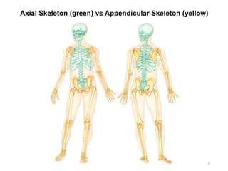
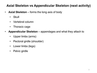
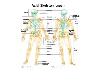
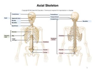
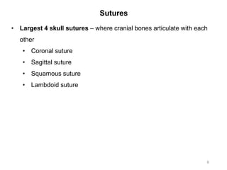
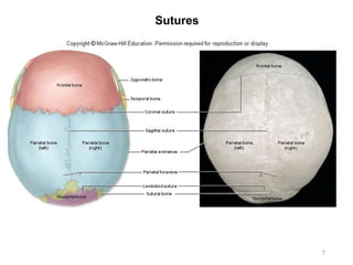
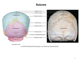
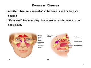
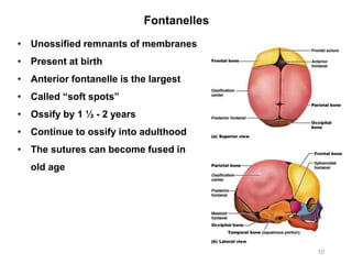
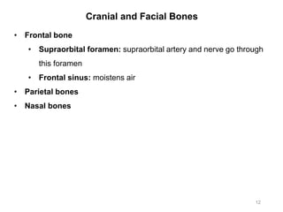
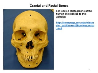
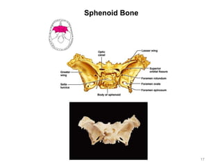
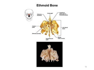
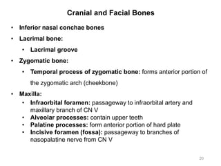
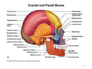
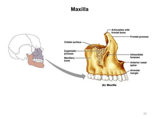
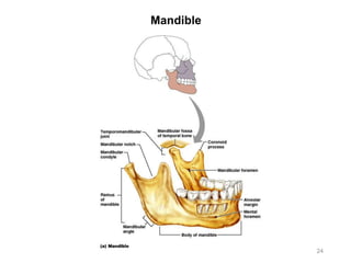
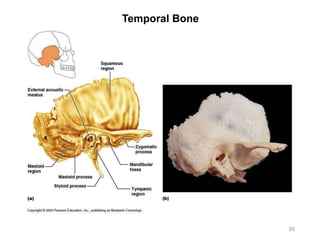
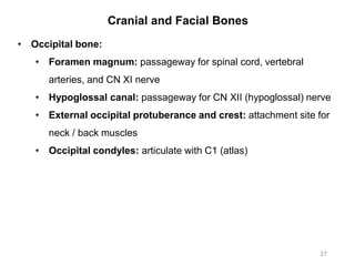
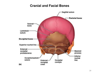
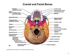
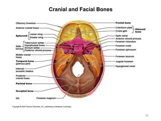
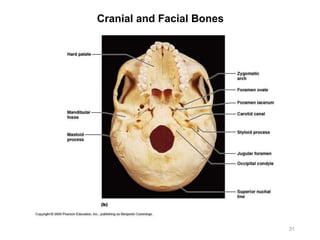
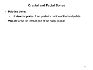
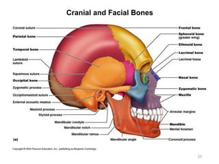
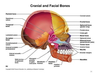
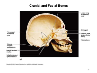
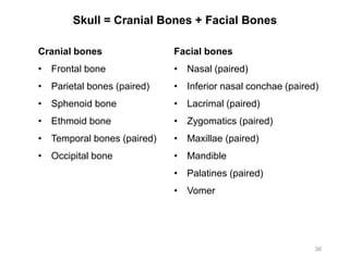
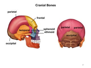
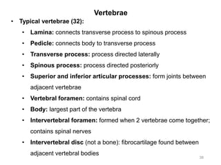
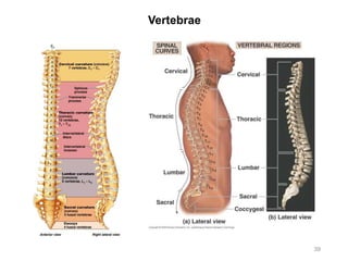
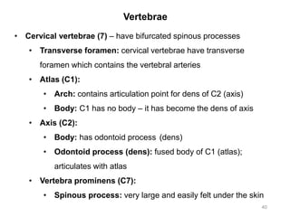
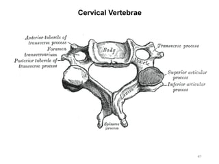
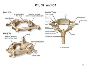
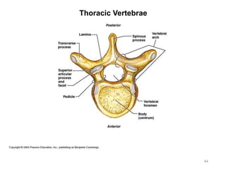
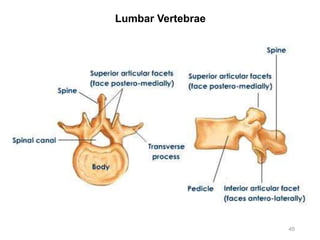
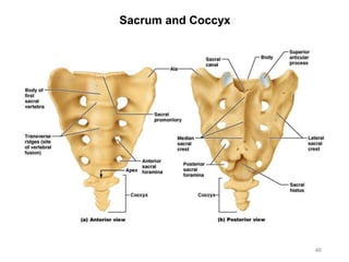
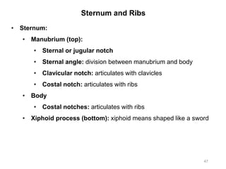
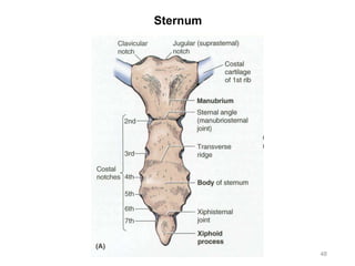
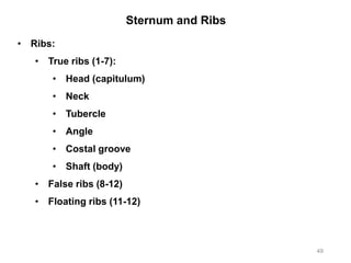
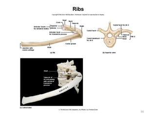
Activity 3:
Axial Skeleton
Chapter 7 – McKinley et al., Human Anatomy, 4e.
Objectives:
• Observe and dissect a fresh long bone from a cow
• Identify the cranial and facial bones and important bone
markings on each
• Identify the vertebrae and other features of the vertebral
column, and important bone markings on each
• Identify ribs and sternum, and important bone markings on
each
1
Compilation: Kathryn Watson & Claudia Gonzales
Last Revision: Mohammad Tomaraei
Axial Skeleton (green) vs Appendicular Skeleton (yellow)
2
Axial Skeleton vs Appendicular Skeleton (next activity)
3
• Axial Skeleton – forms the long axis of body
• Skull
• Vertebral column
• Thoracic cage
• Appendicular Skeleton – appendages and what they attach to
• Upper limbs (arms)
• Pectoral girdle (shoulder)
• Lower limbs (legs)
• Pelvic girdle
Axial Skeleton (green)
4
Axial Skeleton
5
Sutures
6
• Largest 4 skull sutures – where cranial bones articulate with each
other
• Coronal suture
• Sagittal suture
• Squamous suture
• Lambdoid suture
Sutures
7
Sutures
8
Paranasal Sinuses
9
• Air-filled chambers named after the bone in which they are
housed
• “Paranasal” because they cluster around and connect to the
nasal cavity
Fontanelles
10
• Unossified remnants of membranes
• Present at birth
• Anterior fontanelle is the largest
• Called “soft spots”
• Ossify by 1 ½ - 2 years
• Continue to ossify into adulthood
• The sutures can become fused in
old age
Cranial and Facial Bones
11
• Frontal bone
• Supraorbital foramen: supraorbital artery and nerve go through
this foramen
• Frontal sinus: moistens air
• Parietal bones
• Nasal bones
Cranial and Facial Bones
12
• Frontal bone
• Supraorbital foramen: supraorbital artery and nerve go through
this foramen
• Frontal sinus: moistens air
• Parietal bones
• Nasal bones
Cranial and Facial Bones
13
Cranial and Facial Bones
14
For labeled photographs of the
human skeleton go to this
website:
http://homepage.smc.edu/wissm
ann_paul/bones/EBbonestutorial
.html
Cranial and Facial Bones
15
• Sphenoid bone:
• Greater wing
• Lesser wing
• Sella turcica: houses the pituitary gland
• Optic foramen / canal: contains the optic nerve (CN II)
• Foramen ovale: contains the mandibular branch of the trigeminal
nerve (CN V)
• Foramen rotundum: contains the maxillary branch of the
trigeminal nerve (CN V)
• Foramen spinosum: contains the middle meningeal vessels
• Foramen lacerum
Cranial and Facial Bones
16
• Sphenoid bone:
• Superior orbital fissure: contains CN III, the ophthalmic branch
of CN V, and CN VI
• Inferior orbital fissure: contains the maxillary branch of CN V
• Sphenoidal sinus: moistens air
• Lateral and medial plates
• Pterygoid plates
Sphenoid Bone
17
Cranial and Facial Bones
18
• Ethmoid bone:
• Perpendicular plate: superior part of the nasal septum
• Superior and middle nasal concha(e): increase surface area for
warming and filtering air
• Cribriform plate and foramina: passageway for olfactory nerves
• Crista galli: attachment site for dura mater to skull
Ethmoid Bone
19
Cranial and Facial Bones
20
• Inferior nasal conchae bones
• Lacrimal bone:
• Lacrimal groove
• Zygomatic bone:
• Temporal process of zygomatic bone: forms anterior portion of
the zygomatic arch (cheekbone)
• Maxilla:
• Infraorbital foramen: passageway to infraorbital artery and
maxillary branch of CN V
• Alveolar processes: contain upper teeth
• Palatine processes: form anterior portion of hard plate
• Incisive foramen (fossa): passageway to branches of
nasopalatine nerve from CN V
Cranial and Facial Bones
21
Maxilla
22
Cranial and Facial Bones
23
• Mandible:
• Body
• Ramus
• Alveolar processes: contain lower teeth
• Angle
• Mental foramen: passageway for the mandibular branch of CN V
and blood vessels
• CoroNOID process: insertion point of the temporalis muscle
• Mandibular condyle or condylar process: forms a joint with the
mandibular fossa of the temporal bone
• Mandibular notch
Mandible
24
Cranial and Facial Bones
25
• Temporal bone:
• Zygomatic process of temporal bone: forms the posterior
portion of the zygomatic arch (cheekbone)
• Squamous region: squamous means flat – flat part of the bone
• Styloid process: attachment for hyoid and tongue muscles
• Mastoid process: insertion for sternocleidomastoid muscle
• External auditory / acoustic meatus: opening of auditory canal
• Petrous part: petrous means rock
• Internal auditory / acoustic meatus: passageway for CN VII and
CN VIII nerves, and blood vessels to inner ear
• Jugular foramen: passageway for internal jugular vein, CN IX,
CN X, and CN XI nerves
• Carotid canal: passageway for internal carotid artery
• Mandibular fossa: forms a joint with mandibular condyle of
mandible
Temporal Bone
26
Cranial and Facial Bones
27
• Occipital bone:
• Foramen magnum: passageway for spinal cord, vertebral
arteries, and CN XI nerve
• Hypoglossal canal: passageway for CN XII (hypoglossal) nerve
• External occipital protuberance and crest: attachment site for
neck / back muscles
• Occipital condyles: articulate with C1 (atlas)
Cranial and Facial Bones
28
Cranial and Facial Bones
29
Cranial and Facial Bones
30
Cranial and Facial Bones
31
Cranial and Facial Bones
32
• Palatine bone:
• Horizontal plates: form posterior portion of the hard palate
• Vomer: forms the inferior part of the nasal septum
Cranial and Facial Bones
33
Cranial and Facial Bones
34
Cranial and Facial Bones
35
Skull = Cranial Bones + Facial Bones
36
Cranial bones
• Frontal bone
• Parietal bones (paired)
• Sphenoid bone
• Ethmoid bone
• Temporal bones (paired)
• Occipital bone
Facial bones
• Nasal (paired)
• Inferior nasal conchae (paired)
• Lacrimal (paired)
• Zygomatics (paired)
• Maxillae (paired)
• Mandible
• Palatines (paired)
• Vomer
Cranial Bones
37
parietal
parietal
frontal
temporal
parietal
occipital
_________sphenoid
_______ ethmoid
occipital
Vertebrae
38
• Typical vertebrae (32):
• Lamina: connects transverse process to spinous process
• Pedicle: connects body to transverse process
• Transverse process: process directed laterally
• Spinous process: process directed posteriorly
• Superior and inferior articular processes: form joints between
adjacent vertebrae
• Vertebral foramen: contains spinal cord
• Body: largest part of the vertebra
• Intervertebral foramen: formed when 2 vertebrae come together;
contains spinal nerves
• Intervertebral disc (not a bone): fibrocartilage found between
adjacent vertebral bodies
Vertebrae
39
Vertebrae
40
• Cervical vertebrae (7) – have bifurcated spinous processes
• Transverse foramen: cervical vertebrae have transverse
foramen which contains the vertebral arteries
• Atlas (C1):
• Arch: contains articulation point for dens of C2 (axis)
• Body: C1 has no body – it has become the dens of axis
• Axis (C2):
• Body: has odontoid process (dens)
• Odontoid process (dens): fused body of C1 (atlas);
articulates with atlas
• Vertebra prominens (C7):
• Spinous process: very large and easily felt under the skin
Cervical Vertebrae
41
C1, C2, and C7
42
Vertebrae
43
• Thoracic vertebrae (12):
• Transverse process: on thoracic vertebrae, transverse
processes contain costal facets for articulation with angle of a rib
• Lumbar vertebrae (5)
• Sacrum (5 bones fused):
• Anterior sacral foramina: contain ventral rami of the sacral
spinal nerves
• Posterior sacral foramina: contain dorsal rami of the sacral
spinal nerves
• Median sacral crest: represents fused spinous processes of
sacral vertebrae
• Auricular surfaces: ear-like processes, articulate with the iliac
bones
• Superior articular processes: articulate with inferior articular
processes of L5
• Coccyx (2-3 bones fused):
• Cornu or horns: small horns that point superiorly
Thoracic Vertebrae
44
Lumbar Vertebrae
45
Sacrum and Coccyx
46
Sternum and Ribs
47
• Sternum:
• Manubrium (top):
• Sternal or jugular notch
• Sternal angle: division between manubrium and body
• Clavicular notch: articulates with clavicles
• Costal notch: articulates with ribs
• Body
• Costal notches: articulates with ribs
• Xiphoid process (bottom): xiphoid means shaped like a sword
Sternum
48
Sternum and Ribs
49
• Ribs:
• True ribs (1-7):
• Head (capitulum)
• Neck
• Tubercle
• Angle
• Costal groove
• Shaft (body)
• False ribs (8-12)
• Floating ribs (11-12)
Ribs
50

Activity 3 - Axial Skeleton

  • 1.
    Activity 3: Axial Skeleton Chapter7 – McKinley et al., Human Anatomy, 4e. Objectives: • Observe and dissect a fresh long bone from a cow • Identify the cranial and facial bones and important bone markings on each • Identify the vertebrae and other features of the vertebral column, and important bone markings on each • Identify ribs and sternum, and important bone markings on each 1 Compilation: Kathryn Watson & Claudia Gonzales Last Revision: Mohammad Tomaraei
  • 2.
    Axial Skeleton (green)vs Appendicular Skeleton (yellow) 2
  • 3.
    Axial Skeleton vsAppendicular Skeleton (next activity) 3 • Axial Skeleton – forms the long axis of body • Skull • Vertebral column • Thoracic cage • Appendicular Skeleton – appendages and what they attach to • Upper limbs (arms) • Pectoral girdle (shoulder) • Lower limbs (legs) • Pelvic girdle
  • 4.
  • 5.
  • 6.
    Sutures 6 • Largest 4skull sutures – where cranial bones articulate with each other • Coronal suture • Sagittal suture • Squamous suture • Lambdoid suture
  • 7.
  • 8.
  • 9.
    Paranasal Sinuses 9 • Air-filledchambers named after the bone in which they are housed • “Paranasal” because they cluster around and connect to the nasal cavity
  • 10.
    Fontanelles 10 • Unossified remnantsof membranes • Present at birth • Anterior fontanelle is the largest • Called “soft spots” • Ossify by 1 ½ - 2 years • Continue to ossify into adulthood • The sutures can become fused in old age
  • 11.
    Cranial and FacialBones 11 • Frontal bone • Supraorbital foramen: supraorbital artery and nerve go through this foramen • Frontal sinus: moistens air • Parietal bones • Nasal bones
  • 12.
    Cranial and FacialBones 12 • Frontal bone • Supraorbital foramen: supraorbital artery and nerve go through this foramen • Frontal sinus: moistens air • Parietal bones • Nasal bones
  • 13.
  • 14.
    Cranial and FacialBones 14 For labeled photographs of the human skeleton go to this website: http://homepage.smc.edu/wissm ann_paul/bones/EBbonestutorial .html
  • 15.
    Cranial and FacialBones 15 • Sphenoid bone: • Greater wing • Lesser wing • Sella turcica: houses the pituitary gland • Optic foramen / canal: contains the optic nerve (CN II) • Foramen ovale: contains the mandibular branch of the trigeminal nerve (CN V) • Foramen rotundum: contains the maxillary branch of the trigeminal nerve (CN V) • Foramen spinosum: contains the middle meningeal vessels • Foramen lacerum
  • 16.
    Cranial and FacialBones 16 • Sphenoid bone: • Superior orbital fissure: contains CN III, the ophthalmic branch of CN V, and CN VI • Inferior orbital fissure: contains the maxillary branch of CN V • Sphenoidal sinus: moistens air • Lateral and medial plates • Pterygoid plates
  • 17.
  • 18.
    Cranial and FacialBones 18 • Ethmoid bone: • Perpendicular plate: superior part of the nasal septum • Superior and middle nasal concha(e): increase surface area for warming and filtering air • Cribriform plate and foramina: passageway for olfactory nerves • Crista galli: attachment site for dura mater to skull
  • 19.
  • 20.
    Cranial and FacialBones 20 • Inferior nasal conchae bones • Lacrimal bone: • Lacrimal groove • Zygomatic bone: • Temporal process of zygomatic bone: forms anterior portion of the zygomatic arch (cheekbone) • Maxilla: • Infraorbital foramen: passageway to infraorbital artery and maxillary branch of CN V • Alveolar processes: contain upper teeth • Palatine processes: form anterior portion of hard plate • Incisive foramen (fossa): passageway to branches of nasopalatine nerve from CN V
  • 21.
  • 22.
  • 23.
    Cranial and FacialBones 23 • Mandible: • Body • Ramus • Alveolar processes: contain lower teeth • Angle • Mental foramen: passageway for the mandibular branch of CN V and blood vessels • CoroNOID process: insertion point of the temporalis muscle • Mandibular condyle or condylar process: forms a joint with the mandibular fossa of the temporal bone • Mandibular notch
  • 24.
  • 25.
    Cranial and FacialBones 25 • Temporal bone: • Zygomatic process of temporal bone: forms the posterior portion of the zygomatic arch (cheekbone) • Squamous region: squamous means flat – flat part of the bone • Styloid process: attachment for hyoid and tongue muscles • Mastoid process: insertion for sternocleidomastoid muscle • External auditory / acoustic meatus: opening of auditory canal • Petrous part: petrous means rock • Internal auditory / acoustic meatus: passageway for CN VII and CN VIII nerves, and blood vessels to inner ear • Jugular foramen: passageway for internal jugular vein, CN IX, CN X, and CN XI nerves • Carotid canal: passageway for internal carotid artery • Mandibular fossa: forms a joint with mandibular condyle of mandible
  • 26.
  • 27.
    Cranial and FacialBones 27 • Occipital bone: • Foramen magnum: passageway for spinal cord, vertebral arteries, and CN XI nerve • Hypoglossal canal: passageway for CN XII (hypoglossal) nerve • External occipital protuberance and crest: attachment site for neck / back muscles • Occipital condyles: articulate with C1 (atlas)
  • 28.
  • 29.
  • 30.
  • 31.
  • 32.
    Cranial and FacialBones 32 • Palatine bone: • Horizontal plates: form posterior portion of the hard palate • Vomer: forms the inferior part of the nasal septum
  • 33.
  • 34.
  • 35.
  • 36.
    Skull = CranialBones + Facial Bones 36 Cranial bones • Frontal bone • Parietal bones (paired) • Sphenoid bone • Ethmoid bone • Temporal bones (paired) • Occipital bone Facial bones • Nasal (paired) • Inferior nasal conchae (paired) • Lacrimal (paired) • Zygomatics (paired) • Maxillae (paired) • Mandible • Palatines (paired) • Vomer
  • 37.
  • 38.
    Vertebrae 38 • Typical vertebrae(32): • Lamina: connects transverse process to spinous process • Pedicle: connects body to transverse process • Transverse process: process directed laterally • Spinous process: process directed posteriorly • Superior and inferior articular processes: form joints between adjacent vertebrae • Vertebral foramen: contains spinal cord • Body: largest part of the vertebra • Intervertebral foramen: formed when 2 vertebrae come together; contains spinal nerves • Intervertebral disc (not a bone): fibrocartilage found between adjacent vertebral bodies
  • 39.
  • 40.
    Vertebrae 40 • Cervical vertebrae(7) – have bifurcated spinous processes • Transverse foramen: cervical vertebrae have transverse foramen which contains the vertebral arteries • Atlas (C1): • Arch: contains articulation point for dens of C2 (axis) • Body: C1 has no body – it has become the dens of axis • Axis (C2): • Body: has odontoid process (dens) • Odontoid process (dens): fused body of C1 (atlas); articulates with atlas • Vertebra prominens (C7): • Spinous process: very large and easily felt under the skin
  • 41.
  • 42.
  • 43.
    Vertebrae 43 • Thoracic vertebrae(12): • Transverse process: on thoracic vertebrae, transverse processes contain costal facets for articulation with angle of a rib • Lumbar vertebrae (5) • Sacrum (5 bones fused): • Anterior sacral foramina: contain ventral rami of the sacral spinal nerves • Posterior sacral foramina: contain dorsal rami of the sacral spinal nerves • Median sacral crest: represents fused spinous processes of sacral vertebrae • Auricular surfaces: ear-like processes, articulate with the iliac bones • Superior articular processes: articulate with inferior articular processes of L5 • Coccyx (2-3 bones fused): • Cornu or horns: small horns that point superiorly
  • 44.
  • 45.
  • 46.
  • 47.
    Sternum and Ribs 47 •Sternum: • Manubrium (top): • Sternal or jugular notch • Sternal angle: division between manubrium and body • Clavicular notch: articulates with clavicles • Costal notch: articulates with ribs • Body • Costal notches: articulates with ribs • Xiphoid process (bottom): xiphoid means shaped like a sword
  • 48.
  • 49.
    Sternum and Ribs 49 •Ribs: • True ribs (1-7): • Head (capitulum) • Neck • Tubercle • Angle • Costal groove • Shaft (body) • False ribs (8-12) • Floating ribs (11-12)
  • 50.