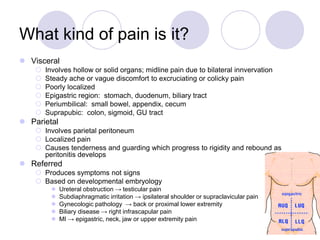
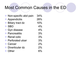
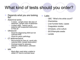
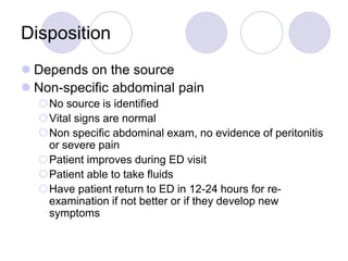
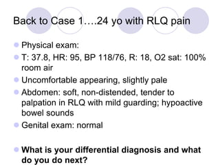
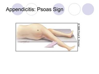
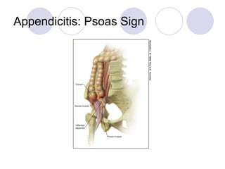
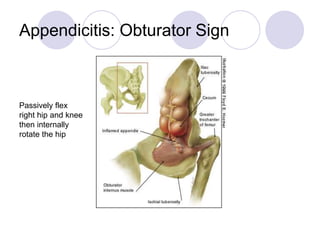
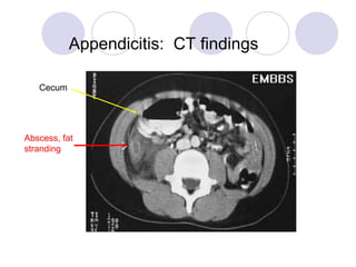
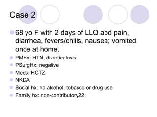
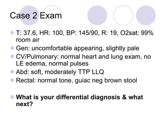
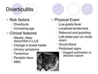
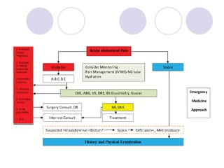
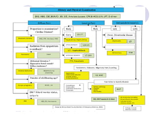
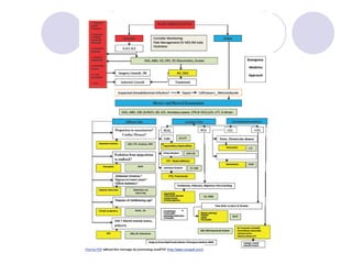
The document discusses a case of acute abdominal pain in a 24-year-old male. It provides details of the patient's history, physical exam findings, and differential diagnosis. The document then reviews approaches to evaluating abdominal pain, including types of pain, history taking, physical exam maneuvers, potential diagnoses, appropriate tests, and disposition planning.
This document provides an overview of acute abdominal pain, including classifications, causes, symptoms, diagnostic tests, and treatment considerations. It describes three types of abdominal pain - visceral, parietal, and referred - and covers common intra-abdominal etiologies like appendicitis, cholecystitis, small bowel obstruction, and ischemic bowel. It also discusses extra-abdominal, toxic, metabolic, and neurogenic causes of abdominal pain and emphasizes the importance of thorough history taking and physical exam in diagnosing the source.
1. A 24-year-old male presents with right lower quadrant abdominal pain radiating to the groin. On exam, he has mild guarding and tenderness in the right lower quadrant. His differential diagnosis includes appendicitis.
2. A 68-year-old female presents with left lower quadrant pain, diarrhea, fevers and nausea. On exam, she has tenderness in the left lower quadrant. Her differential diagnosis includes diverticulitis.
3. The document discusses the history, exam, differential diagnosis, diagnostic workup, and treatment of common causes of acute abdominal pain such as appendicitis and diverticulitis.
1. The goal is to rule out life-threatening causes of abdominal pain such as a ruptured abdominal aortic aneurysm or bowel perforation.
2. A urine pregnancy test should be obtained in all women of childbearing age to exclude ectopic pregnancy.
3. The history and physical exam aim to determine the location and characteristics of the pain to narrow the differential diagnosis and guide testing.
This is a lecture by Dr. Joseph House from the Ghana Emergency Medicine Collaborative. To download the editable version (in PPT), to access additional learning modules, or to learn more about the project, see http://openmi.ch/em-gemc. Unless otherwise noted, this material is made available under the terms of the Creative Commons Attribution Share Alike-3.0 License: http://creativecommons.org/licenses/by-sa/3.0/.
the presention about abdominal pain and it s different cause we talking briefly about medical and surgical causes
and the presention mainly about the vascular causes for abdominal pain
i hope its helpful for you
- Md. Dulal, a 42-year-old male shopkeeper, presented with 6 months of recurrent upper abdominal pain aggravated by fatty foods. Examination found right hypochondriac tenderness.
- Tests showed thickened gallbladder and cholelithiasis on ultrasound. Provisional diagnosis was chronic cholecystitis given recurrent pain related to fatty foods and tenderness in the right upper quadrant.
- Differential diagnoses considered were chronic peptic ulcer and pancreatitis but were less likely given absence of hematemesis, melena, or projectile vomiting. Management options discussed were open or laparoscopic cholecystectomy.
The document defines an acute abdomen as a rapid onset of severe abdominal symptoms that may indicate a life-threatening condition. It lists many potential causes including appendicitis, cholecystitis, pancreatitis, and others. It describes assessing patients with an acute abdomen through history, examination, and potential emergency department care and investigations like blood tests, imaging, and laparoscopy. It highlights some red flags to watch for and includes three case studies.
1. Abdominal pain is a common presenting symptom that can be caused by many intra-abdominal and extra-abdominal processes.
2. A thorough history and physical exam is important to help determine the cause, including assessing location, character, and timing of pain.
3. Differential diagnosis depends on factors like location of pain and patient characteristics, and may include conditions like appendicitis, cholecystitis, pancreatitis, or diverticulitis. Laboratory tests, imaging, and other evaluations can help make the diagnosis.
This document discusses recurrent abdominal pain (RAP) in children. It provides information on the epidemiology, clinical profile, classification, pathophysiology, etiology, alarm symptoms and signs, diagnosis, investigations, and treatment of RAP. Treatment involves ruling out organic causes, reassurance, allowing normal activity, addressing stressors, and may include pharmacological interventions, dietary modifications, and behavioral therapies. The goal is to help the child return to normal activities and improve their pain over time.
A 22-year-old woman presents with severe lower abdominal pain. The differential diagnosis includes appendicitis, pelvic inflammatory disease, ovarian cyst, and ectopic pregnancy. Given her age, a pregnancy test and pelvic exam are important to evaluate for potential gynecologic causes of her pain.
Abdominal pain is one of the common symptoms for 17 more diseases. This ppt will help you to get awareness of Abdominal pain and its causes, signs and symptoms, treatment, natural remedies, and medical advice, diagnosis investigation, abdominal pain faqs etc.
writes I have a good salary, am married, and have two children. My whole life I've been drawn to prescription and have always enjoyed it. However, I have a unattached in English literature, so I've always put it as unattainable to become a doctor. Now, once again, I'm bearing in mind doing one of the post bac premed programs out there and going for it.
The document discusses acute epigastric pain, dividing it into causes such as acute gastritis, exacerbation of duodenal ulcer, biliary colic, acute cholecystitis, and acute pancreatitis. For each cause, it describes the typical history, examination findings, diagnostic tests, and treatment approach. For example, it notes that acute gastritis is often caused by H. pylori or NSAIDs, while acute cholecystitis presents with right upper quadrant tenderness and Murphy's sign on examination. Ultrasound is useful for gallstones, while lipase checks for pancreatitis. Treatment focuses on conservative measures, though cholecystectomy may be considered for cholecystitis.
This document discusses abdominal pain, providing information on:
1. Common causes of abdominal pain including conditions like pancreatitis, appendicitis, and diverticulitis. Abdominal pain accounts for 35% of presentations and can be somatic, visceral, or referred.
2. Key aspects of the history including location, aggravating/relieving factors, and risk factors. Special considerations for evaluating elderly patients and children are also outlined.
3. Imaging tests for evaluating common conditions causing abdominal pain and their sensitivities and specificities. Ultrasound, CT, and CTA are discussed.
4. Several case examples are presented to demonstrate differential diagnoses and initial workups for patients presenting with
1) Between one-third to one-half of children admitted to the hospital for acute abdominal pain have non-specific abdominal pain, while another one-third have acute appendicitis.
2) A careful history, physical exam, and observation are important for evaluating acute abdominal pain in children. Additional tests like ultrasound, urine analysis, and bloodwork may also help diagnose conditions like UTIs, gastrointestinal issues, or appendicitis.
3) Non-specific abdominal pain is poorly localized, not worsened by movement, and rarely accompanied by guarding. It is usually self-limiting within 24 hours and may be caused by viral infections or transient intussusception.
An 85-year-old man presented with 5 days of abdominal pain and worsening mental status. On exam, he had a suprapubic mass. Labs showed elevated creatinine. A renal ultrasound found right hydronephrosis. Inserting a Foley catheter drained 2500cc of urine and resolved the mass. He was diagnosed with urinary retention from an enlarged prostate and treated with catheterization, hydration, and medications for his prostate and urinary symptoms.
This document provides an overview of the management approach for acute abdomen. It begins with definitions and epidemiology, noting that acute abdomen is undiagnosed abdominal pain lasting less than 7-10 days. It then covers the surgical and medical causes, with etiological classifications including inflammatory/infective, perforation, obstruction, infarction, and spontaneous bleeding. Differential diagnoses are provided for different age groups. The management approach involves clinical evaluation, resuscitation, diagnostic tools like ultrasound and CT scan, and categorizing patients based on urgency of condition.
SYMPTOMS & SIGNS IN GIT PROBLEMS
• Dear Viewers
• Greetings from “ Surgical Educator”
• I am uploading a PPT presentation on symptoms and signs in GI problems
• What are the questions you have to ask the patients for each problem in GIT is explained
• How to examine and elicit various signs in abdomen is also explained
• I hope this PPT presentation will be very useful to you
• You can watch all my surgery teaching videocasts in the following links
• surgicaleducator.blogspot.com youtube.com/c/surgicaleducator
1. Acute abdominal pain has many potential causes that can be categorized by organ system affected or type of pain experienced.
2. A thorough history and physical exam is important to determine the likely cause and guide appropriate testing or treatment.
3. Common pediatric causes include gastroenteritis, appendicitis, urinary tract infections, while adolescent females should also be evaluated for gynecological issues.
Acute abdominal pain sarah Alotibi and samiyah aljohaniさ ん
This document discusses acute abdominal pain, including:
1. It defines acute abdomen as a rapid onset of severe abdominal symptoms that may indicate life-threatening pathology. Major causes include appendicitis, cholecystitis, and bowel obstruction.
2. It provides guidance on assessing and examining patients with acute abdominal pain, including evaluating vital signs, performing physical exams like auscultation and palpation, and considering differential diagnoses based on pain location.
3. It outlines a nursing care plan for patients with acute abdominal pain, which includes pain assessment, non-pharmacological interventions, medication administration, monitoring, education, and expected outcomes like pain relief and management of side effects.
Diagnosis And Management Of Acute Abdominal PainDimitri Raptis
This document discusses the diagnosis and management of acute abdominal pain (AAP). It defines AAP and lists some of the most common causes. Over 1000 causes exist and the initial diagnosis is inaccurate in 20-40% of cases. A thorough history, physical exam, and selective use of basic blood tests and imaging studies are important for diagnosis. Early laparoscopy may help diagnose unclear cases and prevent unnecessary laparotomies. Proper initial management focuses on resuscitation, analgesia and seeking senior help to guide further evaluation and treatment.
This document presents the case of an 18-year-old female patient with intermittent epigastric pain for 9 days. Physical examination revealed direct tenderness in the epigastric area and Murphy's sign was positive. Blood tests showed leukocytosis. Ultrasound showed gallbladder hydrops and cholecystolithiasis. The patient was diagnosed with acute cholecystitis and underwent an emergency open cholecystectomy. Her postoperative course was uncomplicated and she was discharged in stable condition.
The document provides information on acute abdomen including its definition, epidemiology, physiology, differential diagnosis by location, history and physical examination findings, important investigations, management principles, and criteria for surgical consultation. Acute abdomen is defined as sudden severe abdominal pain lasting less than 24 hours that often requires urgent diagnosis and some causes need surgical treatment. The differential diagnosis considers location of pain and includes conditions like appendicitis, diverticulitis, bowel obstruction, pancreatitis and others. Key aspects of evaluation involve history, physical exam, labs, imaging and identifying high-risk patients who may require emergent surgery.
Investigations in the case of abdominal painAbino David
This document discusses investigations for abdominal pain. It outlines various examinations of feces, vomitus, ascitic fluid, and radiological tests that can help diagnose the underlying cause of abdominal pain. Macroscopic and microscopic examination of feces can provide clues to conditions like malabsorption, steatorrhea, dysentery, or bleeding. Chemical tests on feces can detect occult blood or measure fat and nitrogen content. Analysis of vomitus may indicate gastric outlet obstruction or gastrocolic fistula. Ascitic fluid appearance and biochemical tests help differentiate between portal hypertension, tuberculosis peritonitis, and malignancy. Radiological investigations include plain films, barium studies, ultrasound, endoscopy, CT, MRI, and laparoscopy
The document discusses the evaluation and management of acute abdominal pain. It describes how acute abdominal pain can be caused by many different intra-abdominal and extra-abdominal conditions ranging from minor to life-threatening. A thorough history, physical exam, and diagnostic testing are needed to make an accurate diagnosis as the cause is often not apparent initially. Common etiologies of acute abdominal pain discussed include appendicitis, cholecystitis, diverticulitis, pancreatitis, bowel obstruction, renal colic, pelvic inflammatory disease, and ectopic pregnancy.
A 24-year-old male presented with one day of abdominal pain localized to the right lower abdomen. On examination, he had tenderness and guarding in the right lower quadrant. The differential diagnosis includes appendicitis. Laboratory tests and a CT scan may be needed to make a definitive diagnosis.
A 68-year-old female presented with two days of left lower quadrant abdominal pain, diarrhea, fever and nausea. She has a history of hypertension and diverticulosis. On examination, she had tenderness in the left lower quadrant. The differential diagnosis includes diverticulitis.
1) The document discusses the evaluation and management of acute abdominal pain, focusing on history taking, physical exam, differential diagnosis, and initial testing.
2) Common causes of acute abdominal pain in the emergency department setting include nonspecific abdominal pain, appendicitis, biliary tract disease, small bowel obstruction, gynecologic issues, and pancreatitis.
3) The history should inquire about the characteristics of the pain, associated symptoms, past medical history, surgical history, medications, and social history. The physical exam involves inspection, auscultation, and palpation of the abdomen along with pertinent system reviews.
4) Initial testing may
This document discusses a case of a 20-year-old man presenting with abdominal pain and diarrhea who was found to have elevated inflammatory markers and free fluid on ultrasound. It then provides a general overview of acute abdomen including common causes, diagnostic approach, important physical exam findings and signs, and appropriate imaging studies.
This document outlines a presentation on the pathophysiology and management of acute abdomen. It begins with definitions of acute abdomen and types of abdominal pain. Pathophysiological mechanisms including luminal obstruction, inflammation, peritonitis, ischemia and non-specific pain are described. Common causes like appendicitis, cholecystitis, bowel obstruction and perforated viscus are listed. Immediately life-threatening diagnoses of perforated viscus, bowel ischemia, ruptured abdominal aortic aneurysm and ruptured ectopic pregnancy are highlighted. Clinical assessment techniques and investigations are outlined. Management principles focusing on ABCs, fluid resuscitation and need for surgery in some cases are emphasized in the take-home message.
Dr. Guy Nicastri, Associate Professor of Surgery and Family Medicine at the Warren Alpert School of Medicine at Brown University takes us through some of the pearls of the Acute Abdomen Examination in the Adult
This document discusses the approach to recurrent abdominal pain in children. It defines acute, subacute, and chronic abdominal pain and discusses recurrent abdominal pain. The most common causes of abdominal pain seen in emergency departments are also summarized. A full history and physical exam are important for evaluating abdominal pain, and diagnostic testing should be guided by symptoms and exam findings. Home care and lifestyle advice are usually sufficient for recurrent abdominal pain in children without concerning alarm symptoms.
The 15-year-old boy presented with gradually worsening lower right abdominal pain for 4 days, along with nausea, vomiting, and loss of appetite. On examination, he had tenderness in the right iliac fossa and rebound tenderness. Acute appendicitis should be considered, as the presentation is consistent with the classic progression of abdominal pain from periumbilical to localized right lower quadrant pain, along with common associated symptoms of anorexia, nausea, and vomiting. Laboratory tests such as a CBC can help evaluate for elevated white blood cells, though the diagnosis of appendicitis is primarily based on the history and physical exam findings.
The document discusses the acute abdomen, which refers to symptoms and signs of acute intra-abdominal disease that are usually best treated with surgery. It defines the different types of abdominal pain and covers the epidemiology, causes, diagnostic evaluation including history, exams, labs, and imaging. High risk patients are identified. The management of acute abdomen in the emergency department is outlined, including when surgery is indicated versus other conservative treatments.
The document discusses the acute abdomen, which refers to symptoms and signs of acute intra-abdominal disease that usually require surgical intervention. It defines the types of abdominal pain and covers the epidemiology, causes, diagnostic evaluation including history, physical exam, labs, and imaging. Evaluation focuses on identifying patients at high risk who may require urgent surgery. The differential diagnosis is broad and includes appendicitis, diverticulitis, cholecystitis, and others. History, exam, and testing can help distinguish surgical from non-surgical causes to guide emergency management and need for operation.
This document provides an introduction to examining the abdomen in clinical skills. It outlines the anatomy and physiology of the abdomen, landmarks, techniques for inspection, auscultation, percussion and palpation. Specific assessment techniques are described for organs like the liver and spleen. Common abdominal disorders and patterns of abdominal pain are also discussed. The goal is to teach medical students how to properly examine the abdomen.
The document provides guidance on assessing the digestive tract through a nursing assessment. It outlines questions to ask about the patient's chief complaint, medical history including medications, family history, and review of systems. The objective assessment includes inspection, auscultation, percussion and light palpation of the abdomen. Various diagnostic tests are also discussed such as radiographic imaging, endoscopy, and ERCP to further evaluate abnormalities.
This document discusses the acute abdomen, including definitions, clinical diagnosis, differential diagnosis, evaluation, and management. An acute abdomen is signs and symptoms of intra-abdominal disease that may require surgery. The clinical diagnosis involves characterizing the pain location, onset, and nature. Broad differential categories include inflammation, obstruction, ischemia, and perforation. Evaluation involves history, physical exam, labs, and imaging like ultrasound or CT scan. Decision for surgery is made for peritonitis, severe unrelenting pain, instability, or suspected intestinal ischemia/strangulation. Common etiologies are perforated ulcer, appendicitis, diverticulitis, bowel obstruction, cholecystitis, ischemic or perforated bowel, and ruptured
This document discusses acute abdomen and provides information on evaluating and diagnosing various potential causes. It defines acute abdomen and outlines the challenges surgeons face. A full history, physical exam, and further investigations are needed to make an exact diagnosis. Common differential diagnoses include appendicitis, peptic ulcer disease, cholecystitis, bowel obstruction, pancreatitis, diverticulitis, renal colic, pelvic inflammatory disease, and ectopic pregnancy. Key diagnostic tests include bloodwork, imaging like CT scans, and ultrasound.
1) Acute abdomen is a common presentation accounting for 4-10% of emergency department visits. 50% have a clear diagnosis while 15-30% require surgical procedures, especially in the elderly.
2) Unique presentations can occur in pediatric and elderly patients, with the elderly having higher rates of misdiagnosis and mortality due to less prominent physical exam findings.
3) A thorough history and physical exam remain important for assessing abdominal pain, though imaging studies can help when the diagnosis is unclear. Close observation is often needed to determine if the condition is surgical or non-surgical.
This document discusses a case of acute abdomen in a 20-year-old man presenting with diffuse abdominal pain and diarrhea for 1 day with a history of abdominal pain 1 month prior. On examination, he was febrile with abdominal tenderness in all quadrants but no rebound or guarding. The document then provides an overview of acute abdomen including common causes, diagnosis, abdominal pain characteristics, physical examination findings, investigations including imaging and differential diagnosis for acute abdomen conditions. Key mimickers of acute appendicitis discussed include mesenteric lymphadenitis, bacterial ileocecitis, and pelvic inflammatory disease.
This document provides information on evaluating and diagnosing acute and chronic abdominal pain. It discusses the history, physical exam, diagnostic studies, and management of various acute conditions like appendicitis, diverticulitis, cholecystitis, and perforated ulcer. It also covers chronic pain syndromes like irritable bowel syndrome and chronic pancreatitis. The goal is to distinguish between organic and functional causes of abdominal pain.
An acute abdomen is severe abdominal pain that comes on suddenly and may require immediate medical care. It can be caused by conditions like appendicitis, diverticulitis, gallstones, or intestinal infections. Diagnosis involves examining the patient's medical history and symptoms, as well as tests like blood tests, imaging scans, and surgery if needed. Treatment depends on the underlying cause but may include pain relievers, surgery, or other medical interventions.
This document discusses the relationship between the gastrointestinal (GI) system and other body systems. It outlines how the neurological, endocrine, respiratory, cardiovascular, muscular, integumentary, digestive, urinary, lymphatic, and reproductive systems interact with and can affect the GI system. It also provides details on assessing the GI system through health history, physical examination, diagnostic tests, and various radiologic and endoscopic procedures.
Cell Therapy Expansion and Challenges in Autoimmune DiseaseHealth Advances
There is increasing confidence that cell therapies will soon play a role in the treatment of autoimmune disorders, but the extent of this impact remains to be seen. Early readouts on autologous CAR-Ts in lupus are encouraging, but manufacturing and cost limitations are likely to restrict access to highly refractory patients. Allogeneic CAR-Ts have the potential to broaden access to earlier lines of treatment due to their inherent cost benefits, however they will need to demonstrate comparable or improved efficacy to established modalities.
In addition to infrastructure and capacity constraints, CAR-Ts face a very different risk-benefit dynamic in autoimmune compared to oncology, highlighting the need for tolerable therapies with low adverse event risk. CAR-NK and Treg-based therapies are also being developed in certain autoimmune disorders and may demonstrate favorable safety profiles. Several novel non-cell therapies such as bispecific antibodies, nanobodies, and RNAi drugs, may also offer future alternative competitive solutions with variable value propositions.
Widespread adoption of cell therapies will not only require strong efficacy and safety data, but also adapted pricing and access strategies. At oncology-based price points, CAR-Ts are unlikely to achieve broad market access in autoimmune disorders, with eligible patient populations that are potentially orders of magnitude greater than the number of currently addressable cancer patients. Developers have made strides towards reducing cell therapy COGS while improving manufacturing efficiency, but payors will inevitably restrict access until more sustainable pricing is achieved.
Despite these headwinds, industry leaders and investors remain confident that cell therapies are poised to address significant unmet need in patients suffering from autoimmune disorders. However, the extent of this impact on the treatment landscape remains to be seen, as the industry rapidly approaches an inflection point.
These lecture slides, by Dr Sidra Arshad, offer a simplified look into the mechanisms involved in the regulation of respiration:
Learning objectives:
1. Describe the organisation of respiratory center
2. Describe the nervous control of inspiration and respiratory rhythm
3. Describe the functions of the dorsal and respiratory groups of neurons
4. Describe the influences of the Pneumotaxic and Apneustic centers
5. Explain the role of Hering-Breur inflation reflex in regulation of inspiration
6. Explain the role of central chemoreceptors in regulation of respiration
7. Explain the role of peripheral chemoreceptors in regulation of respiration
8. Explain the regulation of respiration during exercise
9. Integrate the respiratory regulatory mechanisms
10. Describe the Cheyne-Stokes breathing
Study Resources:
1. Chapter 42, Guyton and Hall Textbook of Medical Physiology, 14th edition
2. Chapter 36, Ganong’s Review of Medical Physiology, 26th edition
3. Chapter 13, Human Physiology by Lauralee Sherwood, 9th edition
- Video recording of this lecture in English language: https://youtu.be/Pt1nA32sdHQ
- Video recording of this lecture in Arabic language: https://youtu.be/uFdc9F0rlP0
- Link to download the book free: https://nephrotube.blogspot.com/p/nephrotube-nephrology-books.html
- Link to NephroTube website: www.NephroTube.com
- Link to NephroTube social media accounts: https://nephrotube.blogspot.com/p/join-nephrotube-on-social-media.html
10 Benefits an EPCR Software should Bring to EMS Organizations Traumasoft LLC
The benefits of an ePCR solution should extend to the whole EMS organization, not just certain groups of people or certain departments. It should provide more than just a form for entering and a database for storing information. It should also include a workflow of how information is communicated, used and stored across the entire organization.
share - Lions, tigers, AI and health misinformation, oh my!.pptxTina Purnat
• Pitfalls and pivots needed to use AI effectively in public health
• Evidence-based strategies to address health misinformation effectively
• Building trust with communities online and offline
• Equipping health professionals to address questions, concerns and health misinformation
• Assessing risk and mitigating harm from adverse health narratives in communities, health workforce and health system
Adhd Medication Shortage Uk - trinexpharmacy.comreignlana06
The UK is currently facing a Adhd Medication Shortage Uk, which has left many patients and their families grappling with uncertainty and frustration. ADHD, or Attention Deficit Hyperactivity Disorder, is a chronic condition that requires consistent medication to manage effectively. This shortage has highlighted the critical role these medications play in the daily lives of those affected by ADHD. Contact : +1 (747) 209 – 3649 E-mail : sales@trinexpharmacy.com
8 Surprising Reasons To Meditate 40 Minutes A Day That Can Change Your Life.pptxHolistified Wellness
We’re talking about Vedic Meditation, a form of meditation that has been around for at least 5,000 years. Back then, the people who lived in the Indus Valley, now known as India and Pakistan, practised meditation as a fundamental part of daily life. This knowledge that has given us yoga and Ayurveda, was known as Veda, hence the name Vedic. And though there are some written records, the practice has been passed down verbally from generation to generation.
Promoting Wellbeing - Applied Social Psychology - Psychology SuperNotesPsychoTech Services
A proprietary approach developed by bringing together the best of learning theories from Psychology, design principles from the world of visualization, and pedagogical methods from over a decade of training experience, that enables you to: Learn better, faster!
1. Acute Abdominal Pain
Alireza Majidi MD
Emergency Medicine Specialist
Faculty Member of SBMU
Shohaday e Tajrish Haspital
2. Case 1
24 yo healthy M with one day hx of abdominal pain. Pain
was generalized at first, now worse in right lower abd &
radiates to his right groin. He has vomited twice today.
Denies any diarrhea, fevers, dysuria or other complaints.
No appetite today. ROS otherwise negative.
PMHx: negative
PSurgHx: negative
Meds: none
Social hx: no alcohol, tobacco or drug use
Family hx: non-contributory
3. Abdominal pain
What else do you want to know?
What is on your differential diagnosis so far?
(healthy male with RLQ abd pain….)
How do you approach the complaint of
abdominal pain in general?
Let’s review in this lecture:
Types of pain
History and physical examination
Labs and imaging
Abdominal pain in special populations (Elderly, HIV)
Clinical pearls to help you in the ED
4. “Tell me more about your pain….”
Site
Onset
Character
Radiation
Alleviating Factor
Timing
Exacerbating factors
Severity
Remember Mnemonic:
SOCRATES
5. What kind of pain is it?
Visceral
Involves hollow or solid organs; midline pain due to bilateral innvervation
Steady ache or vague discomfort to excruciating or colicky pain
Poorly localized
Epigastric region: stomach, duodenum, biliary tract
Periumbilical: small bowel, appendix, cecum
Suprapubic: colon, sigmoid, GU tract
Parietal
Involves parietal peritoneum
Localized pain
Causes tenderness and guarding which progress to rigidity and rebound as
peritonitis develops
Referred
Produces symptoms not signs
Based on developmental embryology
Ureteral obstruction → testicular pain
Subdiaphragmatic irritation → ipsilateral shoulder or supraclavicular pain
Gynecologic pathology → back or proximal lower extremity
Biliary disease → right infrascapular pain
MI → epigastric, neck, jaw or upper extremity pain
7. And don’t forget the history
GI
Past abdominal surgeries, h/o GB disease, ulcers; FamHx IBD
GU
Past surgeries, h/o kidney stones, pyelonephritis, UTI
Gyn
Last menses, sexual activity, contraception, h/o PID or STDs, h/o
ovarian cysts, past gynecological surgeries, pregnancies
Vascular
h/o MI, heart disease, a-fib, anticoagulation, CHF, PVD, Fam Hx of AAA
Other medical history
DM, organ transplant, HIV/AIDS, cancer
Social
Tobacco, drugs – Especially cocaine, alcohol
Medications
NSAIDs, H2 blockers, PPIs, immunosuppression, coumadin
8. Moving on to the Physical Exam
General
Pallor, diaphoresis, general appearance, level of distress or discomfort, is the patient lying
still or moving around in the bed
Vital Signs
Orthostatic VS when volume depletion is suspected
Cardiac
Arrhythmias
Lungs
Pneumonia
Abdomen
Look for distention, scars, masses
Auscultate – hyperactive or obstructive BS increase likelihood of SBO fivefold – otherwise
not very helpful
Palpate for tenderness, masses, aortic aneurysm, organomegaly, rebound, guarding, rigidity
Percuss for tympany
Look for hernias!
rectal exam
Back
CVA tenderness
Pelvic exam
CMT
Vaginal discharge – Culture
Adenexal mass or fullness
9. Abdominal Findings
Guarding
Voluntary
Contraction of abdominal musculature in anticipation of palpation
Diminish by having patient flex knees
Involuntary
Reflex spasm of abdominal muscles
aka: rigidity
Suggests peritoneal irritation
Rebound
Present in 1 of 4 patients without peritonitis
Pain referred to the point of maximum tenderness when palpating an
adjacent quadrant is suggestive of peritonitis
Rovsing’s sign in appendicitis
Rectal exam
Little evidence that tenderness adds any useful information beyond
abdominal examination
Gross blood or melena indicates a GIB
10. Differential Diagnosis
It’s Huge!
Use history and physical exam to narrow it down
Rule out life-threatening pathology
Half the time you will send the patient home with a diagnosis of nonspecific
abdominal pain (NSAP or Abdominal Pain – NOS)
90% will be better or asymptomatic at 2-3 weeks
12. Most Common Causes in the ED
Non-specific abd pain 34%
Appendicitis 28%
Biliary tract dz 10%
SBO 4%
Gyn disease 4%
Pancreatitis 3%
Renal colic 3%
Perforated ulcer 3%
Cancer 2%
Diverticular dz 2%
Other 6%
13. What kind of tests should you order?
Depends what you are looking
for!
Abdominal series
3 views: upright chest, flat view of
abdomen, upright view of abdomen
Limited utility: restrict use to
patients with suspected obstruction
or free air
Ultrasound
Good for diagnosing AAA but not
ruptured AAA
Good for pelvic pathology
CT abdomen/pelvis
Noncontrast for free air, renal colic,
ruptured AAA, (bowel obstruction)
Contrast study for abscess,
infection, inflammation, unknown
cause
MRI
Most often used when unable to
obtain CT due to contrast issue
Labs
CBC: “What’s the white count?”
Chemistries
Liver function tests, Lipase
Coagulation studies
Urinalysis, urine culture
GC/Chlamydia swabs
Lactate
14. Disposition
Depends on the source
Non-specific abdominal pain
No source is identified
Vital signs are normal
Non specific abdominal exam, no evidence of peritonitis
or severe pain
Patient improves during ED visit
Patient able to take fluids
Have patient return to ED in 12-24 hours for re-
examination if not better or if they develop new
symptoms
15. Back to Case 1….24 yo with RLQ pain
Physical exam:
T: 37.8, HR: 95, BP 118/76, R: 18, O2 sat: 100%
room air
Uncomfortable appearing, slightly pale
Abdomen: soft, non-distended, tender to
palpation in RLQ with mild guarding; hypoactive
bowel sounds
Genital exam: normal
What is your differential diagnosis and what
do you do next?
16. Appendicitis
Classic presentation
Periumbilical pain
Anorexia, nausea, vomiting
Pain localizes to RLQ
Occurs only in ½ to 2/3 of patients
26% of appendices are retrocecal
and cause pain in the flank; 4%
are in the RUQ
A pelvic appendix can cause
suprapubic pain, dysuria
Males may have pain in the
testicles
Findings
Depends on duration of symptoms
Rebound, voluntary guarding,
rigidity, tenderness on rectal exam
Psoas sign
Obturator sign
Fever (a late finding)
Urinalysis abnormal in 19-40%
CBC is not sensitive or specific
Abdominal xrays
Appendiceal fecalith or gas,
localized ileus, blurred right psoas
muscle, free air
CT scan
Pericecal inflammation, abscess,
periappendiceal phlegmon, fluid
collection, localized fat stranding
21. Appendicitis
Diagnosis
WBC
Clinical appendicitis – call
your surgeon
Maybe appendicitis - CT
scan
Not likely appendicitis –
observe for 6-12 hours or
re-examination in 12
hours
Treatment
NPO
IVFs
Preoperative antibiotics –
decrease the incidence of
postoperative wound
infections
Cover anaerobes, gram-
negative and enterococci
Zosyn 3.375 grams IV or
Unasyn 3 grams IV
Analgesia
22. Case 2
68 yo F with 2 days of LLQ abd pain,
diarrhea, fevers/chills, nausea; vomited
once at home.
PMHx: HTN, diverticulosis
PSurgHx: negative
Meds: HCTZ
NKDA
Social hx: no alcohol, tobacco or drug use
Family hx: non-contributory22
23. Case 2 Exam
T: 37.6, HR: 100, BP: 145/90, R: 19, O2sat: 99%
room air
Gen: uncomfortable appearing, slightly pale
CV/Pulmonary: normal heart and lung exam, no
LE edema, normal pulses
Abd: soft, moderately TTP LLQ
Rectal: normal tone, guiac neg brown stool
What is your differential diagnosis & what
next?
24. Diverticulitis
Risk factors
Diverticula
Increasing age
Clinical features
Steady, deep
discomfort in LLQ
Change in bowel habits
Urinary symptoms
Tenesmus
Paralytic ileus
SBO
Physical Exam
Low-grade fever
Localized tenderness
Rebound and guarding
Left-sided pain on rectal
exam
Occult blood
Peritoneal signs
Suggest perforation or
abscess rupture
25. Diverticulitis
Diagnosis
CT scan (IV and oral
contrast)
Pericolic fat stranding
Diverticula
Thickened bowel wall
Peridiverticular
abscess
Leukocytosis present in
only 36% of patients
Treatment
Fluids
Correct electrolyte
abnormalities
NPO
Abx: gentamicin AND
metronidazole OR
clindamycin OR
levaquin/flagyl
For outpatients (non-toxic)
liquid diet x 48 hours
cipro and flagyl
26. Case 3
46 yo M with hx of alcohol abuse with 3
days of severe upper abd pain, vomiting,
subjective fevers.
Med Hx: negative
Surg Hx: negative
Meds: none; Allergies: NKDA
Social hx: homeless, heavy alcohol use,
smokes 2ppd, no drug use
27. Case 3 Exam
Vital signs: T: 37.4, HR: 115, BP: 98/65, R: 22, O2sat:
95% room air
General: ill-appearing, appears in pain
CV: tachycardic, normal heart sounds, pulses normal
Lungs: clear
Abdomen: mildly distended, moderately TTP epigastric,
+voluntary guarding
Rectal: heme neg stool
What is your differential diagnosis & what next?
28. Pancreatitis
Risk Factors
Alcohol
Gallstones
Drugs
Amiodarone, antivirals,
diuretics, NSAIDs,
antibiotics, more…..
Severe hyperlipidemia
Idiopathic
Clinical Features
Epigastric pain
Constant, boring pain
Radiates to back
Severe
N/V
bloating
Physical Findings
Low-grade fevers
Tachycardia, hypotension
Respiratory symptoms
Atelectasis
Pleural effusion
Peritonitis – a late finding
Ileus
Cullen sign*
Bluish discoloration around
the umbilicus
Grey Turner sign*
Bluish discoloration of the
flanks
*Signs of hemorrhagic pancreatitis
29. Pancreatitis
Diagnosis
Lipase
Elevated more than 2
times normal
Sensitivity and specificity
>90%
Amylase
Nonspecific
Don’t bother…
RUQ US if etiology unknown
CT scan
Insensitive in early or mild
disease
NOT necessary to
diagnose pancreatitis
Useful to evaluate for
complications
Treatment
NPO
IV fluid resuscitation
Maintain urine output of
100 mL/hr
NGT if severe, persistent
nausea
No antibiotics unless severe
disease
E coli, Klebsiella,
enterococci,
staphylococci,
pseudomonas
Imipenem or cipro with
metronidazole
Mild disease, tolerating oral
fluids
Discharge on liquid diet
Follow up in 24-48 hours
All others, admit
30. Case 4
72 yo M with hx of CAD on aspirin and Plavix
with several days of dull upper abd pain and
now with worsening pain “in entire abdomen”
today. Some relief with food until today, now
worse after eating lunch.
Med Hx: CAD, HTN, CHF
Surg Hx: appendectomy
Meds: Aspirin, Plavix, Metoprolol, Lasix
Social hx: smokes 1ppd, denies alcohol or drug
use, lives alone
31. Case 4 Exam
T: 99.1, HR: 70, BP: 90/45, R: 22, O2sat: 96%
room air
General: elderly, thin male, ill-appearing
CV: normal
Lungs: clear
Abd: mildly distended and diffusely tender to
palpation, +rebound and guarding
Rectal: blood-streaked heme + brown stool
What is your differential diagnosis & what
next?
32. Peptic Ulcer Disease
Risk Factors
H. pylori
NSAIDs
Smoking
Hereditary
Clinical Features
Burning epigastric pain
Sharp, dull, achy, or “empty” or
“hungry” feeling
Relieved by milk, food, or antacids
Awakens the patient at night
Nausea, retrosternal pain and
belching are NOT related to PUD
Atypical presentations in the
elderly
Physical Findings
Epigastric tenderness
Severe, generalized pain
may indicate perforation
with peritonitis
Occult or gross blood per
rectum or NGT if bleeding
33. Peptic Ulcer Disease
Diagnosis
Rectal exam for occult blood
CBC
Anemia from chronic blood
loss
LFTs
Evaluate for GB, liver and
pancreatic disease
Definitive diagnosis is by EGD
or upper GI barium study
Treatment
Empiric treatment
Avoid tobacco, NSAIDs,
aspirin
PPI or H2 blocker
Immediate referral to GI if:
>45 years
Weight loss
Long h/o symptoms
Anemia
Persistent anorexia or
vomiting
Early satiety
GIB
35. Perforated Peptic Ulcer
Abrupt onset of severe epigastric pain
followed by peritonitis
IV, oxygen, monitor
CBC, T&C, Lipase
Acute abdominal x-ray series
Lack of free air does NOT rule out perforation
Broad-spectrum antibiotics
Surgical consultation
36. Case 5
35 yo healthy F to ED c/o nausea and vomiting
since yesterday along with generalized
abdominal pain. No fevers/chills, +anorexia. Last
stool 2 days ago.
Med Hx: negative
Surg Hx: s/p hysterectomy (for fibroids)
Meds: none, Allergies: NKDA
Social Hx: denies alcohol, tobacco or drug use
Family Hx: non-contributory
37. Case 5 Exam
T: 36.9, HR: 100, BP: 130/85, R: 22, O2 sat:
97% room air
General: mildly obese female, vomiting
CV: normal
Lungs: clear
Abd: moderately distended, mild TTP diffusely,
hypoactive bowel sounds, no rebound or
guarding
What is your differential and what next?
39. Bowel Obstruction
Mechanical or nonmechanical
causes
#1 - Adhesions from previous
surgery
#2 - Groin hernia incarceration
Clinical Features
Crampy, intermittent pain
Periumbilical or diffuse
Inability to have BM or flatus
N/V
Abdominal bloating
Sensation of fullness, anorexia
Physical Findings
Distention
Tympany
Absent, high pitched or
tinkling bowel sound or
“rushes”
Abdominal tenderness:
diffuse, localized, or
minimal
40. Bowel Obstruction
Diagnosis
CBC and electrolytes
electrolyte abnormalities
WBC >20,000 suggests bowel
necrosis, abscess or
peritonitis
Abdominal x-ray series
Flat, upright, and chest x-ray
Air-fluid levels, dilated loops of
bowel
Lack of gas in distal bowel and
rectum
CT scan
Identify cause of obstruction
Delineate partial from
complete obstruction
Treatment
Fluid resuscitation
NGT
Analgesia
Surgical consult
Hospital observation for ileus
OR for complete obstruction
Peri-operative antibiotics
• Zosyn or unasyn
41. Case 6
48 yo obese F with one day hx of upper
abd pain after eating, does not radiate, is
intermittent cramping pain, +N/V, no
diarrhea, subjective fevers. No prior similar
symptoms.
Med hx: denies
Surg hx: denies
No meds or allergies
Social hx: no alcohol, tobacco or drug use
42. Case 6 Exam
T: 100.4, HR: 96, BP: 135/76, R: 18, O2 sat:
100% room air
General: moderately obese, no acute distress
CV: normal
Lungs: clear
Abd: moderately TTP RUQ, +Murphy’s sign,
non-distended, normal bowel sounds
What is your differential and what next?
43. Cholecystitis
Clinical Features
RUQ or epigastric pain
Radiation to the back or
shoulders
Dull and achy → sharp
and localized
Pain lasting longer than
6 hours
N/V/anorexia
Fever, chills
Physical Findings
Epigastric or RUQ pain
Murphy’s sign
Patient appears ill
Peritoneal signs
suggest perforation
44. Cholecystitis
Diagnosis
CBC, LFTs, Lipase
Elevated alkaline
phosphatase
Elevated lipase suggests
gallstone pancreatitis
RUQ US
Thicken gallbladder wall
Pericholecystic fluid
Gallstones or sludge
Sonographic murphy sign
HIDA scan
more sensitive & specific
than US
H&P and laboratory findings
have a poor predictive value –
if you suspect it, get the US
Treatment
Surgical consult
IV fluids
Correct electrolyte
abnormalities
Analgesia
Antibiotics
Ceftriaxone 1 gram IV
If septic, broaden coverage
to zosyn, unasyn,
imipenem or add anaerobic
coverage to ceftriaxone
NGT if intractable vomiting
45. Case 7
34 yo healthy M with 4 hour hx of sudden onset
left flank pain, +nausea/vomiting; no prior hx of
similar symptoms; no fevers/chills. +difficulty
urinating, no hematuria. Feels like has to urinate
but cannot.
PMHx: neg
Surg Hx: neg
Meds: none, Allergies: NKDA
Social hx: occasional alcohol, denies tobacco or
drug use
Family hx: non-contributory
46. Case 7 Exam
T: 98.9, HR: 110, BP: 150/90, R: 20, O2 sat: 99% room
air
General: writhing around on stretcher in pain,
+diaphoretic
CV: tachycardic, heart sounds normal
Lungs: clear
Abd: soft; non-tender
Back: mild left CVA tenderness
Genital exam: normal
Neuro exam: normal
What is your differential diagnosis and what next?
47. Renal Colic
Clinical Features
Acute onset of severe,
dull, achy visceral pain
Flank pain
Radiates to abdomen or
groin including testicles
N/V and sometimes
diaphoresis
Fever is unusual
Waxing and waning
symptoms
Physical Findings
non tender or mild
tenderness to palpation
Anxious, pacing,
writhing in bed – unable
to sit still
48. Renal Colic
Diagnosis
Urinalysis
RBCs
WBCs suggest infection or
other etiology for pain (ie
appendicitis)
CBC
If infection suspected
BUN/Creatinine
In older patients
If patient has single kidney
If severe obstruction is
suspected
CT scan
In older patients or patients
with comorbidities (DM,
SCD)
Not necessary in young
patients or patients with h/o
stones that pass
spontaneously
Treatment
IV fluid boluses
Analgesia
Narcotics
NSAIDS
• If no renal insufficiency
Strain all urine
Follow up with urology in 1-2
weeks
If stone > 5mm, consider
admission and urology consult
If toxic appearing or infection
found
IV antibiotics
Urologic consult
49. Just a few more to go….hang in there
Ovarian torsion
Testicular torsion
GI bleeding
Abd pain in the Elderly
50. Ovarian Torsion
Acute onset severe pelvic pain
May wax and wane
Possible hx of ovarian cysts
Menstrual cycle: midcycle also
possibly in pregnancy
Can have variable exam:
acute, rigid abdomen,
peritonitis
Fever
Tachycardia
Decreased bowel sounds
May look just like Appendicitis
Obtain ultrasound
Labs
CBC, beta-hCG,
electrolytes, T&S
IV fluids
NPO
Pain medications
GYN consult
51. Testicular Torsion
Sudden onset of severe
testicular pain
If torsion is repaired within 6
hours of the initial insult,
salvage rates of 80-100% are
typical. These rates decline
to nearly 0% at 24 hours.
Approximately 5-10% of torsed
testes spontaneously detorse,
but the risk of retorsion at a
later date remains high.
Most occur in males less than
20yrs old but 10% of affected
patients are older than 30
years.
Detorsion
Emergent urology consult
Ultrasound with doppler
52. Abdominal Pain in the Elderly
Mortality rate for
abdominal pain in the
elderly is 11-14%
Perception of pain is
altered
Altered reporting of pain:
stoicism, fear,
communication problems
Most common causes:
Cholecystitis
Appendicitis
Bowel obstruction
Diverticulitis
Perforated peptic ulcer
Don’t miss these:
AAA, ruptured AAA
Mesenteric ischemia
Myocardial ischemia
Aortic dissection
53. Appendicitis – do not exclude it because of prolonged
symptoms. Only 20% will have fever, N/V, RLQ pain and
↑WBC
Acute cholecystitis – most common surgical emergency
in the elderly.
Perforated peptic ulcer – only 50% report a sudden
onset of pain. In one series, missed diagnosis of PPU
was leading cause of death.
Mesenteric ischemia – we make the diagnosis only 25%
of the time. Early diagnosis improves chances of
survival. Overall survival is 30%.
Increased frequency of abdominal aortic aneurysms
AAA may look like renal colic in elderly patients
Abdominal Pain in the Elderly
54. Mesenteric Ischemia
Consider this diagnosis in all elderly patients with risk factors
Atrial fibrillation, recent MI
Atherosclerosis, CHF, digoxin therapy
Hypercoagulability, prior DVT, liver disease
Severe pain, often refractory to analgesics
Relatively normal abdominal exam
Embolic source: sudden onset (more gradual if thrombosis)
Nausea, vomiting and anorexia are common
50% will have diarrhea
Eventually stools will be guiaic-positive
Metabolic acidosis and extreme leukocytosis when advanced
disease is present (bowel necrosis)
Diagnosis requires mesenteric angiography or CT angiography
55. Abdominal Aortic Aneurysm
Risk increases with age, women >70, men >55
Abdominal pain in 70-80% (not back pain!)
Back pain in 50%
Sudden onset of significant pain
Atypical locations of pain: hips, inguinal area, external genitalia
Syncope can occur
Hypotension may be present
Palpation of a tender, enlarged aorta on exam is an important finding
May present with hematuria
Suspect it in any older patient with back, flank or abdominal pain especially
with a renal colic presentation
Ultrasound can reveal the presence of a AAA but is not helpful for rupture.
CT abd/pelvis without contrast for stable patients. High suspicion in an
unstable patient requires surgical consult and emergent surgery.
56. GI Bleeding
Upper
Proximal to Ligament of Treitz
Peptic ulcer disease most common
Erosive gastritis
Esophagitis
Esophageal and gastric varices
Mallory-Weiss tear
Lower
Hemorrhoids most common
Diverticulosis
Angiodysplasia
57. Medical History
Common Presentation:
Hematemesis (source proximal to right colon)
Coffee-ground emesis
Melena
Hematochezia (distal colorectal source)
High level of suspicion with
Hypotension
Tachycardia
Angina
Syncope
Weakness
Confusion
Cardiac arrest
58. Labs and Imaging
Type and crossmatch: Most important!
Other studies: CBC, BUN, creatinine, electrolyte, coagulation studies,
LFTs
Initial Hct often will not reflect the actual amount of blood
loss
Abdominal and chest x-rays of limited value for source of
bleed
Nasogastric (NG) tube
Gastric lavage
Angiography
Bleeding scan
Endoscopy/colonoscopy
59. Management in the ED
ABCs of Resuscitation
AIRWAY:
Consider definitive airway to prevent aspiration
of blood
BREATHING
Supplemental Oxygen
Continuous pulse oximetry
60. Management in ED
Circulation
Cardiac monitoring
Volume replacement
Crystalloids
2 large-bore intravenous lines (18g or larger)
Blood Products
General guidelines for transfusion
• Active bleeding
• Failure to improve perfusion and vital signs after the infusion of
2 L of crystalloid
• Lower threshold in the elderly
NOT BASED ON INITIAL HEMATOCRIT ALONE
Coagulation factors replaced as needed
Urinary catheter with hypotension to monitor output
61. Management
Early GI consult for severe bleeds
Therapeutic Endoscopy: band ligation or
injection sclerotherapy
Also….electrocoagulation, heater probes, and lasers
Drug Therapy: somatostatin, octreotide,
vasopressin, PPIs
Balloon tamponade: adjunct or
temporizing measure
Surgery: if all else fails
62. Disposition
ADMIT
Certain patients with lower GI bleeding may be discharged for
Outpatient work-up
Patients are risk stratified by clinical and endoscopic
criteria
Independent predictors of adverse outcomes in upper GI
bleeding (Corley and colleagues):
Initial hematocrit < 30 %
Initial SBP < 100 mm Hg
Red blood in the NG lavage
History of cirrhosis or ascites on examination
History of vomiting red blood
63. Abdominal Pain Clinical Pearls
Significant abdominal tenderness should never be attributed to
gastroenteritis
Incidence of gastroenteritis in the elderly is very low
Always perform genital examinations when lower abdominal pain is present
– in males and females, in young and old
In older patients with renal colic symptoms, exclude AAA
Severe pain should be taken as an indicator of serious disease
Pain awakening the patient from sleep should always be considered
signficant
Sudden, severe pain suggests serious disease
Pain almost always precedes vomiting in surgical causes; converse is true
for most gastroenteritis and NSAP
Acute cholecystitis is the most common surgical emergency in the elderly
A lack of free air on a chest xray does NOT rule out perforation
Signs and symptoms of PUD, gastritis, reflux and nonspecific dyspepsia
have significant overlap
If the pain of biliary colic lasts more than 6 hours, suspect early cholecystitis