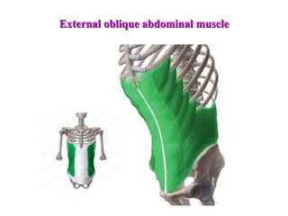
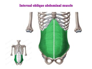
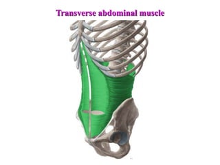
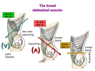
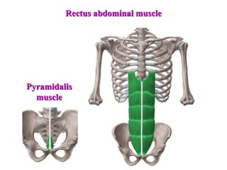
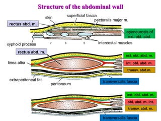
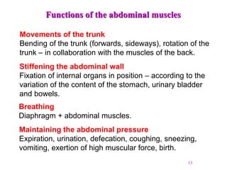
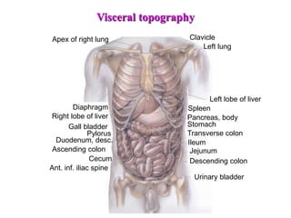
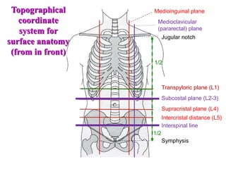
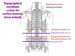
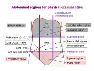
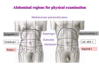
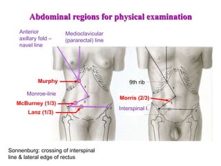
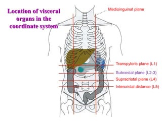
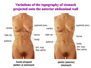
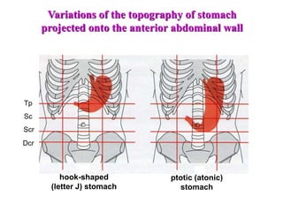
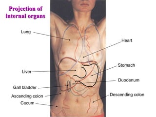
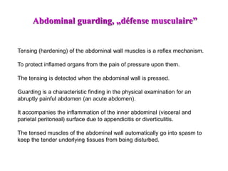
Surface Anatomy of the
Abdomen, Peritoneal
Relations
Department of Anatomy, Histology and Embryology
Semmelweis University
March 5, 2019
Dr. Ágoston Szél
Abdominal wall
External oblique abdominal muscle
Internal oblique abdominal muscle
Transverse abdominal muscle
6
ext.obl.
abd. m.
int. obl.
abd. m.
transv.
abd. m.
inguinal lig.
inguinal
lig.
The broad
abdominal muscles
iliac crest
lumbar
fascia
pubic
tubercle
(v)
(v) lumbar
fascia
inguinal lig.
Rectus abdominal muscle
Pyramidalis
muscle
8
ext. obl. abd. m.
int. obl. abd. m.
transv. abd.m.
ext. obl. abd. m.
obl. abd. m. int.
transv. abd. m.
transversalis fascia
peritoneum
linea alba
skin superficial fascia
rectus abd. m.
intercostal muscles
pectoralis major m.
extraperitoneal fat
aponeurosis of
ext. obl. abd.
xyphoid process
Structure of the abdominal wall
rectus abd. m.
transversalis fascia
urinary bladder
vagina
rectum
rectus abdominis
muscle
inferior epigastric
a&v.
lateral
abdominal
muscles
external iliac
artery.
Rectus sheath
superficial fascia
fatty layer (Camper’s
fascia)
position of penis
spermatic cord
position of scrotum
fascia lata
line of fusion
membranous layer
(Scarpa’s fascia)
aponeurosis of external
oblique muscle
Arrangement of the fatty and membranous layers of the
superficial fascia of the abdominal wall
*fatty layer
(Camper’s fascia)
*membranous layer
(Scarpa’s fascia)
perineal
membrane
dartos muscle
symphysis
urethra
Arrangement of fasciae in the lower abdominal wall
*superficial fascia
**Colles’s fascia
**membranous layer of
the superficial fascia
external spermatic,
cremasteric, internal
spermatic fasciae
12
Ext. obl. Abd.
Adductors
Rhomboideus
Serratus ant.
Pectoralis major
Int. obl. abd.
Gluteus maximus
Sartorius
Insertion of abdominal muscles in the
loop system of the body musculature
13
Functions of the abdominal muscles
Movements of the trunk
Bending of the trunk (forwards, sideways), rotation of the
trunk – in collaboration with the muscles of the back.
Stiffening the abdominal wall
Fixation of internal organs in position – according to the
variation of the content of the stomach, urinary bladder
and bowels.
Maintaining the abdominal pressure
Expiration, urination, defecation, coughing, sneezing,
vomiting, exertion of high muscular force, birth.
Breathing
Diaphragm + abdominal muscles.
Visceral
topography
Transverse colon
Urinary bladder
Descending colon
Ileum
Jejunum
Stomach
Pancreas, body
Spleen
Left lung
Clavicle
Cecum
Ascending colon
Right lobe of liver
Gall bladder
Pylorus
Duodenum, desc.
Ant. inf. iliac spine
Apex of right lung
Diaphragm
Left lobe of liver
Visceral topography
Medioinguinal plane
Transpyloric plane (L1)
Subcostal plane (L2-3)
Supracristal plane (L4)
Intercristal distance (L5)
Symphysis
Jugular notch
Interspinal line
Medioclavicular
(pararectal) plane
Topographical
coordinate
system for
surface anatomy
(from in front)
1/2
1/2
Medioinguinal plane
Transpyloric plane (L1)
Supracristal plane (L4)
Intercristal distance (L5)
Vertebra prominens
Subcostal plane (L2-3)
Interspinal line
Topographical
coordinate
system for
surface anatomy
(from behind)
EPIGASTRIUM
HYPOGASTRIUM
Medioclavicular
(pararectal) plane
Subcostal plane
Interspinal plane
Hypochondriac region
Epigastric region
Lateral abd. region
Umbilical region
Inguinal region
Pubic region
umb.
McBurney (1/2-1/3)
Lanz (1/3)
Abdominal regions for physical examinantion
MESOGASTRIUM
Ant. sup. iliac spine
Subcostal
Interspinal
Mediclavicular (pararectal) plane
Diaphragm Hypochondriac r.
Epigastric r.
Lat. abd. r.
Umbilical r.
Inguinal r.
Pubic r.
Abdominal regions for physical examination
Interspinal l.
Monroe-line
Medioclavicular
(pararectal) line
Anterior
axillary fold –
navel line
Murphy
McBurney (1/3)
Lanz (1/3)
9th rib
Abdominal regions for physical examination
Morris (2/3)
Sonnenburg: crossing of interspinal
line & lateral edge of rectus
Medioinguinal plane
Transpyloric plane (L1)
Supracristal plane (L4)
Intercristal distance (L5)
Subcostal plane (L2-3)
Location of visceral
organs in the
coordinate system
pylorus
pylorus
10th rib
10th rib
cardia cardia
xyphoid proc.
fornix
fornix
body
body
ant. sup.
iliac spine
hook-shaped
(letter J) stomach
ptotic (atonic)
stomach
Variations of the topography of stomach
projected onto the anterior abdominal wall
ant. sup.
iliac spine
xyphoid proc.
Tp
Sc
Scr
Dcr
Variations of the topography of stomach
projected onto the anterior abdominal wall
hook-shaped
(letter J) stomach
ptotic (atonic)
stomach
Cecum
Liver
Gall bladder
Lung
Heart
Stomach
Descending colon
Ascending colon
Duodenum
Projection of
internal organs
25
Diaphragm (C4)
Heart (T3-4)
Esophagus (T4-5)
Stomach (T8)
Liver, gall bladder (T8-11)
Small intestines (T10)
Large intestines T11)
Urinary bladder (T11-L1)
Testis, kidney (T10-L1)
Head-zones: irradiation of visceral pain
Tensing (hardening) of the abdominal wall muscles is a reflex mechanism.
To protect inflamed organs from the pain of pressure upon them.
The tensing is detected when the abdominal wall is pressed.
Guarding is a characteristic finding in the physical examination for an
abruptly painful abdomen (an acute abdomen).
It accompanies the inflammation of the inner abdominal (visceral and
parietal peritoneal) surface due to appendicitis or diverticulitis.
The tensed muscles of the abdominal wall automatically go into spasm to
keep the tender underlying tissues from being disturbed.
Abdominal guarding, „défense musculaire”
spine-navel
(Monroe’s) line
McBurney’s point
(1/2-1/3), Morris (2/3)
Lanz’s point (1/3)
ant. sup.
iliac spine
interspinal line
inguinal lig.
pelvic position (a)
anteroparietal position (b)
Ilioinguinal position (c)
lumbar position (d)
retrocecal position (e)
Position of appendix
Pelvic
position (a)
Ilioinguinal
position (c)
Lumbar position (d)
Anteroparietal
position (b)
Retrocecal
position (e)
Position of appendix
Change of
position of
appendix during
pregnancy
Tipical pressure
points in
appendicitis
Rovsing-signal:
upon vigorously
pressing the
abdomen counter-
clockwise,
the pain felt on
the appendix
region becomes
more intensive.
internal iliac artery
external iliac artery
median sacral artery
bifurcation of aorta (L4)
inf. mesenteric artery
psoas major muscle
gonadal arteries
left crus
aortic hiatus (Th12-L1)
inf. phrenic artery
esophageal hiatus (Th11)
median arcuate ligament
common iliac artery
bifurcation
(sacroiliac joint)
coeliac trunk
suprarenal artery
sup. mesenteric artery
renal artery
lumbar arteries
Branches of abdominal aorta
inferior vena cava
(Th12) coeliac trunk
(L1) superior
mesenteric artery
(L3) inferior
mesenteric artery
abdominal aorta
external iliac a.&v.
common iliac a&v.
anterior superior
iliac spine
The prjection of the abdominal aorta and branches
internal iliac a.&v.
Projection of colon
transverse colon
left colic flexure
right colic flexure
ascending colon descending colon
colon sigmoideum
rectum
appendix
cecum
ileum
medioinguinal plane
distantia cristarum
medioclavicular plane
cecum
Left lobe
Right lobe
Gall bladder
5th rib
8th rib
Medioinguinal plane
Transpyloric plane
Projection of liver and gall bladder
Gall bladder
Superior part (L1)
Descending part (L1-3)
Inferior part (L3) Ascending part (L3-2)
Duodenojejunal flexure (L1-2)
Tail (L1)
Body (L1-2)
Head (L2)
Projection of duodenum and pancreas
Jejunum: rather
vertical, in the upper
left quadrant
Ileum: rather
horizontal, in the
lower right
quadrant
Duodenum: horse-
shoe shaped, in
the upper right
quadrant
Position of small intestine
Skeletotopy of the spleen (from in front)
ribs
spleen (rib
8/9th-11th)
left lung
liver
stomach
transverse colon
diaphragm
costo-diaphrag-
matic recess
Topography of spleen (as seen from the left)
transpyloric plane (L1)
supracristal plane (L4)
intercristal plane (L5)
subcostal plane (L2-3)
medioinguinal plane
Topography of
the lung,
pleura, kidney
and spleen
left: 12th rib:
1/3-2/3
right: 12th rib:
upper pole
crista iliaca
post. sup. iliac spine
right kidney
12th rib
11th rib
left kidney
Posterior projection of kidneys
The peritoneum
Bibliography
• Lippert H: Lehrbuch Anatomie, Urban & Fischer, München, 2000
• Mac Kinnon P, Morris J: Oxford Lehrbuch der klinischen Anatomie,
Hans Huber, Bern, 1997
• Moore KL, Dalley AF: Clinically Oriented Anatomy, Lippincott, 1999
• Putz R, Pabst R: Sobotta. Alliter, Budapest, 2004.
• Patel R: Applied Peritoneal Anatomy, www.myESRorg.
• Renz-Polster H, Krautzig S, Braun J: Innere Medizin, Urban &
Fischer, München, 2004
• Snell RS, Clinical Anatomy, Little, Brown & Co, Boston, 1995
• Tirkes T et al: Peritoneal Anatomy. Gastrointestinal Imaging,
https://doi.org/10.1148/rg.322115032.
• https://www.kenhub.com/en/start/anatomy.

Has-felületi-topográfiája_angol veterinary.ppt

  • 1.
    Surface Anatomy ofthe Abdomen, Peritoneal Relations Department of Anatomy, Histology and Embryology Semmelweis University March 5, 2019 Dr. Ágoston Szél
  • 2.
  • 3.
  • 4.
  • 5.
  • 6.
    6 ext.obl. abd. m. int. obl. abd.m. transv. abd. m. inguinal lig. inguinal lig. The broad abdominal muscles iliac crest lumbar fascia pubic tubercle (v) (v) lumbar fascia inguinal lig.
  • 7.
  • 8.
    8 ext. obl. abd.m. int. obl. abd. m. transv. abd.m. ext. obl. abd. m. obl. abd. m. int. transv. abd. m. transversalis fascia peritoneum linea alba skin superficial fascia rectus abd. m. intercostal muscles pectoralis major m. extraperitoneal fat aponeurosis of ext. obl. abd. xyphoid process Structure of the abdominal wall rectus abd. m. transversalis fascia
  • 9.
    urinary bladder vagina rectum rectus abdominis muscle inferiorepigastric a&v. lateral abdominal muscles external iliac artery. Rectus sheath
  • 10.
    superficial fascia fatty layer(Camper’s fascia) position of penis spermatic cord position of scrotum fascia lata line of fusion membranous layer (Scarpa’s fascia) aponeurosis of external oblique muscle Arrangement of the fatty and membranous layers of the superficial fascia of the abdominal wall
  • 11.
    *fatty layer (Camper’s fascia) *membranouslayer (Scarpa’s fascia) perineal membrane dartos muscle symphysis urethra Arrangement of fasciae in the lower abdominal wall *superficial fascia **Colles’s fascia **membranous layer of the superficial fascia external spermatic, cremasteric, internal spermatic fasciae
  • 12.
    12 Ext. obl. Abd. Adductors Rhomboideus Serratusant. Pectoralis major Int. obl. abd. Gluteus maximus Sartorius Insertion of abdominal muscles in the loop system of the body musculature
  • 13.
    13 Functions of theabdominal muscles Movements of the trunk Bending of the trunk (forwards, sideways), rotation of the trunk – in collaboration with the muscles of the back. Stiffening the abdominal wall Fixation of internal organs in position – according to the variation of the content of the stomach, urinary bladder and bowels. Maintaining the abdominal pressure Expiration, urination, defecation, coughing, sneezing, vomiting, exertion of high muscular force, birth. Breathing Diaphragm + abdominal muscles.
  • 14.
  • 15.
    Transverse colon Urinary bladder Descendingcolon Ileum Jejunum Stomach Pancreas, body Spleen Left lung Clavicle Cecum Ascending colon Right lobe of liver Gall bladder Pylorus Duodenum, desc. Ant. inf. iliac spine Apex of right lung Diaphragm Left lobe of liver Visceral topography
  • 16.
    Medioinguinal plane Transpyloric plane(L1) Subcostal plane (L2-3) Supracristal plane (L4) Intercristal distance (L5) Symphysis Jugular notch Interspinal line Medioclavicular (pararectal) plane Topographical coordinate system for surface anatomy (from in front) 1/2 1/2
  • 17.
    Medioinguinal plane Transpyloric plane(L1) Supracristal plane (L4) Intercristal distance (L5) Vertebra prominens Subcostal plane (L2-3) Interspinal line Topographical coordinate system for surface anatomy (from behind)
  • 18.
    EPIGASTRIUM HYPOGASTRIUM Medioclavicular (pararectal) plane Subcostal plane Interspinalplane Hypochondriac region Epigastric region Lateral abd. region Umbilical region Inguinal region Pubic region umb. McBurney (1/2-1/3) Lanz (1/3) Abdominal regions for physical examinantion MESOGASTRIUM Ant. sup. iliac spine
  • 19.
    Subcostal Interspinal Mediclavicular (pararectal) plane DiaphragmHypochondriac r. Epigastric r. Lat. abd. r. Umbilical r. Inguinal r. Pubic r. Abdominal regions for physical examination
  • 20.
    Interspinal l. Monroe-line Medioclavicular (pararectal) line Anterior axillaryfold – navel line Murphy McBurney (1/3) Lanz (1/3) 9th rib Abdominal regions for physical examination Morris (2/3) Sonnenburg: crossing of interspinal line & lateral edge of rectus
  • 21.
    Medioinguinal plane Transpyloric plane(L1) Supracristal plane (L4) Intercristal distance (L5) Subcostal plane (L2-3) Location of visceral organs in the coordinate system
  • 22.
    pylorus pylorus 10th rib 10th rib cardiacardia xyphoid proc. fornix fornix body body ant. sup. iliac spine hook-shaped (letter J) stomach ptotic (atonic) stomach Variations of the topography of stomach projected onto the anterior abdominal wall ant. sup. iliac spine xyphoid proc.
  • 23.
    Tp Sc Scr Dcr Variations of thetopography of stomach projected onto the anterior abdominal wall hook-shaped (letter J) stomach ptotic (atonic) stomach
  • 24.
  • 25.
    25 Diaphragm (C4) Heart (T3-4) Esophagus(T4-5) Stomach (T8) Liver, gall bladder (T8-11) Small intestines (T10) Large intestines T11) Urinary bladder (T11-L1) Testis, kidney (T10-L1) Head-zones: irradiation of visceral pain
  • 26.
    Tensing (hardening) ofthe abdominal wall muscles is a reflex mechanism. To protect inflamed organs from the pain of pressure upon them. The tensing is detected when the abdominal wall is pressed. Guarding is a characteristic finding in the physical examination for an abruptly painful abdomen (an acute abdomen). It accompanies the inflammation of the inner abdominal (visceral and parietal peritoneal) surface due to appendicitis or diverticulitis. The tensed muscles of the abdominal wall automatically go into spasm to keep the tender underlying tissues from being disturbed. Abdominal guarding, „défense musculaire”
  • 27.
    spine-navel (Monroe’s) line McBurney’s point (1/2-1/3),Morris (2/3) Lanz’s point (1/3) ant. sup. iliac spine interspinal line inguinal lig. pelvic position (a) anteroparietal position (b) Ilioinguinal position (c) lumbar position (d) retrocecal position (e) Position of appendix
  • 28.
    Pelvic position (a) Ilioinguinal position (c) Lumbarposition (d) Anteroparietal position (b) Retrocecal position (e) Position of appendix
  • 29.
  • 30.
  • 31.
    Rovsing-signal: upon vigorously pressing the abdomencounter- clockwise, the pain felt on the appendix region becomes more intensive.
  • 32.
    internal iliac artery externaliliac artery median sacral artery bifurcation of aorta (L4) inf. mesenteric artery psoas major muscle gonadal arteries left crus aortic hiatus (Th12-L1) inf. phrenic artery esophageal hiatus (Th11) median arcuate ligament common iliac artery bifurcation (sacroiliac joint) coeliac trunk suprarenal artery sup. mesenteric artery renal artery lumbar arteries Branches of abdominal aorta
  • 33.
    inferior vena cava (Th12)coeliac trunk (L1) superior mesenteric artery (L3) inferior mesenteric artery abdominal aorta external iliac a.&v. common iliac a&v. anterior superior iliac spine The prjection of the abdominal aorta and branches internal iliac a.&v.
  • 34.
    Projection of colon transversecolon left colic flexure right colic flexure ascending colon descending colon colon sigmoideum rectum appendix cecum ileum medioinguinal plane distantia cristarum medioclavicular plane cecum
  • 35.
    Left lobe Right lobe Gallbladder 5th rib 8th rib Medioinguinal plane Transpyloric plane Projection of liver and gall bladder Gall bladder
  • 36.
    Superior part (L1) Descendingpart (L1-3) Inferior part (L3) Ascending part (L3-2) Duodenojejunal flexure (L1-2) Tail (L1) Body (L1-2) Head (L2) Projection of duodenum and pancreas
  • 37.
    Jejunum: rather vertical, inthe upper left quadrant Ileum: rather horizontal, in the lower right quadrant Duodenum: horse- shoe shaped, in the upper right quadrant Position of small intestine
  • 38.
    Skeletotopy of thespleen (from in front) ribs
  • 39.
    spleen (rib 8/9th-11th) left lung liver stomach transversecolon diaphragm costo-diaphrag- matic recess Topography of spleen (as seen from the left)
  • 40.
    transpyloric plane (L1) supracristalplane (L4) intercristal plane (L5) subcostal plane (L2-3) medioinguinal plane Topography of the lung, pleura, kidney and spleen left: 12th rib: 1/3-2/3 right: 12th rib: upper pole
  • 41.
    crista iliaca post. sup.iliac spine right kidney 12th rib 11th rib left kidney Posterior projection of kidneys
  • 42.
  • 43.
    Bibliography • Lippert H:Lehrbuch Anatomie, Urban & Fischer, München, 2000 • Mac Kinnon P, Morris J: Oxford Lehrbuch der klinischen Anatomie, Hans Huber, Bern, 1997 • Moore KL, Dalley AF: Clinically Oriented Anatomy, Lippincott, 1999 • Putz R, Pabst R: Sobotta. Alliter, Budapest, 2004. • Patel R: Applied Peritoneal Anatomy, www.myESRorg. • Renz-Polster H, Krautzig S, Braun J: Innere Medizin, Urban & Fischer, München, 2004 • Snell RS, Clinical Anatomy, Little, Brown & Co, Boston, 1995 • Tirkes T et al: Peritoneal Anatomy. Gastrointestinal Imaging, https://doi.org/10.1148/rg.322115032. • https://www.kenhub.com/en/start/anatomy.