Peritoneum
To -MBBS 2ND Year
Dr Laxman Khanal
Assistant Professor (Department of Human Anatomy)
05-09-2016
For getting through annual
examination !!!
Q. Peritoneum derived from.
a. Ectoderm b. Mesoderm
c. Endoderm d. All three germ layer
Q. Peritoneal fluid is derived from.
a. Plasma b. Interstitial fluid
c. Plasma and interstitial fluid d. Intracellular fluid
Q. All of the peritoneal folds are double layered except.
a. mesentery b. Lesser omentum
c. Greater omentum d. Transverse mesocolon
Today’s objective
• To define peritoneum
• Formation and importance of peritoneum.
• To define peritoneal and retroperitoneal
organs.
• Define different modification of peritoneum.
Introduction
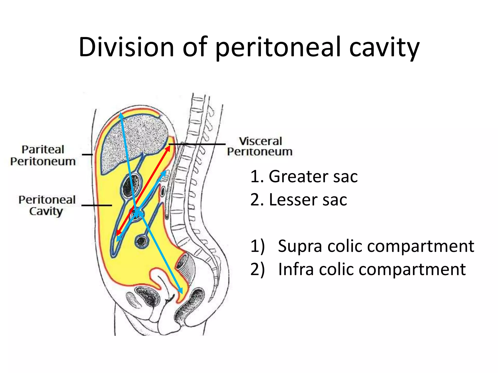
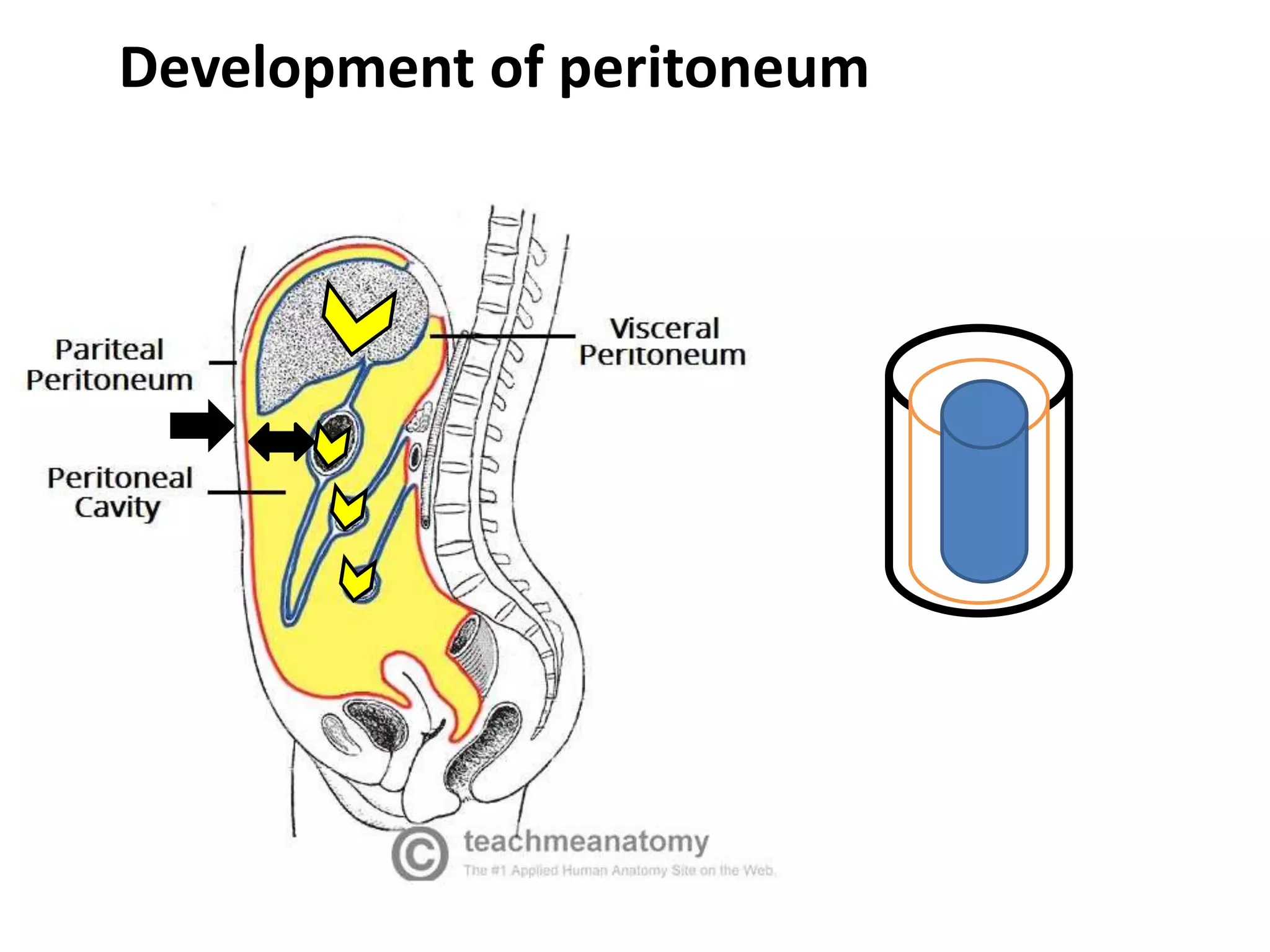
• The peritoneum is a thin serous membrane that
line the walls of the abdominal and pelvic cavities
and cover the organs within these cavities.
• Peritoneal cavity is the potential space between
the parietal and visceral layer of peritoneum.
• Cavity is divided into 2 main sacs- greater sac and
lesser sac.
• Small recesses of peritoneal cavity also present
which are separated from main cavity by fold of
peritoneum ( site for internal hernia).
Sex differences
in male
• Closed sac
• Lined by mesothelium or squamous epithelium
in female
•Its not closed sac , communicate through uterine tubes.
•Peritoneum covering the ovary is cuboidal epithelium.
•Peritoneum of the fimbria is lined by ciliated columner
epithelium.
Function
• Movement of viscera
• Protection of viscera
• Absorptive function- used for dialysis purpose
and Ventriculoperitoneal shunts.
• Regeneration
• Storage of fat
Normal amount of peritoneal fluid is 50 ml
Innervations
• Innervations of Parietal peritoneum is same as
that of body wall (intercostal nerve and phrenic
nerve). It is pain sensitive.
• Innervations of the Viscera peritoneum is same as
that of viscera ( autonomic nerve).
Pain of foregut referred to the Epigastric region
Pain of midgut to the umbilical region
Pain of hindgut to the hypogastric region
Division of peritoneal cavity
1. Greater sac
2. Lesser sac
1) Supra colic compartment
2) Infra colic compartment
Development of peritoneum
At the end of 3rd week lateral plate mesoderm split
into 2 layer, somatic layer and splanchnic layer.
Understand the path from retroperitoneal area to the peritoneal
area.
Ventral mesentery exists only in the region of the terminal
part of the esophagus, the stomach, and the upper part of
the duodenum, is derived from the septum transversum.
Relationship between viscera and
peritoneum
• Peritoneal organ
• Retroperitoneal organ
1. Primary
2. Secondary
 No organ found in peritoneal cavity.
 Mesentery hangs the peritoneal organ to the
body wall.
zygosis
• Duodenum-2nd and 3rd part
• Pancreas (except tail)
• Ascending colon
• Descending colon
• Upper rectum
Secondary
retroperitoneal organ
{Foregut
Mid gut and Hindgut
Lesser omentum
Falciform ligament
More caudal part forms the greater omentum
VDRL
Structures formed by peritoneum
• Omentum – Stomach
• Mesentery
• Transverse mesocolon
• Sigmoid mesocolon
• Meso appendix
• Ligaments
• Folds, recesses and fossa
Derivatives of ventral mesogastrium
Sigmoid mesocolon
• It is a broad fan shaped fold of
peritoneum which suspends the
coils of jejunum and ileum from
posterior abdominal wall.
• Root of mesentery is 15 cm long.
• Extends from D-J flexure to the
ileocaecal junction.
Mesentery
Mesoappendix
• Extends from terminal part of ileum to appendix.
• Appendicular artery runs in free margin of it.
Cont. in Peritoneum II

Peritoneum i

  • 1.
    Peritoneum To -MBBS 2NDYear Dr Laxman Khanal Assistant Professor (Department of Human Anatomy) 05-09-2016
  • 2.
    For getting throughannual examination !!!
  • 3.
    Q. Peritoneum derivedfrom. a. Ectoderm b. Mesoderm c. Endoderm d. All three germ layer Q. Peritoneal fluid is derived from. a. Plasma b. Interstitial fluid c. Plasma and interstitial fluid d. Intracellular fluid Q. All of the peritoneal folds are double layered except. a. mesentery b. Lesser omentum c. Greater omentum d. Transverse mesocolon
  • 4.
    Today’s objective • Todefine peritoneum • Formation and importance of peritoneum. • To define peritoneal and retroperitoneal organs. • Define different modification of peritoneum.
  • 5.
    Introduction • The peritoneumis a thin serous membrane that line the walls of the abdominal and pelvic cavities and cover the organs within these cavities. • Peritoneal cavity is the potential space between the parietal and visceral layer of peritoneum. • Cavity is divided into 2 main sacs- greater sac and lesser sac. • Small recesses of peritoneal cavity also present which are separated from main cavity by fold of peritoneum ( site for internal hernia).
  • 7.
    Sex differences in male •Closed sac • Lined by mesothelium or squamous epithelium in female •Its not closed sac , communicate through uterine tubes. •Peritoneum covering the ovary is cuboidal epithelium. •Peritoneum of the fimbria is lined by ciliated columner epithelium.
  • 9.
    Function • Movement ofviscera • Protection of viscera • Absorptive function- used for dialysis purpose and Ventriculoperitoneal shunts. • Regeneration • Storage of fat Normal amount of peritoneal fluid is 50 ml
  • 10.
    Innervations • Innervations ofParietal peritoneum is same as that of body wall (intercostal nerve and phrenic nerve). It is pain sensitive. • Innervations of the Viscera peritoneum is same as that of viscera ( autonomic nerve). Pain of foregut referred to the Epigastric region Pain of midgut to the umbilical region Pain of hindgut to the hypogastric region
  • 11.
    Division of peritonealcavity 1. Greater sac 2. Lesser sac 1) Supra colic compartment 2) Infra colic compartment
  • 12.
  • 14.
    At the endof 3rd week lateral plate mesoderm split into 2 layer, somatic layer and splanchnic layer.
  • 15.
    Understand the pathfrom retroperitoneal area to the peritoneal area.
  • 16.
    Ventral mesentery existsonly in the region of the terminal part of the esophagus, the stomach, and the upper part of the duodenum, is derived from the septum transversum.
  • 17.
    Relationship between visceraand peritoneum • Peritoneal organ • Retroperitoneal organ 1. Primary 2. Secondary  No organ found in peritoneal cavity.  Mesentery hangs the peritoneal organ to the body wall. zygosis
  • 18.
    • Duodenum-2nd and3rd part • Pancreas (except tail) • Ascending colon • Descending colon • Upper rectum Secondary retroperitoneal organ
  • 19.
  • 20.
  • 22.
    More caudal partforms the greater omentum
  • 23.
  • 25.
    Structures formed byperitoneum • Omentum – Stomach • Mesentery • Transverse mesocolon • Sigmoid mesocolon • Meso appendix • Ligaments • Folds, recesses and fossa
  • 26.
  • 34.
  • 35.
    • It isa broad fan shaped fold of peritoneum which suspends the coils of jejunum and ileum from posterior abdominal wall. • Root of mesentery is 15 cm long. • Extends from D-J flexure to the ileocaecal junction. Mesentery
  • 37.
    Mesoappendix • Extends fromterminal part of ileum to appendix. • Appendicular artery runs in free margin of it.
  • 39.