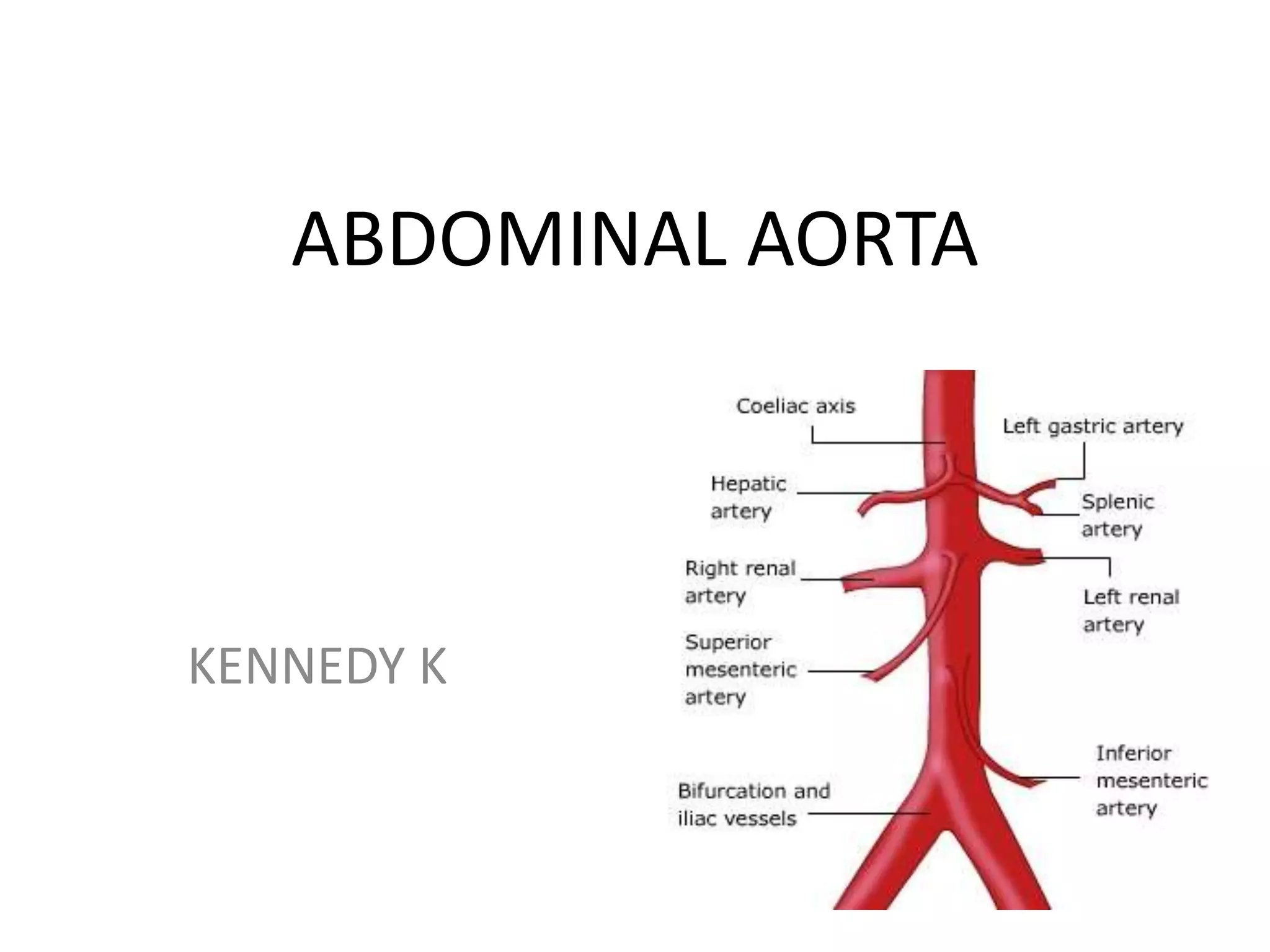
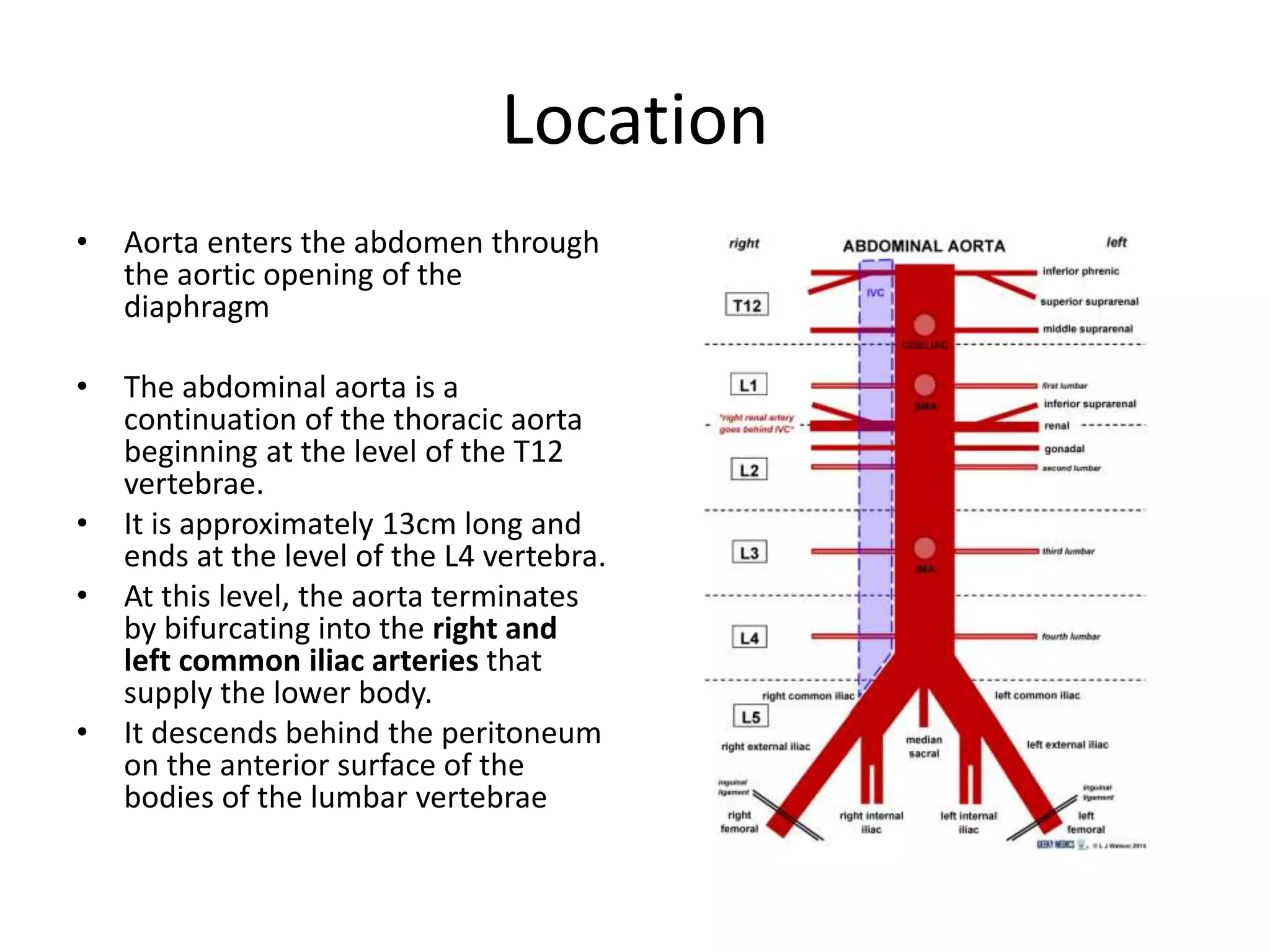
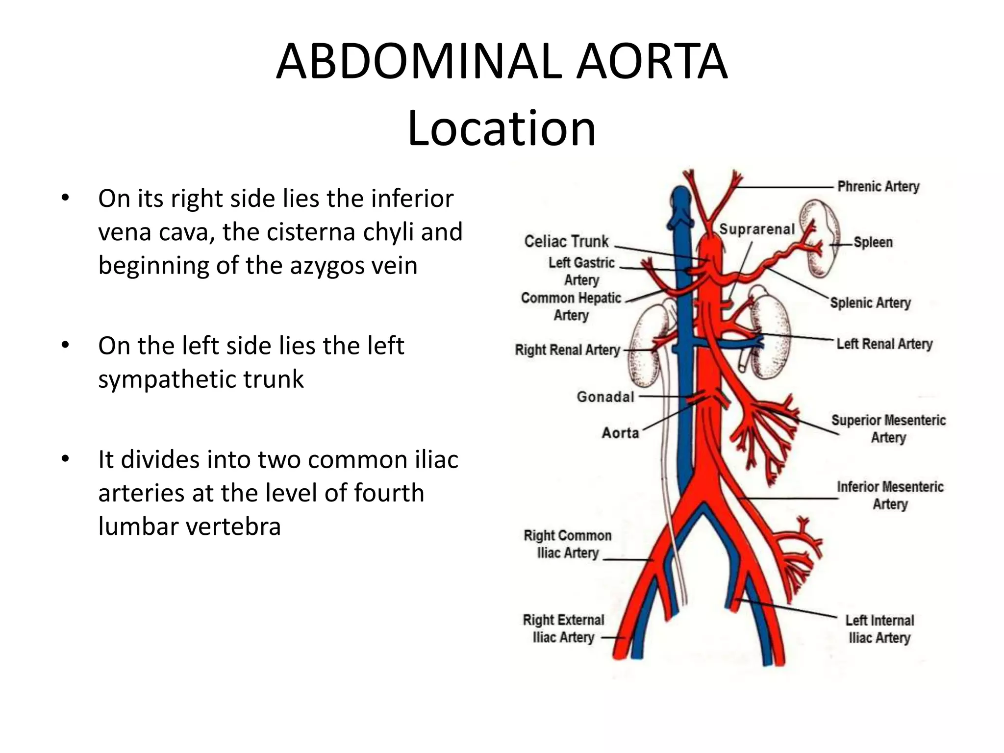
ABDOMINAL AORTA
KENNEDY K
Location
• Aorta enters the abdomen through
the aortic opening of the
diaphragm
• The abdominal aorta is a
continuation of the thoracic aorta
beginning at the level of the T12
vertebrae.
• It is approximately 13cm long and
ends at the level of the L4 vertebra.
• At this level, the aorta terminates
by bifurcating into the right and
left common iliac arteries that
supply the lower body.
• It descends behind the peritoneum
on the anterior surface of the
bodies of the lumbar vertebrae
ABDOMINAL AORTA
Location
• On its right side lies the inferior
vena cava, the cisterna chyli and
beginning of the azygos vein
• On the left side lies the left
sympathetic trunk
• It divides into two common iliac
arteries at the level of fourth
lumbar vertebra
Abdominal Aorta
Branches
• In descending order:
1. Inferior phrenic arteries: Paired parietal arteries arising posteriorly
at the level of T12. They supply the diaphragm.
2. Coeliac artery: A large, unpaired visceral artery arising anteriorly at
the level of L1. It is also known as the celiac trunk and supplies the
liver, stomach, abdominal oesophagus, spleen, the superior
duodenum and the superior pancreas.
3. Superior mesenteric artery: A large, unpaired visceral artery
arising anteriorly, just below the celiac artery. It supplies the distal
duodenum, jejuno-ileum, ascending colon and part of the
transverse colon. It arises at the lower level of L1.
4. Middle suprarenal arteries: Small paired visceral arteries that arise
either side posteriorly at the level of L1 to supply the adrenal
glands.
Abdominal Aorta…
5. Renal arteries: Paired visceral arteries that arise laterally at the level
between L1 and L2. They supply the kidneys.
6. Gonadal arteries: Paired visceral arteries that arise laterally at the
level of L2. Note that the male gonadal artery is referred to as the
testicular artery and in females, the ovarian artery.
7. Inferior mesenteric artery: A large, unpaired visceral artery that
arises anteriorly at the level of L3. It supplies the large intestine
from the splenic flexure to the upper part of the rectum.
8. Median sacral artery: An unpaired parietal artery that arises
posteriorly at the level of L4 to supply the coccyx, lumbar
vertebrae and the sacrum.
9. Lumbar arteries: There are four pairs of parietal lumbar arteries that
arise posterolaterally between the levels of L1 and L4 to supply
the abdominal wall and spinal cord.
ABDOMINAL AORTA
Branches
• Three anterior visceral
branches:
– celiac artery,
– superior and
– inferior mesenteric arteries
• Three lateral visceral
branches:
– suprarenal artery,
– renal artery,
– testicular or ovarian artery
ABDOMINAL AORTA
Branches
• Five lateral abdominal wall branches:
– the inferior phrenic artery and
– four lumbar arteries
• Three terminal branches:
– two common iliac and
– the median sacral artery
COELIC TRUNK
• The coeliac trunk is the second branch of the abdominal aorta (the
first branches are the paired inferior phrenic arteries).
• It arises from the anterior aspect of the aorta, at the aortic hiatus of
the diaphragm (T12 level).
• After emerging from the aorta, the coeliac trunk extends
approximately 1cm before dividing into three major branches:
• left gastric,
• splenic and
• common hepatic arteries.
• Of these branches, two go left and one goes to the right-hand side.
• Collectively, they are the major arterial supply to the stomach,
spleen, liver, gall bladder, abdominal oesophagus, pancreas and
duodenum.
.
MAJOR BRANCHES OF COELIAC TRUNK:
1. Left Gastric Artery
• The left gastric artery is
the smallest of the three
branches.
• It ascends across the
diaphragm, giving rise
to oesophageal
branches, before
continuing anteriorly
along the lesser curvature
of the stomach.
• Here, it anastomoses
with the right gastric
artery
2. Splenic Artery
• Arises from the coeliac trunk just inferior to the left gastric artery.
• It then travels left towards the spleen, running posterior to the
stomach and along the superior margin of the pancreas.
• During its course, it is contained within the splenorenal ligament.
• It terminates into five branches which supply the segments of the
spleen.
• splenic artery also gives rise to several important vessels:
– Left gastroepiploic: supplies the greater curvature of the
stomach. Anastomoses with the right gastroepiploic artery.
– Short gastrics: 5-7 small branches supplying the fundus of the
stomach.
– Pancreatic branches: supply the body and tail of the pancreas.
.
3. Common Hepatic Artery
• The common hepatic artery is the sole arterial supply to
the liver and the only branch of the coeliac artery to pass
to the right.
• As it travels past the superior aspect of the duodenum, it
divides into its two terminal branches
– the proper hepatic and
– gastroduodenal arteries.
• Each of these arteries has multiple branches and
variation in the arrangement of these branches is
common.
.
Proper Hepatic
• The proper hepatic artery ascends through the lesser
omentum towards the liver. It gives rise to:
– Right gastric: supplies the pylorus and lesser
curvature of the stomach.
– Right and left hepatic: divide inferior to the porta
hepatis and supply their respective lobes of the liver.
– Cystic: branch of the right hepatic artery – supplies
the gall bladder.
.
Gastroduodenal
• The gastroduodenal artery descends posterior to the
superior portion of the duodenum. Its branches are:
– Right gastroepiploic: supplies the greater curvature of
the stomach. Found between the layers of the greater
omentum, which it also supplies.
– Superior pancreaticoduodenal: divides into an
anterior and posterior branch, which supplies the
head of the pancreas.
Anastomoses
Stomach
• The stomach is the only organ to receive arterial supply from all three
branches of the coeliac trunk. This is achieved through a system of
anastomoses along the greater (gastroepiploic arteries) and lesser
(gastric arteries) curvatures.
Pancreas
• The pancreaticoduodenal arcade is a network of arteries that
surround and supply the head of the pancreas.
• There are two main arteries – each has an anterior and posterior
branch, that anastomose (e.g. anterior to anterior) forming a ring
structure:
– Superior pancreaticoduodenal– a branch of the gastroduodenal
artery.
– Inferior pancreaticoduodenal – branch of superior mesenteric
artery (SMA)
.
.
2. The Superior Mesenteric Artery (SMA)
• It arises from the abdominal aorta, and supplies
arterial blood to the organs of the midgut – which
spans from the major duodenal papilla (of the
duodenum) to the proximal 2/3 of the transverse
colon.
• It arises anteriorly from the abdominal aorta at the
level of the L1 vertebrae, immediately inferior to the
origin of the coeliac trunk.
Branches of Superior Mesenteric
Artery
1. inferior pancreaticoduodenal artery
2. Numerous arteries that supply the jejunum
and ileum.
3. Middle and Right Colic Arteries
4. ileocolic artery
.
Major Branches of SMA
1. Inferior Pancreaticoduodenal Artery
• It forms anterior and posterior vessels, which
anastamose with branches of the superior
pancreaticoduodenal artery (derived from the
coeliac trunk).
• This network supplies the inferior region of
the head of the pancreas, the uncinate
process, and the duodenum.
2. Jejunal and Ileal Arteries
• The superior mesenteric artery gives rise to numerous
arteries that supply the jejunum and ileum.
• The arteries pass between the layers of the mesentery
and form anastamotic arcades – from which smaller,
straight arteries (known as the “vasa recta”) arise to
supply the organs.
• The jejunal blood supply is characterised by a smaller
number of arterial arcades, but longer vasa recta.
• In contrast, the ileal blood supply is marked by more
arterial arcades with shorter vasa recta
3. Middle and Right Colic Arteries
• The right and middle colic arteries arise from
the right side of the superior mesenteric
artery:
– Middle colic artery – supplies the transverse
colon.
– Right colic artery – supplies the ascending colon.
4. Ileocolic Artery
• The ileocolic artery is the final major branch of
the superior meseneric artery.
• It passes inferiorly and to the right, giving rise
to branches to the ascending colon, appendix,
cecum, and ileum.
• In cases of appendectomy, the appendicular
artery is ligated.
The Inferior Mesenteric Artery (IMA)
• The inferior mesenteric artery (IMA) is a major branch
of the abdominal aorta.
• It supplies arterial blood to the organs of the hindgut –
the distal 1/3 of the transverse colon, splenic flexure,
descending colon, sigmoid colon and rectum.
Anatomical Position
• It arises at L3, near the inferior border of the
duodenum, 3-4 cm above where the aorta bifurcates
into the common iliac arteries.
• As the artery arises from the aorta, it descends
anteriorly to its parent vessel, before moving to the left
side.
• It is a retroperitoneal structure.
Branches of Inferior Mesenteric Artery
• .
.
BRANCHES OF IMA
1. Left Colic Artery
• It supplies the distal 1/3 of the transverse colon and
the descending colon.
• After arising from its parent artery, it travels anteriorly
to the psoas major muscle, left ureter and left internal
spermatic vessels, before dividing into ascending and
descending branches:
– Ascending branch – crosses the left kidney anteriorly,
before entering the mesentry of the transverse colon,
moving superiorly. It supplies the distal 1/3 of the
transverse colon, and the upper aspect of the descending
colon.
– Descending branch – moves inferiorly to supply the lower
part of the descending colon. It anastamoses with the
superior sigmoid artery.
Branches of inferior mesenteric artery
2. Sigmoid Arteries
• The sigmoid arteries supply the descending
colon and the sigmoid colon.
• There are typically 2-4 branches, with the
uppermost branch termed the superior
sigmoid artery.
• They run inferiorly, obliquely and to the left,
crossing over the psoas major, left ureter and
left internal spermatic vessels.
3. Superior Rectal Artery
• The superior rectal artery is a continuation of the
inferior mesenteric artery, supplying the rectum.
• It descends into the pelvis, crossing the left
common iliac artery and vein.
• At the S3 vertebral level, the artery divides into
two terminal branches – one supplying each side
of the rectum.
• Within the walls of the rectum, smaller divisions
of these branches eventually communicate with
the middle and inferior rectal arteries.
Renal arteries
• Are two large trunks, which arise from the side of the aorta, immediately
below the superior mesenteric artery.
• Each is directed across the crus of the diaphragm, so as to form nearly a
right angle with the aorta.
• Before reaching the hilus of the kidney, each artery divides into four or five
branches; the greater number of these lie between the renal vein and
ureter, the vein being in front, the ureter behind, but one or more
branches are usually situated behind the ureter.
• Each vessel gives off some small inferior suprarenal branches to the
suprarenal gland, the ureter, and the surrounding cellular tissue and
muscles.
• One or two accessory renal arteries are frequently found, more especially
on the left side they usually arise from the aorta, and may come off above
or below the main artery, the former being the more common position.
• Instead of entering the kidney at the hilus, they usually pierce the upper
or lower part of the gland.
GONAND ARTERIES
• They are two slender vessels of considerable
length, and arise from the front of the aorta a
little below the renal arteries.
• These are:
– ovarian arteries
– internal spermatic arteries
inferior phrenic arteries
• two small vessels, which supply the diaphragm but present much
variety in their origin.
• They may arise separately from the front of the aorta, immediately
above the celiac artery, or by a common trunk, which may spring
either from the aorta or from the celiac artery.
• The medial branch curves forward, and anastomoses with its fellow
of the opposite side, and with the musculophrenic and
pericardiacophrenic arteries.
• The lateral branch passes toward the side of the thorax, and
anastomoses with the lower intercostal arteries, and with the
musculophrenic.
• The lateral branch of the right phrenic gives off a few vessels to the
inferior vena cava; and the left one, some branches to the
esophagus. Each vessel gives off superior suprarenal branches to
the suprarenal gland of its own side.
• The spleen and the liver also receive a few twigs from the left and
right vessels respectively.
The lumbar arteries
• are in series with the intercostals.
• They are usually four in number on either
side, and arise from the back of the aorta,
opposite the bodies of the upper four lumbar
vertebræ.
THANK YOU

2 b. ABDOMINAL AORTA1.ppt

  • 1.
  • 2.
    Location • Aorta entersthe abdomen through the aortic opening of the diaphragm • The abdominal aorta is a continuation of the thoracic aorta beginning at the level of the T12 vertebrae. • It is approximately 13cm long and ends at the level of the L4 vertebra. • At this level, the aorta terminates by bifurcating into the right and left common iliac arteries that supply the lower body. • It descends behind the peritoneum on the anterior surface of the bodies of the lumbar vertebrae
  • 4.
    ABDOMINAL AORTA Location • Onits right side lies the inferior vena cava, the cisterna chyli and beginning of the azygos vein • On the left side lies the left sympathetic trunk • It divides into two common iliac arteries at the level of fourth lumbar vertebra
  • 5.
    Abdominal Aorta Branches • Indescending order: 1. Inferior phrenic arteries: Paired parietal arteries arising posteriorly at the level of T12. They supply the diaphragm. 2. Coeliac artery: A large, unpaired visceral artery arising anteriorly at the level of L1. It is also known as the celiac trunk and supplies the liver, stomach, abdominal oesophagus, spleen, the superior duodenum and the superior pancreas. 3. Superior mesenteric artery: A large, unpaired visceral artery arising anteriorly, just below the celiac artery. It supplies the distal duodenum, jejuno-ileum, ascending colon and part of the transverse colon. It arises at the lower level of L1. 4. Middle suprarenal arteries: Small paired visceral arteries that arise either side posteriorly at the level of L1 to supply the adrenal glands.
  • 6.
    Abdominal Aorta… 5. Renalarteries: Paired visceral arteries that arise laterally at the level between L1 and L2. They supply the kidneys. 6. Gonadal arteries: Paired visceral arteries that arise laterally at the level of L2. Note that the male gonadal artery is referred to as the testicular artery and in females, the ovarian artery. 7. Inferior mesenteric artery: A large, unpaired visceral artery that arises anteriorly at the level of L3. It supplies the large intestine from the splenic flexure to the upper part of the rectum. 8. Median sacral artery: An unpaired parietal artery that arises posteriorly at the level of L4 to supply the coccyx, lumbar vertebrae and the sacrum. 9. Lumbar arteries: There are four pairs of parietal lumbar arteries that arise posterolaterally between the levels of L1 and L4 to supply the abdominal wall and spinal cord.
  • 7.
    ABDOMINAL AORTA Branches • Threeanterior visceral branches: – celiac artery, – superior and – inferior mesenteric arteries • Three lateral visceral branches: – suprarenal artery, – renal artery, – testicular or ovarian artery
  • 8.
    ABDOMINAL AORTA Branches • Fivelateral abdominal wall branches: – the inferior phrenic artery and – four lumbar arteries • Three terminal branches: – two common iliac and – the median sacral artery
  • 9.
    COELIC TRUNK • Thecoeliac trunk is the second branch of the abdominal aorta (the first branches are the paired inferior phrenic arteries). • It arises from the anterior aspect of the aorta, at the aortic hiatus of the diaphragm (T12 level). • After emerging from the aorta, the coeliac trunk extends approximately 1cm before dividing into three major branches: • left gastric, • splenic and • common hepatic arteries. • Of these branches, two go left and one goes to the right-hand side. • Collectively, they are the major arterial supply to the stomach, spleen, liver, gall bladder, abdominal oesophagus, pancreas and duodenum.
  • 10.
  • 11.
    MAJOR BRANCHES OFCOELIAC TRUNK: 1. Left Gastric Artery • The left gastric artery is the smallest of the three branches. • It ascends across the diaphragm, giving rise to oesophageal branches, before continuing anteriorly along the lesser curvature of the stomach. • Here, it anastomoses with the right gastric artery
  • 12.
    2. Splenic Artery •Arises from the coeliac trunk just inferior to the left gastric artery. • It then travels left towards the spleen, running posterior to the stomach and along the superior margin of the pancreas. • During its course, it is contained within the splenorenal ligament. • It terminates into five branches which supply the segments of the spleen. • splenic artery also gives rise to several important vessels: – Left gastroepiploic: supplies the greater curvature of the stomach. Anastomoses with the right gastroepiploic artery. – Short gastrics: 5-7 small branches supplying the fundus of the stomach. – Pancreatic branches: supply the body and tail of the pancreas.
  • 13.
  • 14.
    3. Common HepaticArtery • The common hepatic artery is the sole arterial supply to the liver and the only branch of the coeliac artery to pass to the right. • As it travels past the superior aspect of the duodenum, it divides into its two terminal branches – the proper hepatic and – gastroduodenal arteries. • Each of these arteries has multiple branches and variation in the arrangement of these branches is common.
  • 15.
    . Proper Hepatic • Theproper hepatic artery ascends through the lesser omentum towards the liver. It gives rise to: – Right gastric: supplies the pylorus and lesser curvature of the stomach. – Right and left hepatic: divide inferior to the porta hepatis and supply their respective lobes of the liver. – Cystic: branch of the right hepatic artery – supplies the gall bladder.
  • 16.
    . Gastroduodenal • The gastroduodenalartery descends posterior to the superior portion of the duodenum. Its branches are: – Right gastroepiploic: supplies the greater curvature of the stomach. Found between the layers of the greater omentum, which it also supplies. – Superior pancreaticoduodenal: divides into an anterior and posterior branch, which supplies the head of the pancreas.
  • 17.
    Anastomoses Stomach • The stomachis the only organ to receive arterial supply from all three branches of the coeliac trunk. This is achieved through a system of anastomoses along the greater (gastroepiploic arteries) and lesser (gastric arteries) curvatures. Pancreas • The pancreaticoduodenal arcade is a network of arteries that surround and supply the head of the pancreas. • There are two main arteries – each has an anterior and posterior branch, that anastomose (e.g. anterior to anterior) forming a ring structure: – Superior pancreaticoduodenal– a branch of the gastroduodenal artery. – Inferior pancreaticoduodenal – branch of superior mesenteric artery (SMA)
  • 18.
  • 19.
  • 20.
    2. The SuperiorMesenteric Artery (SMA) • It arises from the abdominal aorta, and supplies arterial blood to the organs of the midgut – which spans from the major duodenal papilla (of the duodenum) to the proximal 2/3 of the transverse colon. • It arises anteriorly from the abdominal aorta at the level of the L1 vertebrae, immediately inferior to the origin of the coeliac trunk.
  • 21.
    Branches of SuperiorMesenteric Artery 1. inferior pancreaticoduodenal artery 2. Numerous arteries that supply the jejunum and ileum. 3. Middle and Right Colic Arteries 4. ileocolic artery
  • 22.
  • 23.
    Major Branches ofSMA 1. Inferior Pancreaticoduodenal Artery • It forms anterior and posterior vessels, which anastamose with branches of the superior pancreaticoduodenal artery (derived from the coeliac trunk). • This network supplies the inferior region of the head of the pancreas, the uncinate process, and the duodenum.
  • 24.
    2. Jejunal andIleal Arteries • The superior mesenteric artery gives rise to numerous arteries that supply the jejunum and ileum. • The arteries pass between the layers of the mesentery and form anastamotic arcades – from which smaller, straight arteries (known as the “vasa recta”) arise to supply the organs. • The jejunal blood supply is characterised by a smaller number of arterial arcades, but longer vasa recta. • In contrast, the ileal blood supply is marked by more arterial arcades with shorter vasa recta
  • 25.
    3. Middle andRight Colic Arteries • The right and middle colic arteries arise from the right side of the superior mesenteric artery: – Middle colic artery – supplies the transverse colon. – Right colic artery – supplies the ascending colon.
  • 26.
    4. Ileocolic Artery •The ileocolic artery is the final major branch of the superior meseneric artery. • It passes inferiorly and to the right, giving rise to branches to the ascending colon, appendix, cecum, and ileum. • In cases of appendectomy, the appendicular artery is ligated.
  • 27.
    The Inferior MesentericArtery (IMA) • The inferior mesenteric artery (IMA) is a major branch of the abdominal aorta. • It supplies arterial blood to the organs of the hindgut – the distal 1/3 of the transverse colon, splenic flexure, descending colon, sigmoid colon and rectum. Anatomical Position • It arises at L3, near the inferior border of the duodenum, 3-4 cm above where the aorta bifurcates into the common iliac arteries. • As the artery arises from the aorta, it descends anteriorly to its parent vessel, before moving to the left side. • It is a retroperitoneal structure.
  • 28.
    Branches of InferiorMesenteric Artery • .
  • 29.
  • 30.
    BRANCHES OF IMA 1.Left Colic Artery • It supplies the distal 1/3 of the transverse colon and the descending colon. • After arising from its parent artery, it travels anteriorly to the psoas major muscle, left ureter and left internal spermatic vessels, before dividing into ascending and descending branches: – Ascending branch – crosses the left kidney anteriorly, before entering the mesentry of the transverse colon, moving superiorly. It supplies the distal 1/3 of the transverse colon, and the upper aspect of the descending colon. – Descending branch – moves inferiorly to supply the lower part of the descending colon. It anastamoses with the superior sigmoid artery.
  • 31.
    Branches of inferiormesenteric artery
  • 32.
    2. Sigmoid Arteries •The sigmoid arteries supply the descending colon and the sigmoid colon. • There are typically 2-4 branches, with the uppermost branch termed the superior sigmoid artery. • They run inferiorly, obliquely and to the left, crossing over the psoas major, left ureter and left internal spermatic vessels.
  • 33.
    3. Superior RectalArtery • The superior rectal artery is a continuation of the inferior mesenteric artery, supplying the rectum. • It descends into the pelvis, crossing the left common iliac artery and vein. • At the S3 vertebral level, the artery divides into two terminal branches – one supplying each side of the rectum. • Within the walls of the rectum, smaller divisions of these branches eventually communicate with the middle and inferior rectal arteries.
  • 34.
    Renal arteries • Aretwo large trunks, which arise from the side of the aorta, immediately below the superior mesenteric artery. • Each is directed across the crus of the diaphragm, so as to form nearly a right angle with the aorta. • Before reaching the hilus of the kidney, each artery divides into four or five branches; the greater number of these lie between the renal vein and ureter, the vein being in front, the ureter behind, but one or more branches are usually situated behind the ureter. • Each vessel gives off some small inferior suprarenal branches to the suprarenal gland, the ureter, and the surrounding cellular tissue and muscles. • One or two accessory renal arteries are frequently found, more especially on the left side they usually arise from the aorta, and may come off above or below the main artery, the former being the more common position. • Instead of entering the kidney at the hilus, they usually pierce the upper or lower part of the gland.
  • 35.
    GONAND ARTERIES • Theyare two slender vessels of considerable length, and arise from the front of the aorta a little below the renal arteries. • These are: – ovarian arteries – internal spermatic arteries
  • 36.
    inferior phrenic arteries •two small vessels, which supply the diaphragm but present much variety in their origin. • They may arise separately from the front of the aorta, immediately above the celiac artery, or by a common trunk, which may spring either from the aorta or from the celiac artery. • The medial branch curves forward, and anastomoses with its fellow of the opposite side, and with the musculophrenic and pericardiacophrenic arteries. • The lateral branch passes toward the side of the thorax, and anastomoses with the lower intercostal arteries, and with the musculophrenic. • The lateral branch of the right phrenic gives off a few vessels to the inferior vena cava; and the left one, some branches to the esophagus. Each vessel gives off superior suprarenal branches to the suprarenal gland of its own side. • The spleen and the liver also receive a few twigs from the left and right vessels respectively.
  • 37.
    The lumbar arteries •are in series with the intercostals. • They are usually four in number on either side, and arise from the back of the aorta, opposite the bodies of the upper four lumbar vertebræ.
  • 38.