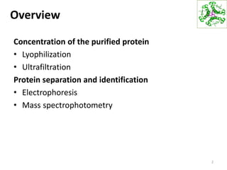
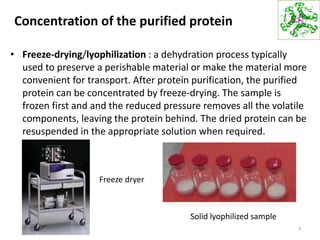
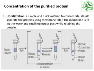
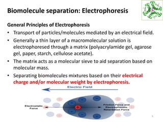
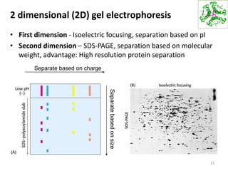
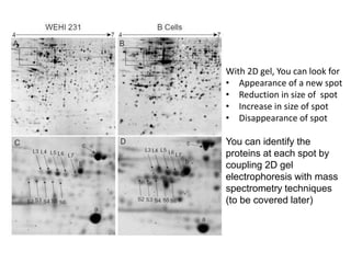
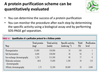
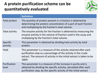
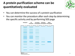
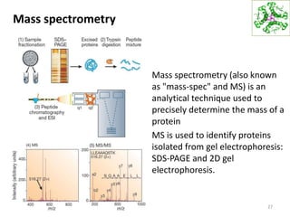
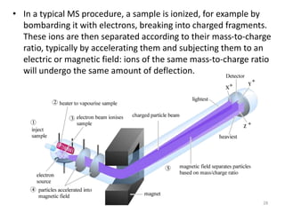
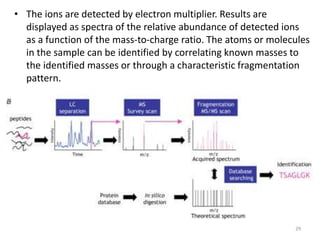
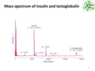
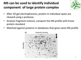
HBC1011 Biochemistry I
Trimester I, 2018/2019
Lecture 10 – Exploring Protein Part II
Ng Chong Han, PhD
MNAR1010, 06-2523751
chng@mmu.edu.my
Overview
Concentration of the purified protein
• Lyophilization
• Ultrafiltration
Protein separation and identification
• Electrophoresis
• Mass spectrophotometry
2
Concentration of the purified protein
• Freeze-drying/lyophilization : a dehydration process typically
used to preserve a perishable material or make the material more
convenient for transport. After protein purification, the purified
protein can be concentrated by freeze-drying. The sample is
frozen first and and the reduced pressure removes all the volatile
components, leaving the protein behind. The dried protein can be
resuspended in the appropriate solution when required.
3
Freeze dryer
Solid lyophilized sample
Concentration of the purified protein
• Ultrafiltration: a simple and quick method to concentrate, desalt,
separate the proteins using membrane filter. The membrane is to
let the water and small molecules pass while retaining the
protein.
4
Biomolecule separation: Electrophoresis
General Principles of Electrophoresis
• Transport of particles/molecules mediated by an electrical field.
• Generally a thin layer of a macromolecular solution is
electrophoresed through a matrix (polyacrylamide gel, agarose
gel, paper, starch, cellulose acetate).
• The matrix acts as a molecular sieve to aid separation based on
molecular mass.
• Separating biomolecules mixtures based on their electrical
charge and/or molecular weight by electrophoresis.
5
Types of electrophoresis
• Polyacrylamide
– Vertical gel: Proteins, DNA sequencing
• Agarose
– Horizontal gel: DNA, plasmids
6
Polyacrylamide gel electrophoresis
7
Reducing: SDS-PAGE gel
Non-reducing: Native gel
Protein
markers
Formation of a polyacrylamide gel by
polymerization
8
Formation of a polyacrylamide gel
The sieving action of a
porous polyacrylamide gel
separates proteins according
to size with the smallest
moving most rapidly.
SDS-PAGE protein separation
• Sodium dodecyl sulfate polyacrylamide gel electrophoresis (SDS-
PAGE)
• Proteins are first heated in a solution consists of detergent SDS
(sodium dodecyl sulfate) (an anionic detergent that disrupts
noncovalent interaction in native protein) often in the presence of
β-mercaptoethanol or dithiothreithol (DTT) (to reduce and break
disulfide bonds).
• This denatures all native protein structures.
• The proteins are then electrophoresed in the presence of SDS.
• All proteins became negatively charged, therefore the proteins
are separated based on the protein size.
9
SDS-PAGE
SDS denatures secondary and non–disulfide–linked tertiary
structures, and additionally applies a negative charge to each
protein in proportion to its mass.
10
SDS-PAGE
• When electric field is applied across the gel, negatively charged
proteins migrate across the gel towards the positive (+) electrode
(anode). Depending on their size, different proteins moves
differently through the gel matrix: small molecules more easily fit,
while larger ones have more difficulty. Smaller biomolecules
travel farther down the gel, while larger ones remain closer to the
point of origin.
11
Protein detection after SDS-PAGE
• Proteins can be visualized by Coomassie blue dye staining after
gel staining.
• In most cases, the separated proteins may be identified by
Western blot or Mass spectrometry (discussed later).
12
Native vs Denaturing gel run
13
• Denaturing gels are run under conditions that disrupt the natural
structure of the analyte, causing it to unfold into a linear chain.
Thus, the mobility of each macromolecule depends only on its
linear length and its mass-to-charge ratio. Thus, the secondary,
tertiary, and quaternary levels of biomolecular structure are
disrupted, leaving only the primary structure to be analyzed.
• Native gels are run in non-denaturing conditions, so that the
analyte's natural structure (secondary, tertiary, and quaternary
levels) is maintained. This allows the physical size of the folded or
assembled complex to affect the mobility, allowing for analysis of
all four levels of the biomolecular structure.
Native vs Denaturing gel run
14
SDS
750KD
250KD
250KD
250KD
Native PAGE gel
SDS PAGE gel
See one band at
250KD
See one band at
750 KD
beta-
mercaptoethanol
Denaturing SDS-PAGE gel
Native vs Denaturing gel run
15
SDS
75Kda
100Kda
150Kda
75 Kda
100 Kda
150 Kda325 Kda
Native gel
beta-
mercaptoethanol
Reducing SDS-PAGE gel
Determination of protein molecular weight
by SDS-PAGE
16
Extrapolate to determine mwt of unknown
Protein
marker
with
known
molecular
weight
Isoelectric focusing
• Isoelectric focusing (IEF) - a
technique for separating
different molecules by
differences in
their isoelectric point (pI).
• Most proteins carry
electrical charges
depending on pH (due to -
NH2 and -COOH groups in
side chains).
17
Charged amino acid
18
Isoelectric focusing
• Ampholytes is used in an electrophoresis gel to generate a stable
pH gradient between the cathode and the anode using
ampholytes.
• At all pH values other than their isoelectric point, proteins will be
charged. If they are positively charged, they will be pulled towards
the more negative end of the gel and if they are negatively
charged, they will be pulled to the more positive end of the gel.
19
Isoelectric focusing
• As the proteins move along a pH gradient, the proteins will
accumulate at the pH region that corresponds to their pI.
• At this point it has no net charge and so migration ceases. As a
result, the proteins become focused into sharp stationary bands
with each protein positioned at its pI.
20
2 dimensional (2D) gel electrophoresis
• First dimension - Isoelectric focusing, separation based on pI
• Second dimension – SDS-PAGE, separation based on molecular
weight, advantage: High resolution protein separation
21
Separate based on charge
Saparatebasedonsize
With 2D gel, You can look for
• Appearance of a new spot
• Reduction in size of spot
• Increase in size of spot
• Disappearance of spot
You can identify the
proteins at each spot by
coupling 2D gel
electrophoresis with mass
spectrometry techniques
(to be covered later)
A protein purification scheme can be
quantitatively evaluated
• You can determine the success of a protein purification
• You can monitor the procedure after each step by determining
the specific activity using a biological assay and by performing
SDS-PAGE gel separation.
23
A protein purification scheme can be
quantitatively evaluated
Definition
Total protein The quantity of protein present in a fraction is obtained by
determining the protein concentration of a part of each fraction
and multiplying by the fraction’s total volume.
Total activity The enzyme activity for the fraction is obtained by measuring the
enzyme activity in the volume of fraction used in the assay and
multiplying by the fraction’s total volume.
Specific
activity
This parameter is obtained by dividing total activity by total
protein.
Yield This parameter is a measure of the activity retained after each
purification step as a percentage of the activity in the crude
extract. The amount of activity in the initial extract is taken to be
100%
Purification
level
This parameter is a measure of the increase in purity and is
obtained by dividing the specific activity, calculated after each
purification step, by the specific activity of the initial extract. 24
A protein purification scheme can be
quantitatively evaluated
• You can determine the success of a protein purification
• You can monitor the procedure after each step by determining
the specific activity and by performing SDS page
25
Isolating and Purifying Proteins
1. Develop an assay to identify and quantify the protein.
2. Select a biological source for the protein and prepare a cell
homogenate.
3. Separate the cell homogenate into fractions by differential
centrifugation.
4. Perform chromatography: ion-exchange, gel filtration, and
affinity.
5. Determine the protein’s purity and molecular size by gel
electrophoresis.
26
Mass spectrometry
• Mass spectrometry (also known
as "mass-spec" and MS) is an
analytical technique used to
precisely determine the mass of a
protein
• MS is used to identify proteins
isolated from gel electrophoresis:
SDS-PAGE and 2D gel
electrophoresis.
27
• In a typical MS procedure, a sample is ionized, for example by
bombarding it with electrons, breaking into charged fragments.
These ions are then separated according to their mass-to-charge
ratio, typically by accelerating them and subjecting them to an
electric or magnetic field: ions of the same mass-to-charge ratio
will undergo the same amount of deflection.
28
• The ions are detected by electron multiplier. Results are
displayed as spectra of the relative abundance of detected ions
as a function of the mass-to-charge ratio. The atoms or molecules
in the sample can be identified by correlating known masses to
the identified masses or through a characteristic fragmentation
pattern.
29
Mass spectrum of insulin and lactoglobulin
30
MS can be used to identify individual
component of large protein complex
• After 2D gel electrophoresis, protein in individual spots are
cleaved using a protease.
• Analyze fragment mixture, compare the MS profile with know
protein standard
• Matched against proteins in databases that gives same MS profile
31
Study questions
1. What methods are routinely used to concentrate proteins?
2. How do proteins separated in SDS-PAGE gel electrophoresis?
3. What are the roles of SDS and beta-mercaptoethanol used in
SDS-PAGE gel electrophoresis?
4. What happens when proteins are separated on isoelectric
focusing gel?
5. How do proteins separated in 2D gel electrophoresis?
6. What method would you use to identify proteins after protein
separation?
32

219159 lecture 10

  • 1.
    HBC1011 Biochemistry I TrimesterI, 2018/2019 Lecture 10 – Exploring Protein Part II Ng Chong Han, PhD MNAR1010, 06-2523751 chng@mmu.edu.my
  • 2.
    Overview Concentration of thepurified protein • Lyophilization • Ultrafiltration Protein separation and identification • Electrophoresis • Mass spectrophotometry 2
  • 3.
    Concentration of thepurified protein • Freeze-drying/lyophilization : a dehydration process typically used to preserve a perishable material or make the material more convenient for transport. After protein purification, the purified protein can be concentrated by freeze-drying. The sample is frozen first and and the reduced pressure removes all the volatile components, leaving the protein behind. The dried protein can be resuspended in the appropriate solution when required. 3 Freeze dryer Solid lyophilized sample
  • 4.
    Concentration of thepurified protein • Ultrafiltration: a simple and quick method to concentrate, desalt, separate the proteins using membrane filter. The membrane is to let the water and small molecules pass while retaining the protein. 4
  • 5.
    Biomolecule separation: Electrophoresis GeneralPrinciples of Electrophoresis • Transport of particles/molecules mediated by an electrical field. • Generally a thin layer of a macromolecular solution is electrophoresed through a matrix (polyacrylamide gel, agarose gel, paper, starch, cellulose acetate). • The matrix acts as a molecular sieve to aid separation based on molecular mass. • Separating biomolecules mixtures based on their electrical charge and/or molecular weight by electrophoresis. 5
  • 6.
    Types of electrophoresis •Polyacrylamide – Vertical gel: Proteins, DNA sequencing • Agarose – Horizontal gel: DNA, plasmids 6
  • 7.
    Polyacrylamide gel electrophoresis 7 Reducing:SDS-PAGE gel Non-reducing: Native gel Protein markers
  • 8.
    Formation of apolyacrylamide gel by polymerization 8 Formation of a polyacrylamide gel The sieving action of a porous polyacrylamide gel separates proteins according to size with the smallest moving most rapidly.
  • 9.
    SDS-PAGE protein separation •Sodium dodecyl sulfate polyacrylamide gel electrophoresis (SDS- PAGE) • Proteins are first heated in a solution consists of detergent SDS (sodium dodecyl sulfate) (an anionic detergent that disrupts noncovalent interaction in native protein) often in the presence of β-mercaptoethanol or dithiothreithol (DTT) (to reduce and break disulfide bonds). • This denatures all native protein structures. • The proteins are then electrophoresed in the presence of SDS. • All proteins became negatively charged, therefore the proteins are separated based on the protein size. 9
  • 10.
    SDS-PAGE SDS denatures secondaryand non–disulfide–linked tertiary structures, and additionally applies a negative charge to each protein in proportion to its mass. 10
  • 11.
    SDS-PAGE • When electricfield is applied across the gel, negatively charged proteins migrate across the gel towards the positive (+) electrode (anode). Depending on their size, different proteins moves differently through the gel matrix: small molecules more easily fit, while larger ones have more difficulty. Smaller biomolecules travel farther down the gel, while larger ones remain closer to the point of origin. 11
  • 12.
    Protein detection afterSDS-PAGE • Proteins can be visualized by Coomassie blue dye staining after gel staining. • In most cases, the separated proteins may be identified by Western blot or Mass spectrometry (discussed later). 12
  • 13.
    Native vs Denaturinggel run 13 • Denaturing gels are run under conditions that disrupt the natural structure of the analyte, causing it to unfold into a linear chain. Thus, the mobility of each macromolecule depends only on its linear length and its mass-to-charge ratio. Thus, the secondary, tertiary, and quaternary levels of biomolecular structure are disrupted, leaving only the primary structure to be analyzed. • Native gels are run in non-denaturing conditions, so that the analyte's natural structure (secondary, tertiary, and quaternary levels) is maintained. This allows the physical size of the folded or assembled complex to affect the mobility, allowing for analysis of all four levels of the biomolecular structure.
  • 14.
    Native vs Denaturinggel run 14 SDS 750KD 250KD 250KD 250KD Native PAGE gel SDS PAGE gel See one band at 250KD See one band at 750 KD beta- mercaptoethanol Denaturing SDS-PAGE gel
  • 15.
    Native vs Denaturinggel run 15 SDS 75Kda 100Kda 150Kda 75 Kda 100 Kda 150 Kda325 Kda Native gel beta- mercaptoethanol Reducing SDS-PAGE gel
  • 16.
    Determination of proteinmolecular weight by SDS-PAGE 16 Extrapolate to determine mwt of unknown Protein marker with known molecular weight
  • 17.
    Isoelectric focusing • Isoelectricfocusing (IEF) - a technique for separating different molecules by differences in their isoelectric point (pI). • Most proteins carry electrical charges depending on pH (due to - NH2 and -COOH groups in side chains). 17
  • 18.
  • 19.
    Isoelectric focusing • Ampholytesis used in an electrophoresis gel to generate a stable pH gradient between the cathode and the anode using ampholytes. • At all pH values other than their isoelectric point, proteins will be charged. If they are positively charged, they will be pulled towards the more negative end of the gel and if they are negatively charged, they will be pulled to the more positive end of the gel. 19
  • 20.
    Isoelectric focusing • Asthe proteins move along a pH gradient, the proteins will accumulate at the pH region that corresponds to their pI. • At this point it has no net charge and so migration ceases. As a result, the proteins become focused into sharp stationary bands with each protein positioned at its pI. 20
  • 21.
    2 dimensional (2D)gel electrophoresis • First dimension - Isoelectric focusing, separation based on pI • Second dimension – SDS-PAGE, separation based on molecular weight, advantage: High resolution protein separation 21 Separate based on charge Saparatebasedonsize
  • 22.
    With 2D gel,You can look for • Appearance of a new spot • Reduction in size of spot • Increase in size of spot • Disappearance of spot You can identify the proteins at each spot by coupling 2D gel electrophoresis with mass spectrometry techniques (to be covered later)
  • 23.
    A protein purificationscheme can be quantitatively evaluated • You can determine the success of a protein purification • You can monitor the procedure after each step by determining the specific activity using a biological assay and by performing SDS-PAGE gel separation. 23
  • 24.
    A protein purificationscheme can be quantitatively evaluated Definition Total protein The quantity of protein present in a fraction is obtained by determining the protein concentration of a part of each fraction and multiplying by the fraction’s total volume. Total activity The enzyme activity for the fraction is obtained by measuring the enzyme activity in the volume of fraction used in the assay and multiplying by the fraction’s total volume. Specific activity This parameter is obtained by dividing total activity by total protein. Yield This parameter is a measure of the activity retained after each purification step as a percentage of the activity in the crude extract. The amount of activity in the initial extract is taken to be 100% Purification level This parameter is a measure of the increase in purity and is obtained by dividing the specific activity, calculated after each purification step, by the specific activity of the initial extract. 24
  • 25.
    A protein purificationscheme can be quantitatively evaluated • You can determine the success of a protein purification • You can monitor the procedure after each step by determining the specific activity and by performing SDS page 25
  • 26.
    Isolating and PurifyingProteins 1. Develop an assay to identify and quantify the protein. 2. Select a biological source for the protein and prepare a cell homogenate. 3. Separate the cell homogenate into fractions by differential centrifugation. 4. Perform chromatography: ion-exchange, gel filtration, and affinity. 5. Determine the protein’s purity and molecular size by gel electrophoresis. 26
  • 27.
    Mass spectrometry • Massspectrometry (also known as "mass-spec" and MS) is an analytical technique used to precisely determine the mass of a protein • MS is used to identify proteins isolated from gel electrophoresis: SDS-PAGE and 2D gel electrophoresis. 27
  • 28.
    • In atypical MS procedure, a sample is ionized, for example by bombarding it with electrons, breaking into charged fragments. These ions are then separated according to their mass-to-charge ratio, typically by accelerating them and subjecting them to an electric or magnetic field: ions of the same mass-to-charge ratio will undergo the same amount of deflection. 28
  • 29.
    • The ionsare detected by electron multiplier. Results are displayed as spectra of the relative abundance of detected ions as a function of the mass-to-charge ratio. The atoms or molecules in the sample can be identified by correlating known masses to the identified masses or through a characteristic fragmentation pattern. 29
  • 30.
    Mass spectrum ofinsulin and lactoglobulin 30
  • 31.
    MS can beused to identify individual component of large protein complex • After 2D gel electrophoresis, protein in individual spots are cleaved using a protease. • Analyze fragment mixture, compare the MS profile with know protein standard • Matched against proteins in databases that gives same MS profile 31
  • 32.
    Study questions 1. Whatmethods are routinely used to concentrate proteins? 2. How do proteins separated in SDS-PAGE gel electrophoresis? 3. What are the roles of SDS and beta-mercaptoethanol used in SDS-PAGE gel electrophoresis? 4. What happens when proteins are separated on isoelectric focusing gel? 5. How do proteins separated in 2D gel electrophoresis? 6. What method would you use to identify proteins after protein separation? 32