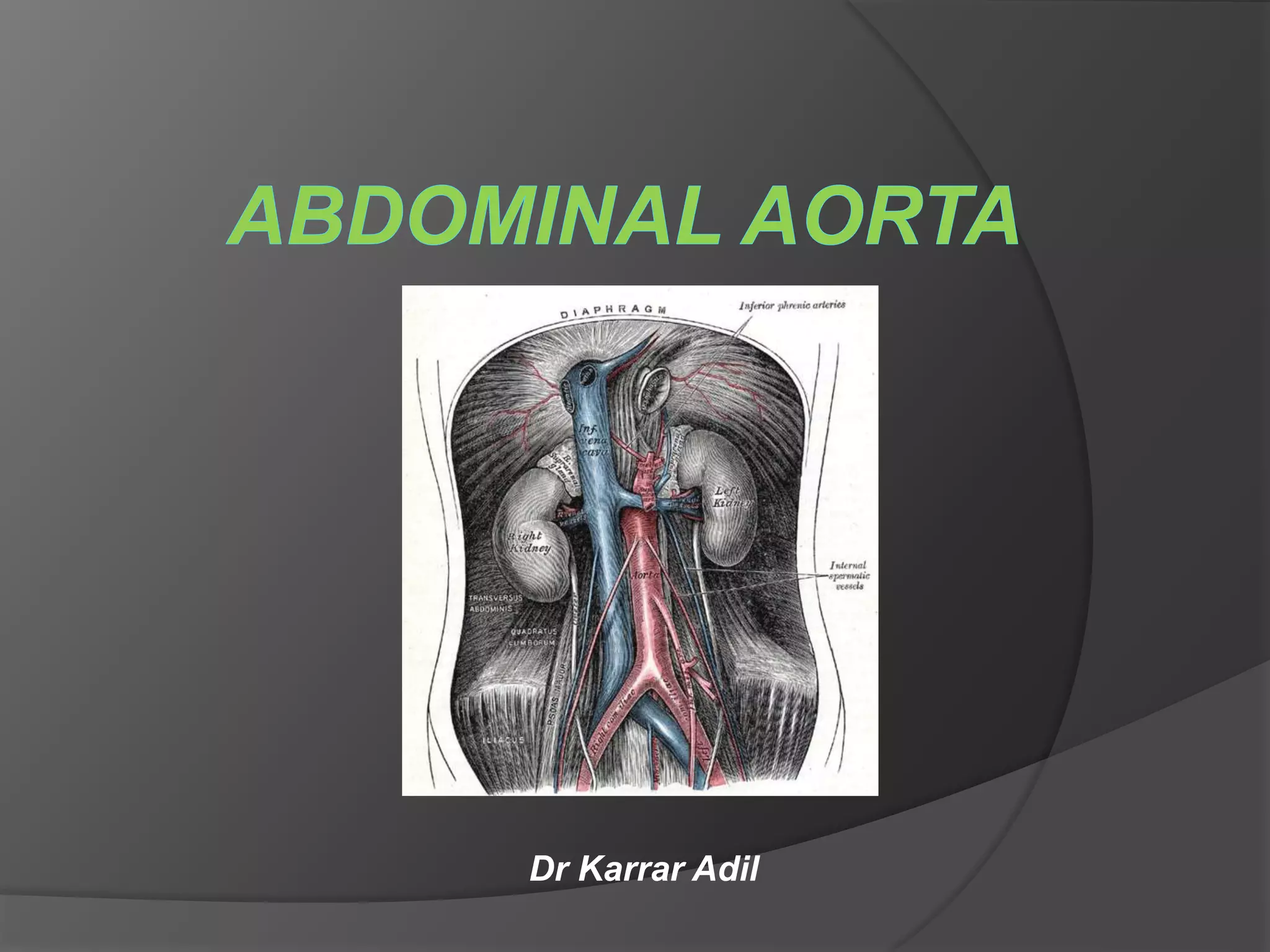
SlideShare a Scribd company logo
Dr Karrar Adil
Anatomy :
 The abdominal aorta is the largest artery in the
abdominal cavity. it is a continuation of the
thoracic aorta.
 is a retroperitoneal structure that begins at the
hiatus of the diaphragm (the level of the T12
vertebra).
 It is approximately 13cm long and ends at the
level of the L4 vertebra. At this level, the aorta
terminates by bifurcating into the right and left
common iliac arteries.
Relations :
• Running parallel to the aorta on its right-hand side : the
inferior vena cava, the cisterna chyli, the azygos vein, and
the para-aortic lymph nodes.
• Running on its left-hand side : the left sympathetic trunk and
the para-aortic lymph nodes.
• Anteriorly : the stomach, duodenum and pancreas.
• It is also crossed anteriorly by the splenic vein and the left
renal vein.
• Posteriorly: it is separated from the lumbar vertebrae by the
anterior longitudinal ligament and left lumbar veins.
Branches :
• Three single anterior visceral branches :
(coeliac trunk, superior mesenteric artery, inferior
mesenteric artery).
• Three paired lateral visceral branches :
(suprarenal, renal, gonadal arteries).
• Five paired lateral abdominal wall branches :
(inferior phrenic and four lumbar arteries).
• Three terminal branches :
(two common iliac arteries and the median sacral artery).
Celiac trunk (T12) :
1- left gastric artery.
2- splenic artery:
 short gastric arteries (6)
 splenic arteries.
 left gastroepiploic a.
 pancreatic arteries.
3- common hepatic artery:
 right gastric a.
 gastroduodenal artery: (right gastroepiploic a. and
superior pancreaticoduodenal a.)
 hepatic artery proper: (right hepatic and left
hepatic a.)
superior mesenteric artery (L1) :
 inferior pancreaticoduodenal a.
 jejunal and ileal arteries
 right colic a.
 middle colic a.
 ileocolic a.
inferior mesenteric artery (L3) :
 left colic a.
 sigmoid arteries (2 or 3)
 superior rectal a.
The abdominal aorta is clinically divided into 2
segments:
• The suprarenal abdominal segment, inferior
to the diaphragm but superior to the renal
arteries.
• The Infrarenal segment, inferior to the renal
arteries and superior to the iliac bifurcation.
Abdominal aortic aneurysm
Definition :
 AAA : is a localised, progressive and
permanent dilatation occurring in any portion of
infra-diaphragmatic aorta. with at least 50%
increase in diameter.
 Most common site is infrarenal aorta.
 Ectasia : dilatation <50% of normal diameter.
Classification :
According to wall :
• true aneurysm : containing the three layers of the
arterial wall (intima, media, adventitia) in the aneurysm
sac.
• false aneurysm (pseudoaneurysm) : having a single
layer of fibrous tissue as the wall of the sac. usually
occurs after trauma.
According to Location :
Infrarenal :
• below the renal arteries
• most common (95%).
• One-third of aneurysms extend into the iliac
arteries
Suprarenal :
• above the renal arteries (5%).
• Isolated suprarenal type is rare; it is usually
associated with thoracic and/or infrarenal types.
• Suprarenal AAA: The aneurysm involves the origins of one or more visceral arteries
but does not extend into the chest.
• Pararenal AAA: The renal arteries arise from the aneurysmal aorta but the aorta at
the level of the superior mesenteric artery is not aneurysmal.
• Juxtarenal AAA: The aneurysm originates just beyond the origins of the renal
arteries. There is no segment of nonaneurysmal aorta distal to the renal arteries, but
the aorta at the level of the renal arteries is not aneurysmal.
• Infrarenal AAA: The aneurysm originates distal to the renal arteries. There is a
segment of nonaneurysmal aorta that extends distal to the origins of the renal
arteries.
According to morphology :
• Fusiform : symmetrical enlargement involving whole
circumference of artery.
• Saccular : affect only part of the arterial circumference. have
higher risk of rupture.
According to size :
• Ectatic or mild dilatation : >2.0 cm and <3.0 cm
• Moderate : 3.0 - 5.0 cm
• Large or severe : >5.0 or 5.5 cm
According to symptoms :
• Asymptomatic.
• Symptomatic.
• Symptomatic ruptured.
Causes :
 Atherosclerosis : most common cause (95%)
 Familial aortic aneurysm (associated with 25% of
AAA). Marfan's, Ehler Danlos syndromes are related
genetically.
 Others :
Syphilis, dissection, trauma, collagen diseases, infection,
arteritis, cystic medial necrosis.
Risk factors :
Age, male gender, white race, smoking and family history.
Asymptomatic Type :
 It is found incidentally either on clinical
examination or on angiography or on
ultrasound.
 Repair is required if diameter is over 5.5 cm on
ultrasound.
Symptomatic without Rupture :
Clinical features :
 back pain or abdominal pain (Most common symptom).
 Abdominal mass which is smooth, soft, non mobile, not
moving with respiration, above the umbilical level, resonant
on percussion.
 Common in males ( 4:1 ); common in smokers.
 GIT, urinary, venous symptoms can also occur.
 Hypertension, diabetes, cardiac problems should be looked
for and dealt with.
 5% present as inflammatory aneurysm adherent
to ureters, left renal vein, inferior vena cava and
duodenum.
 Aortocaval fistula presents as high output cardiac
failure with continuous bruit in abdomen and
lower limb edema.
 Aortoenteric fistula is due to erosion of aneurysm
into 4th part of duodenum presenting as
gastrointestinal (GI) bleed, malaena, shock.
Symptomatic Ruptured Aortic
Aneurysm :
 Rupture is most common and most lethal complication.
 AAA rupture most commonly in left retroperitoneum.
 It may be anterior rupture (20%) into the free peritoneal
cavity causing severe shock and death very early.
 or posterior rupture (80%) with formation of
retroperitoneal haematoma of large size causing severe
back pain, hypotension, shock, absence of femoral
pulses and with a palpable mass in the abdomen.
Diagnosis :
 Plain X-ray detects AAA in up to 70% cases.
 CT angiogram (Ix of choice)
 MRI and MRA is Ix of choice for diagnosis in
patients with renal insufficiency.
 Ultrasound (preferred method of screening).
Management :
Medical Management :
It is done in :
 low-risk abdominal aortic aneurysm (age below 70 years; active
physically without cardiac, respiratory, renal impairment and non
inflammatory aneurysm);
 if aneurysm size is <5 cm.
 if growth rate is <0.5 cm/year.
It includes risk factor modifications :
 stopping smoking.
 control of blood pressure and cholesterol.
 NSAIDs & tetracycline may have potential to reduce aneurysmal
growth by inhibiting MMP (Matrix metalloproteinase).
Indications of repair of AAA :
 Diameter 5.5 cm or more in men.
 Symptomatic aneurysm.
 For women and patients with greater than average rupture risk, AAA
diameter 4.5 to 5.0 cm.
 Rate of expansion >1 cm/year
 Atypical aneurysms (dissecting, pseudoaneurysm, mycotic, saccular
and penetrating ulcer) regardless of size.
Open surgical repair :
Transperitoneal & Retroperitoneal approach .
endo-aneurysmorrhaphy with intraluminal graft placement.
Thank you …

More Related Content

What's hot

Diagnostic Imaging of Pulmonary Vasculature
Diagnostic Imaging of Pulmonary VasculatureDiagnostic Imaging of Pulmonary Vasculature
Diagnostic Imaging of Pulmonary Vasculature
Mohamed M.A. Zaitoun
 
Diagnostic Imaging of Stroke
Diagnostic Imaging of StrokeDiagnostic Imaging of Stroke
Diagnostic Imaging of Stroke
Mohamed M.A. Zaitoun
 
Cardiac chamber enlargement
Cardiac chamber enlargementCardiac chamber enlargement
Cardiac chamber enlargementairwave12
 
Radiographic Presentation of Congenital Heart Disease
Radiographic Presentation of Congenital Heart DiseaseRadiographic Presentation of Congenital Heart Disease
Radiographic Presentation of Congenital Heart Disease
Tarique Ajij
 
Radiological anatomy of the abdominal aorta
Radiological anatomy of the abdominal aortaRadiological anatomy of the abdominal aorta
Radiological anatomy of the abdominal aorta
TaiwoOjeremi
 
Congenital heart disease and vascular abnormality(x-ray findings)
Congenital heart disease and vascular abnormality(x-ray findings)Congenital heart disease and vascular abnormality(x-ray findings)
Congenital heart disease and vascular abnormality(x-ray findings)
z2jeetendra
 
Vascular ring and sling
Vascular ring and slingVascular ring and sling
Vascular ring and sling
Jyotindra Singh
 
New: Radiology 5th year, all lectures/chest (Dr. Abeer)
New: Radiology 5th year, all lectures/chest (Dr. Abeer)New: Radiology 5th year, all lectures/chest (Dr. Abeer)
New: Radiology 5th year, all lectures/chest (Dr. Abeer)
College of Medicine, Sulaymaniyah
 
Imaging modalities of diaphragm
Imaging modalities of diaphragmImaging modalities of diaphragm
Imaging modalities of diaphragm
Arif S
 
Mediastinal mass
Mediastinal massMediastinal mass
Mediastinal mass
Vikram Patil
 
ECG/EKG changes in COPD
ECG/EKG changes in COPDECG/EKG changes in COPD
ECG/EKG changes in COPD
prithvi2911
 
Vascular rings & slings
Vascular rings & slings Vascular rings & slings
Vascular rings & slings
Jyotindra Singh
 
Coa pathophysiology
Coa pathophysiologyCoa pathophysiology
Coa pathophysiology
India CTVS
 
Mapca 1
Mapca 1Mapca 1
Ultrasound of Vascular anomalies by Oscar M. Navarro
Ultrasound of Vascular anomalies by Oscar M. NavarroUltrasound of Vascular anomalies by Oscar M. Navarro
Ultrasound of Vascular anomalies by Oscar M. Navarro
Dr. Naveed Quetta
 
Interpretation of chest x ray
Interpretation of chest x rayInterpretation of chest x ray
Interpretation of chest x ray
gowri shanker
 
Mitral valve anatomy - ppt by kunwar sidharth
Mitral valve    anatomy - ppt by kunwar sidharthMitral valve    anatomy - ppt by kunwar sidharth
Mitral valve anatomy - ppt by kunwar sidharth
kunwar sidharth
 
Role of Doppler in Liver Cirrhosis & Portal Hypertension
Role of Doppler in Liver Cirrhosis & Portal HypertensionRole of Doppler in Liver Cirrhosis & Portal Hypertension
Role of Doppler in Liver Cirrhosis & Portal Hypertension
nishit viradia
 

What's hot (20)

Diagnostic Imaging of Pulmonary Vasculature
Diagnostic Imaging of Pulmonary VasculatureDiagnostic Imaging of Pulmonary Vasculature
Diagnostic Imaging of Pulmonary Vasculature
 
Diagnostic Imaging of Stroke
Diagnostic Imaging of StrokeDiagnostic Imaging of Stroke
Diagnostic Imaging of Stroke
 
Cardiac chamber enlargement
Cardiac chamber enlargementCardiac chamber enlargement
Cardiac chamber enlargement
 
Radiographic Presentation of Congenital Heart Disease
Radiographic Presentation of Congenital Heart DiseaseRadiographic Presentation of Congenital Heart Disease
Radiographic Presentation of Congenital Heart Disease
 
A Case of Mediastinal Mass
A Case of Mediastinal MassA Case of Mediastinal Mass
A Case of Mediastinal Mass
 
Radiological anatomy of the abdominal aorta
Radiological anatomy of the abdominal aortaRadiological anatomy of the abdominal aorta
Radiological anatomy of the abdominal aorta
 
Congenital heart disease and vascular abnormality(x-ray findings)
Congenital heart disease and vascular abnormality(x-ray findings)Congenital heart disease and vascular abnormality(x-ray findings)
Congenital heart disease and vascular abnormality(x-ray findings)
 
Vascular ring and sling
Vascular ring and slingVascular ring and sling
Vascular ring and sling
 
New: Radiology 5th year, all lectures/chest (Dr. Abeer)
New: Radiology 5th year, all lectures/chest (Dr. Abeer)New: Radiology 5th year, all lectures/chest (Dr. Abeer)
New: Radiology 5th year, all lectures/chest (Dr. Abeer)
 
Imaging modalities of diaphragm
Imaging modalities of diaphragmImaging modalities of diaphragm
Imaging modalities of diaphragm
 
Mediastinal mass
Mediastinal massMediastinal mass
Mediastinal mass
 
Pulmonary nodule
Pulmonary nodulePulmonary nodule
Pulmonary nodule
 
ECG/EKG changes in COPD
ECG/EKG changes in COPDECG/EKG changes in COPD
ECG/EKG changes in COPD
 
Vascular rings & slings
Vascular rings & slings Vascular rings & slings
Vascular rings & slings
 
Coa pathophysiology
Coa pathophysiologyCoa pathophysiology
Coa pathophysiology
 
Mapca 1
Mapca 1Mapca 1
Mapca 1
 
Ultrasound of Vascular anomalies by Oscar M. Navarro
Ultrasound of Vascular anomalies by Oscar M. NavarroUltrasound of Vascular anomalies by Oscar M. Navarro
Ultrasound of Vascular anomalies by Oscar M. Navarro
 
Interpretation of chest x ray
Interpretation of chest x rayInterpretation of chest x ray
Interpretation of chest x ray
 
Mitral valve anatomy - ppt by kunwar sidharth
Mitral valve    anatomy - ppt by kunwar sidharthMitral valve    anatomy - ppt by kunwar sidharth
Mitral valve anatomy - ppt by kunwar sidharth
 
Role of Doppler in Liver Cirrhosis & Portal Hypertension
Role of Doppler in Liver Cirrhosis & Portal HypertensionRole of Doppler in Liver Cirrhosis & Portal Hypertension
Role of Doppler in Liver Cirrhosis & Portal Hypertension
 

Similar to Abdominal aorta.pptx

ANUERYSMS ,AV FISTULAS ,ARTERISTIS ,RAYNAUDS DISEASE -1.pptx
ANUERYSMS ,AV FISTULAS ,ARTERISTIS ,RAYNAUDS DISEASE -1.pptxANUERYSMS ,AV FISTULAS ,ARTERISTIS ,RAYNAUDS DISEASE -1.pptx
ANUERYSMS ,AV FISTULAS ,ARTERISTIS ,RAYNAUDS DISEASE -1.pptx
musayansa
 
AORTIC ANEURYSM.pptx
AORTIC ANEURYSM.pptxAORTIC ANEURYSM.pptx
AORTIC ANEURYSM.pptx
Dr.Mohammed Shanil.P
 
Diseases of the aorta
Diseases of the aortaDiseases of the aorta
Diseases of the aorta
Mohamed Ashraf
 
Abdominal arota
Abdominal arotaAbdominal arota
Abdominal arota
Sourabh Ahlawat
 
Vascular imaging ppt
Vascular imaging pptVascular imaging ppt
Vascular imaging ppt
Praveen Kumar
 
Neha diwan presentation on aortic aneurysm
Neha diwan presentation on aortic aneurysmNeha diwan presentation on aortic aneurysm
Neha diwan presentation on aortic aneurysm
NEHAADIWAN
 
Radiological approach to aortic aneurysm and acute diseases
Radiological approach to aortic aneurysm and acute diseasesRadiological approach to aortic aneurysm and acute diseases
Radiological approach to aortic aneurysm and acute diseases
Milan Silwal
 
Abdominal aortic aneurysm
Abdominal aortic aneurysm Abdominal aortic aneurysm
Abdominal aortic aneurysm
Kurnool Medical College, Kurnool
 
Aortic aneurysm
Aortic aneurysmAortic aneurysm
Aortic aneurysm
mohamedrafi112
 
ANEURYSMS , TYPES AND THERE MANAGEMENT.pptx
ANEURYSMS , TYPES  AND  THERE MANAGEMENT.pptxANEURYSMS , TYPES  AND  THERE MANAGEMENT.pptx
ANEURYSMS , TYPES AND THERE MANAGEMENT.pptx
Bipul Thakur
 
Aortic aneurysm imaging
Aortic aneurysm imagingAortic aneurysm imaging
Aortic aneurysm imaging
Sanal Kumar
 
Imaging of aortic pathologies
Imaging of aortic pathologies Imaging of aortic pathologies
Imaging of aortic pathologies
Pankaj Kaira
 
supra vena cava obstruction (SVCO)
supra vena cava obstruction (SVCO)supra vena cava obstruction (SVCO)
supra vena cava obstruction (SVCO)
Surgeon Ibrahim
 
USG of Aorta and Coeliac axis
USG of Aorta and Coeliac axisUSG of Aorta and Coeliac axis
USG of Aorta and Coeliac axis
Dr. Yash Kumar Achantani
 
Aortic aneurysm dr jeevraj
Aortic aneurysm  dr jeevrajAortic aneurysm  dr jeevraj
Aortic aneurysm dr jeevraj
jeevraj24
 
testicular tumors
testicular tumorstesticular tumors
testicular tumors
DrAyush Garg
 
USMLE CVS 001 Mediastinum anatomy medical chest .pdf
USMLE   CVS  001 Mediastinum anatomy medical chest .pdfUSMLE   CVS  001 Mediastinum anatomy medical chest .pdf
USMLE CVS 001 Mediastinum anatomy medical chest .pdf
AHMED ASHOUR
 
RADIOIMAGING IN MEDIASTINAL MASSES YN.pptx
RADIOIMAGING IN MEDIASTINAL MASSES YN.pptxRADIOIMAGING IN MEDIASTINAL MASSES YN.pptx
RADIOIMAGING IN MEDIASTINAL MASSES YN.pptx
dypradio
 
aneurysm-210530112039.pdf
aneurysm-210530112039.pdfaneurysm-210530112039.pdf
aneurysm-210530112039.pdf
DamanpreetSinghSidhu
 

Similar to Abdominal aorta.pptx (20)

ANUERYSMS ,AV FISTULAS ,ARTERISTIS ,RAYNAUDS DISEASE -1.pptx
ANUERYSMS ,AV FISTULAS ,ARTERISTIS ,RAYNAUDS DISEASE -1.pptxANUERYSMS ,AV FISTULAS ,ARTERISTIS ,RAYNAUDS DISEASE -1.pptx
ANUERYSMS ,AV FISTULAS ,ARTERISTIS ,RAYNAUDS DISEASE -1.pptx
 
AORTIC ANEURYSM.pptx
AORTIC ANEURYSM.pptxAORTIC ANEURYSM.pptx
AORTIC ANEURYSM.pptx
 
Diseases of the aorta
Diseases of the aortaDiseases of the aorta
Diseases of the aorta
 
Ruptured aortic aneurysms
Ruptured aortic aneurysmsRuptured aortic aneurysms
Ruptured aortic aneurysms
 
Abdominal arota
Abdominal arotaAbdominal arota
Abdominal arota
 
Vascular imaging ppt
Vascular imaging pptVascular imaging ppt
Vascular imaging ppt
 
Neha diwan presentation on aortic aneurysm
Neha diwan presentation on aortic aneurysmNeha diwan presentation on aortic aneurysm
Neha diwan presentation on aortic aneurysm
 
Radiological approach to aortic aneurysm and acute diseases
Radiological approach to aortic aneurysm and acute diseasesRadiological approach to aortic aneurysm and acute diseases
Radiological approach to aortic aneurysm and acute diseases
 
Abdominal aortic aneurysm
Abdominal aortic aneurysm Abdominal aortic aneurysm
Abdominal aortic aneurysm
 
Aortic aneurysm
Aortic aneurysmAortic aneurysm
Aortic aneurysm
 
ANEURYSMS , TYPES AND THERE MANAGEMENT.pptx
ANEURYSMS , TYPES  AND  THERE MANAGEMENT.pptxANEURYSMS , TYPES  AND  THERE MANAGEMENT.pptx
ANEURYSMS , TYPES AND THERE MANAGEMENT.pptx
 
Aortic aneurysm imaging
Aortic aneurysm imagingAortic aneurysm imaging
Aortic aneurysm imaging
 
Imaging of aortic pathologies
Imaging of aortic pathologies Imaging of aortic pathologies
Imaging of aortic pathologies
 
supra vena cava obstruction (SVCO)
supra vena cava obstruction (SVCO)supra vena cava obstruction (SVCO)
supra vena cava obstruction (SVCO)
 
USG of Aorta and Coeliac axis
USG of Aorta and Coeliac axisUSG of Aorta and Coeliac axis
USG of Aorta and Coeliac axis
 
Aortic aneurysm dr jeevraj
Aortic aneurysm  dr jeevrajAortic aneurysm  dr jeevraj
Aortic aneurysm dr jeevraj
 
testicular tumors
testicular tumorstesticular tumors
testicular tumors
 
USMLE CVS 001 Mediastinum anatomy medical chest .pdf
USMLE   CVS  001 Mediastinum anatomy medical chest .pdfUSMLE   CVS  001 Mediastinum anatomy medical chest .pdf
USMLE CVS 001 Mediastinum anatomy medical chest .pdf
 
RADIOIMAGING IN MEDIASTINAL MASSES YN.pptx
RADIOIMAGING IN MEDIASTINAL MASSES YN.pptxRADIOIMAGING IN MEDIASTINAL MASSES YN.pptx
RADIOIMAGING IN MEDIASTINAL MASSES YN.pptx
 
aneurysm-210530112039.pdf
aneurysm-210530112039.pdfaneurysm-210530112039.pdf
aneurysm-210530112039.pdf
 

More from karrar adil

DISEASES OF THE UMBILICUS.pptx
DISEASES OF THE UMBILICUS.pptxDISEASES OF THE UMBILICUS.pptx
DISEASES OF THE UMBILICUS.pptx
karrar adil
 
CLASSIFICATION OF BILE DUCT INJURY.pptx
CLASSIFICATION OF BILE DUCT INJURY.pptxCLASSIFICATION OF BILE DUCT INJURY.pptx
CLASSIFICATION OF BILE DUCT INJURY.pptx
karrar adil
 
HYDATID CYST OF LIVER.pptx
HYDATID CYST OF LIVER.pptxHYDATID CYST OF LIVER.pptx
HYDATID CYST OF LIVER.pptx
karrar adil
 
Hepatocellular carcinoma.pptx
Hepatocellular carcinoma.pptxHepatocellular carcinoma.pptx
Hepatocellular carcinoma.pptx
karrar adil
 
Pancreatic pseudocyst.pptx
Pancreatic pseudocyst.pptxPancreatic pseudocyst.pptx
Pancreatic pseudocyst.pptx
karrar adil
 
PHYLLOIDES TUMOUR.pptx
PHYLLOIDES TUMOUR.pptxPHYLLOIDES TUMOUR.pptx
PHYLLOIDES TUMOUR.pptx
karrar adil
 
ACUTE PANCREATITIS.pptx
ACUTE PANCREATITIS.pptxACUTE PANCREATITIS.pptx
ACUTE PANCREATITIS.pptx
karrar adil
 
terminology of liver resections.pptx
terminology of liver resections.pptxterminology of liver resections.pptx
terminology of liver resections.pptx
karrar adil
 
Ascites.pptx
Ascites.pptxAscites.pptx
Ascites.pptx
karrar adil
 
infantile HPS.pptx
infantile HPS.pptxinfantile HPS.pptx
infantile HPS.pptx
karrar adil
 
ANORECTAL MALFORMATIONS
ANORECTAL MALFORMATIONSANORECTAL MALFORMATIONS
ANORECTAL MALFORMATIONS
karrar adil
 
Malignant Peritoneal Mesothelioma
Malignant Peritoneal MesotheliomaMalignant Peritoneal Mesothelioma
Malignant Peritoneal Mesothelioma
karrar adil
 
Achalasia
AchalasiaAchalasia
Achalasia
karrar adil
 
Hemostasis
HemostasisHemostasis
Hemostasis
karrar adil
 
Metabolic acidosis and metabloic alkalosis
Metabolic acidosis and metabloic alkalosisMetabolic acidosis and metabloic alkalosis
Metabolic acidosis and metabloic alkalosis
karrar adil
 
Inguinal hernia
Inguinal herniaInguinal hernia
Inguinal hernia
karrar adil
 

More from karrar adil (16)

DISEASES OF THE UMBILICUS.pptx
DISEASES OF THE UMBILICUS.pptxDISEASES OF THE UMBILICUS.pptx
DISEASES OF THE UMBILICUS.pptx
 
CLASSIFICATION OF BILE DUCT INJURY.pptx
CLASSIFICATION OF BILE DUCT INJURY.pptxCLASSIFICATION OF BILE DUCT INJURY.pptx
CLASSIFICATION OF BILE DUCT INJURY.pptx
 
HYDATID CYST OF LIVER.pptx
HYDATID CYST OF LIVER.pptxHYDATID CYST OF LIVER.pptx
HYDATID CYST OF LIVER.pptx
 
Hepatocellular carcinoma.pptx
Hepatocellular carcinoma.pptxHepatocellular carcinoma.pptx
Hepatocellular carcinoma.pptx
 
Pancreatic pseudocyst.pptx
Pancreatic pseudocyst.pptxPancreatic pseudocyst.pptx
Pancreatic pseudocyst.pptx
 
PHYLLOIDES TUMOUR.pptx
PHYLLOIDES TUMOUR.pptxPHYLLOIDES TUMOUR.pptx
PHYLLOIDES TUMOUR.pptx
 
ACUTE PANCREATITIS.pptx
ACUTE PANCREATITIS.pptxACUTE PANCREATITIS.pptx
ACUTE PANCREATITIS.pptx
 
terminology of liver resections.pptx
terminology of liver resections.pptxterminology of liver resections.pptx
terminology of liver resections.pptx
 
Ascites.pptx
Ascites.pptxAscites.pptx
Ascites.pptx
 
infantile HPS.pptx
infantile HPS.pptxinfantile HPS.pptx
infantile HPS.pptx
 
ANORECTAL MALFORMATIONS
ANORECTAL MALFORMATIONSANORECTAL MALFORMATIONS
ANORECTAL MALFORMATIONS
 
Malignant Peritoneal Mesothelioma
Malignant Peritoneal MesotheliomaMalignant Peritoneal Mesothelioma
Malignant Peritoneal Mesothelioma
 
Achalasia
AchalasiaAchalasia
Achalasia
 
Hemostasis
HemostasisHemostasis
Hemostasis
 
Metabolic acidosis and metabloic alkalosis
Metabolic acidosis and metabloic alkalosisMetabolic acidosis and metabloic alkalosis
Metabolic acidosis and metabloic alkalosis
 
Inguinal hernia
Inguinal herniaInguinal hernia
Inguinal hernia
 

Recently uploaded

ARTIFICIAL INTELLIGENCE IN HEALTHCARE.pdf
ARTIFICIAL INTELLIGENCE IN  HEALTHCARE.pdfARTIFICIAL INTELLIGENCE IN  HEALTHCARE.pdf
ARTIFICIAL INTELLIGENCE IN HEALTHCARE.pdf
Anujkumaranit
 
Phone Us ❤85270-49040❤ #ℂall #gIRLS In Surat By Surat @ℂall @Girls Hotel With...
Phone Us ❤85270-49040❤ #ℂall #gIRLS In Surat By Surat @ℂall @Girls Hotel With...Phone Us ❤85270-49040❤ #ℂall #gIRLS In Surat By Surat @ℂall @Girls Hotel With...
Phone Us ❤85270-49040❤ #ℂall #gIRLS In Surat By Surat @ℂall @Girls Hotel With...
Savita Shen $i11
 
heat stroke and heat exhaustion in children
heat stroke and heat exhaustion in childrenheat stroke and heat exhaustion in children
heat stroke and heat exhaustion in children
SumeraAhmad5
 
Hemodialysis: Chapter 3, Dialysis Water Unit - Dr.Gawad
Hemodialysis: Chapter 3, Dialysis Water Unit - Dr.GawadHemodialysis: Chapter 3, Dialysis Water Unit - Dr.Gawad
Hemodialysis: Chapter 3, Dialysis Water Unit - Dr.Gawad
NephroTube - Dr.Gawad
 
How to Give Better Lectures: Some Tips for Doctors
How to Give Better Lectures: Some Tips for DoctorsHow to Give Better Lectures: Some Tips for Doctors
How to Give Better Lectures: Some Tips for Doctors
LanceCatedral
 
ANATOMY AND PHYSIOLOGY OF URINARY SYSTEM.pptx
ANATOMY AND PHYSIOLOGY OF URINARY SYSTEM.pptxANATOMY AND PHYSIOLOGY OF URINARY SYSTEM.pptx
ANATOMY AND PHYSIOLOGY OF URINARY SYSTEM.pptx
Swetaba Besh
 
Pulmonary Thromboembolism - etilogy, types, medical- Surgical and nursing man...
Pulmonary Thromboembolism - etilogy, types, medical- Surgical and nursing man...Pulmonary Thromboembolism - etilogy, types, medical- Surgical and nursing man...
Pulmonary Thromboembolism - etilogy, types, medical- Surgical and nursing man...
VarunMahajani
 
ARTHROLOGY PPT NCISM SYLLABUS AYURVEDA STUDENTS
ARTHROLOGY PPT NCISM SYLLABUS AYURVEDA STUDENTSARTHROLOGY PPT NCISM SYLLABUS AYURVEDA STUDENTS
ARTHROLOGY PPT NCISM SYLLABUS AYURVEDA STUDENTS
Dr. Vinay Pareek
 
basicmodesofventilation2022-220313203758.pdf
basicmodesofventilation2022-220313203758.pdfbasicmodesofventilation2022-220313203758.pdf
basicmodesofventilation2022-220313203758.pdf
aljamhori teaching hospital
 
Ozempic: Preoperative Management of Patients on GLP-1 Receptor Agonists
Ozempic: Preoperative Management of Patients on GLP-1 Receptor Agonists  Ozempic: Preoperative Management of Patients on GLP-1 Receptor Agonists
Ozempic: Preoperative Management of Patients on GLP-1 Receptor Agonists
Saeid Safari
 
micro teaching on communication m.sc nursing.pdf
micro teaching on communication m.sc nursing.pdfmicro teaching on communication m.sc nursing.pdf
micro teaching on communication m.sc nursing.pdf
Anurag Sharma
 
Charaka Samhita Sutra sthana Chapter 15 Upakalpaniyaadhyaya
Charaka Samhita Sutra sthana Chapter 15 UpakalpaniyaadhyayaCharaka Samhita Sutra sthana Chapter 15 Upakalpaniyaadhyaya
Charaka Samhita Sutra sthana Chapter 15 Upakalpaniyaadhyaya
Dr KHALID B.M
 
Cervical & Brachial Plexus By Dr. RIG.pptx
Cervical & Brachial Plexus By Dr. RIG.pptxCervical & Brachial Plexus By Dr. RIG.pptx
Cervical & Brachial Plexus By Dr. RIG.pptx
Dr. Rabia Inam Gandapore
 
Antiulcer drugs Advance Pharmacology .pptx
Antiulcer drugs Advance Pharmacology .pptxAntiulcer drugs Advance Pharmacology .pptx
Antiulcer drugs Advance Pharmacology .pptx
Rohit chaurpagar
 
Report Back from SGO 2024: What’s the Latest in Cervical Cancer?
Report Back from SGO 2024: What’s the Latest in Cervical Cancer?Report Back from SGO 2024: What’s the Latest in Cervical Cancer?
Report Back from SGO 2024: What’s the Latest in Cervical Cancer?
bkling
 
MANAGEMENT OF ATRIOVENTRICULAR CONDUCTION BLOCK.pdf
MANAGEMENT OF ATRIOVENTRICULAR CONDUCTION BLOCK.pdfMANAGEMENT OF ATRIOVENTRICULAR CONDUCTION BLOCK.pdf
MANAGEMENT OF ATRIOVENTRICULAR CONDUCTION BLOCK.pdf
Jim Jacob Roy
 
Flu Vaccine Alert in Bangalore Karnataka
Flu Vaccine Alert in Bangalore KarnatakaFlu Vaccine Alert in Bangalore Karnataka
Flu Vaccine Alert in Bangalore Karnataka
addon Scans
 
Superficial & Deep Fascia of the NECK.pptx
Superficial & Deep Fascia of the NECK.pptxSuperficial & Deep Fascia of the NECK.pptx
Superficial & Deep Fascia of the NECK.pptx
Dr. Rabia Inam Gandapore
 
New Directions in Targeted Therapeutic Approaches for Older Adults With Mantl...
New Directions in Targeted Therapeutic Approaches for Older Adults With Mantl...New Directions in Targeted Therapeutic Approaches for Older Adults With Mantl...
New Directions in Targeted Therapeutic Approaches for Older Adults With Mantl...
i3 Health
 
Charaka Samhita Sutra Sthana 9 Chapter khuddakachatuspadadhyaya
Charaka Samhita Sutra Sthana 9 Chapter khuddakachatuspadadhyayaCharaka Samhita Sutra Sthana 9 Chapter khuddakachatuspadadhyaya
Charaka Samhita Sutra Sthana 9 Chapter khuddakachatuspadadhyaya
Dr KHALID B.M
 

Recently uploaded (20)

ARTIFICIAL INTELLIGENCE IN HEALTHCARE.pdf
ARTIFICIAL INTELLIGENCE IN  HEALTHCARE.pdfARTIFICIAL INTELLIGENCE IN  HEALTHCARE.pdf
ARTIFICIAL INTELLIGENCE IN HEALTHCARE.pdf
 
Phone Us ❤85270-49040❤ #ℂall #gIRLS In Surat By Surat @ℂall @Girls Hotel With...
Phone Us ❤85270-49040❤ #ℂall #gIRLS In Surat By Surat @ℂall @Girls Hotel With...Phone Us ❤85270-49040❤ #ℂall #gIRLS In Surat By Surat @ℂall @Girls Hotel With...
Phone Us ❤85270-49040❤ #ℂall #gIRLS In Surat By Surat @ℂall @Girls Hotel With...
 
heat stroke and heat exhaustion in children
heat stroke and heat exhaustion in childrenheat stroke and heat exhaustion in children
heat stroke and heat exhaustion in children
 
Hemodialysis: Chapter 3, Dialysis Water Unit - Dr.Gawad
Hemodialysis: Chapter 3, Dialysis Water Unit - Dr.GawadHemodialysis: Chapter 3, Dialysis Water Unit - Dr.Gawad
Hemodialysis: Chapter 3, Dialysis Water Unit - Dr.Gawad
 
How to Give Better Lectures: Some Tips for Doctors
How to Give Better Lectures: Some Tips for DoctorsHow to Give Better Lectures: Some Tips for Doctors
How to Give Better Lectures: Some Tips for Doctors
 
ANATOMY AND PHYSIOLOGY OF URINARY SYSTEM.pptx
ANATOMY AND PHYSIOLOGY OF URINARY SYSTEM.pptxANATOMY AND PHYSIOLOGY OF URINARY SYSTEM.pptx
ANATOMY AND PHYSIOLOGY OF URINARY SYSTEM.pptx
 
Pulmonary Thromboembolism - etilogy, types, medical- Surgical and nursing man...
Pulmonary Thromboembolism - etilogy, types, medical- Surgical and nursing man...Pulmonary Thromboembolism - etilogy, types, medical- Surgical and nursing man...
Pulmonary Thromboembolism - etilogy, types, medical- Surgical and nursing man...
 
ARTHROLOGY PPT NCISM SYLLABUS AYURVEDA STUDENTS
ARTHROLOGY PPT NCISM SYLLABUS AYURVEDA STUDENTSARTHROLOGY PPT NCISM SYLLABUS AYURVEDA STUDENTS
ARTHROLOGY PPT NCISM SYLLABUS AYURVEDA STUDENTS
 
basicmodesofventilation2022-220313203758.pdf
basicmodesofventilation2022-220313203758.pdfbasicmodesofventilation2022-220313203758.pdf
basicmodesofventilation2022-220313203758.pdf
 
Ozempic: Preoperative Management of Patients on GLP-1 Receptor Agonists
Ozempic: Preoperative Management of Patients on GLP-1 Receptor Agonists  Ozempic: Preoperative Management of Patients on GLP-1 Receptor Agonists
Ozempic: Preoperative Management of Patients on GLP-1 Receptor Agonists
 
micro teaching on communication m.sc nursing.pdf
micro teaching on communication m.sc nursing.pdfmicro teaching on communication m.sc nursing.pdf
micro teaching on communication m.sc nursing.pdf
 
Charaka Samhita Sutra sthana Chapter 15 Upakalpaniyaadhyaya
Charaka Samhita Sutra sthana Chapter 15 UpakalpaniyaadhyayaCharaka Samhita Sutra sthana Chapter 15 Upakalpaniyaadhyaya
Charaka Samhita Sutra sthana Chapter 15 Upakalpaniyaadhyaya
 
Cervical & Brachial Plexus By Dr. RIG.pptx
Cervical & Brachial Plexus By Dr. RIG.pptxCervical & Brachial Plexus By Dr. RIG.pptx
Cervical & Brachial Plexus By Dr. RIG.pptx
 
Antiulcer drugs Advance Pharmacology .pptx
Antiulcer drugs Advance Pharmacology .pptxAntiulcer drugs Advance Pharmacology .pptx
Antiulcer drugs Advance Pharmacology .pptx
 
Report Back from SGO 2024: What’s the Latest in Cervical Cancer?
Report Back from SGO 2024: What’s the Latest in Cervical Cancer?Report Back from SGO 2024: What’s the Latest in Cervical Cancer?
Report Back from SGO 2024: What’s the Latest in Cervical Cancer?
 
MANAGEMENT OF ATRIOVENTRICULAR CONDUCTION BLOCK.pdf
MANAGEMENT OF ATRIOVENTRICULAR CONDUCTION BLOCK.pdfMANAGEMENT OF ATRIOVENTRICULAR CONDUCTION BLOCK.pdf
MANAGEMENT OF ATRIOVENTRICULAR CONDUCTION BLOCK.pdf
 
Flu Vaccine Alert in Bangalore Karnataka
Flu Vaccine Alert in Bangalore KarnatakaFlu Vaccine Alert in Bangalore Karnataka
Flu Vaccine Alert in Bangalore Karnataka
 
Superficial & Deep Fascia of the NECK.pptx
Superficial & Deep Fascia of the NECK.pptxSuperficial & Deep Fascia of the NECK.pptx
Superficial & Deep Fascia of the NECK.pptx
 
New Directions in Targeted Therapeutic Approaches for Older Adults With Mantl...
New Directions in Targeted Therapeutic Approaches for Older Adults With Mantl...New Directions in Targeted Therapeutic Approaches for Older Adults With Mantl...
New Directions in Targeted Therapeutic Approaches for Older Adults With Mantl...
 
Charaka Samhita Sutra Sthana 9 Chapter khuddakachatuspadadhyaya
Charaka Samhita Sutra Sthana 9 Chapter khuddakachatuspadadhyayaCharaka Samhita Sutra Sthana 9 Chapter khuddakachatuspadadhyaya
Charaka Samhita Sutra Sthana 9 Chapter khuddakachatuspadadhyaya
 

Abdominal aorta.pptx

  • 2. Anatomy :  The abdominal aorta is the largest artery in the abdominal cavity. it is a continuation of the thoracic aorta.  is a retroperitoneal structure that begins at the hiatus of the diaphragm (the level of the T12 vertebra).  It is approximately 13cm long and ends at the level of the L4 vertebra. At this level, the aorta terminates by bifurcating into the right and left common iliac arteries.
  • 3. Relations : • Running parallel to the aorta on its right-hand side : the inferior vena cava, the cisterna chyli, the azygos vein, and the para-aortic lymph nodes. • Running on its left-hand side : the left sympathetic trunk and the para-aortic lymph nodes. • Anteriorly : the stomach, duodenum and pancreas. • It is also crossed anteriorly by the splenic vein and the left renal vein. • Posteriorly: it is separated from the lumbar vertebrae by the anterior longitudinal ligament and left lumbar veins.
  • 4. Branches : • Three single anterior visceral branches : (coeliac trunk, superior mesenteric artery, inferior mesenteric artery). • Three paired lateral visceral branches : (suprarenal, renal, gonadal arteries). • Five paired lateral abdominal wall branches : (inferior phrenic and four lumbar arteries). • Three terminal branches : (two common iliac arteries and the median sacral artery).
  • 5. Celiac trunk (T12) : 1- left gastric artery. 2- splenic artery:  short gastric arteries (6)  splenic arteries.  left gastroepiploic a.  pancreatic arteries. 3- common hepatic artery:  right gastric a.  gastroduodenal artery: (right gastroepiploic a. and superior pancreaticoduodenal a.)  hepatic artery proper: (right hepatic and left hepatic a.)
  • 6. superior mesenteric artery (L1) :  inferior pancreaticoduodenal a.  jejunal and ileal arteries  right colic a.  middle colic a.  ileocolic a. inferior mesenteric artery (L3) :  left colic a.  sigmoid arteries (2 or 3)  superior rectal a.
  • 7.
  • 8. The abdominal aorta is clinically divided into 2 segments: • The suprarenal abdominal segment, inferior to the diaphragm but superior to the renal arteries. • The Infrarenal segment, inferior to the renal arteries and superior to the iliac bifurcation.
  • 10. Definition :  AAA : is a localised, progressive and permanent dilatation occurring in any portion of infra-diaphragmatic aorta. with at least 50% increase in diameter.  Most common site is infrarenal aorta.  Ectasia : dilatation <50% of normal diameter.
  • 11. Classification : According to wall : • true aneurysm : containing the three layers of the arterial wall (intima, media, adventitia) in the aneurysm sac. • false aneurysm (pseudoaneurysm) : having a single layer of fibrous tissue as the wall of the sac. usually occurs after trauma.
  • 12. According to Location : Infrarenal : • below the renal arteries • most common (95%). • One-third of aneurysms extend into the iliac arteries Suprarenal : • above the renal arteries (5%). • Isolated suprarenal type is rare; it is usually associated with thoracic and/or infrarenal types.
  • 13. • Suprarenal AAA: The aneurysm involves the origins of one or more visceral arteries but does not extend into the chest. • Pararenal AAA: The renal arteries arise from the aneurysmal aorta but the aorta at the level of the superior mesenteric artery is not aneurysmal. • Juxtarenal AAA: The aneurysm originates just beyond the origins of the renal arteries. There is no segment of nonaneurysmal aorta distal to the renal arteries, but the aorta at the level of the renal arteries is not aneurysmal. • Infrarenal AAA: The aneurysm originates distal to the renal arteries. There is a segment of nonaneurysmal aorta that extends distal to the origins of the renal arteries.
  • 14. According to morphology : • Fusiform : symmetrical enlargement involving whole circumference of artery. • Saccular : affect only part of the arterial circumference. have higher risk of rupture.
  • 15. According to size : • Ectatic or mild dilatation : >2.0 cm and <3.0 cm • Moderate : 3.0 - 5.0 cm • Large or severe : >5.0 or 5.5 cm According to symptoms : • Asymptomatic. • Symptomatic. • Symptomatic ruptured.
  • 16. Causes :  Atherosclerosis : most common cause (95%)  Familial aortic aneurysm (associated with 25% of AAA). Marfan's, Ehler Danlos syndromes are related genetically.  Others : Syphilis, dissection, trauma, collagen diseases, infection, arteritis, cystic medial necrosis. Risk factors : Age, male gender, white race, smoking and family history.
  • 17. Asymptomatic Type :  It is found incidentally either on clinical examination or on angiography or on ultrasound.  Repair is required if diameter is over 5.5 cm on ultrasound.
  • 18. Symptomatic without Rupture : Clinical features :  back pain or abdominal pain (Most common symptom).  Abdominal mass which is smooth, soft, non mobile, not moving with respiration, above the umbilical level, resonant on percussion.  Common in males ( 4:1 ); common in smokers.  GIT, urinary, venous symptoms can also occur.  Hypertension, diabetes, cardiac problems should be looked for and dealt with.
  • 19.  5% present as inflammatory aneurysm adherent to ureters, left renal vein, inferior vena cava and duodenum.  Aortocaval fistula presents as high output cardiac failure with continuous bruit in abdomen and lower limb edema.  Aortoenteric fistula is due to erosion of aneurysm into 4th part of duodenum presenting as gastrointestinal (GI) bleed, malaena, shock.
  • 20. Symptomatic Ruptured Aortic Aneurysm :  Rupture is most common and most lethal complication.  AAA rupture most commonly in left retroperitoneum.  It may be anterior rupture (20%) into the free peritoneal cavity causing severe shock and death very early.  or posterior rupture (80%) with formation of retroperitoneal haematoma of large size causing severe back pain, hypotension, shock, absence of femoral pulses and with a palpable mass in the abdomen.
  • 21. Diagnosis :  Plain X-ray detects AAA in up to 70% cases.  CT angiogram (Ix of choice)  MRI and MRA is Ix of choice for diagnosis in patients with renal insufficiency.  Ultrasound (preferred method of screening).
  • 22. Management : Medical Management : It is done in :  low-risk abdominal aortic aneurysm (age below 70 years; active physically without cardiac, respiratory, renal impairment and non inflammatory aneurysm);  if aneurysm size is <5 cm.  if growth rate is <0.5 cm/year. It includes risk factor modifications :  stopping smoking.  control of blood pressure and cholesterol.  NSAIDs & tetracycline may have potential to reduce aneurysmal growth by inhibiting MMP (Matrix metalloproteinase).
  • 23. Indications of repair of AAA :  Diameter 5.5 cm or more in men.  Symptomatic aneurysm.  For women and patients with greater than average rupture risk, AAA diameter 4.5 to 5.0 cm.  Rate of expansion >1 cm/year  Atypical aneurysms (dissecting, pseudoaneurysm, mycotic, saccular and penetrating ulcer) regardless of size. Open surgical repair : Transperitoneal & Retroperitoneal approach . endo-aneurysmorrhaphy with intraluminal graft placement.