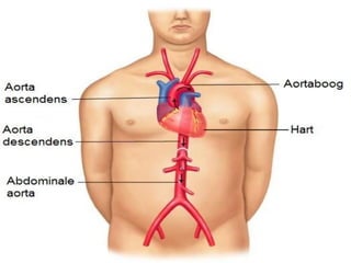
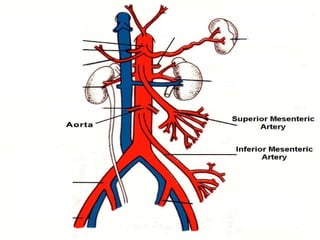
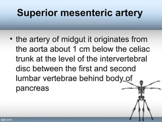
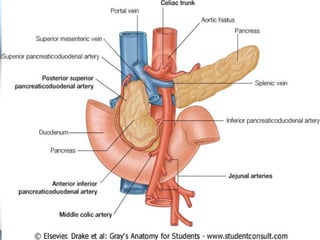
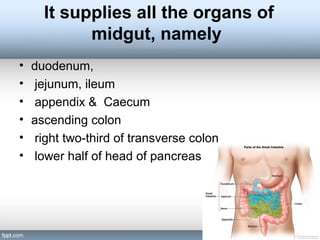
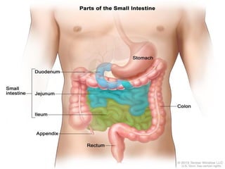
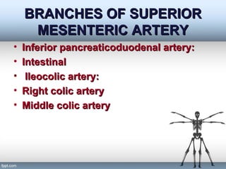
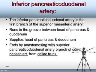
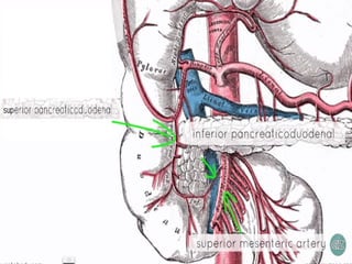
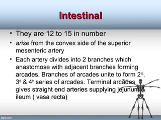
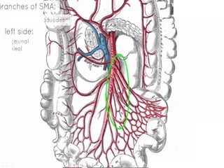
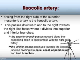
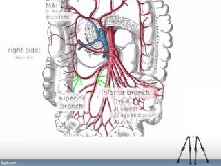
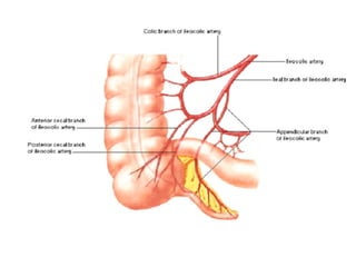
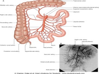
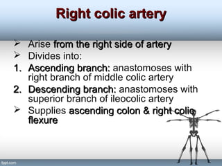
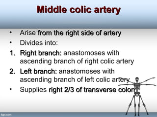
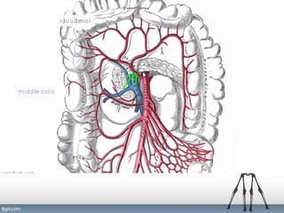
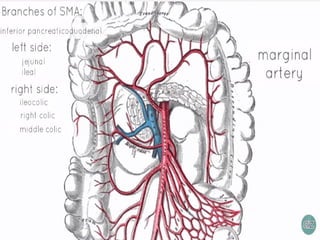
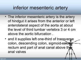
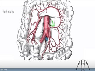
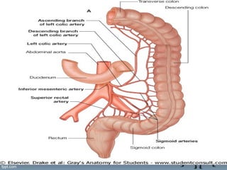
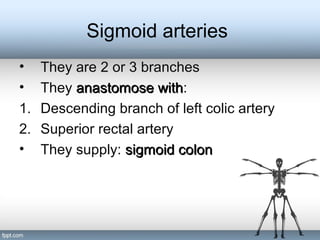
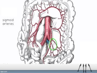
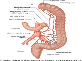
The document describes the arterial blood supply of the gastrointestinal tract. It details the branches and territories of the superior and inferior mesenteric arteries. The superior mesenteric artery supplies the midgut, including parts of the small intestine, right colon, and pancreas. It gives off branches like the intestinal, ileocolic, right colic, and middle colic arteries. The inferior mesenteric artery supplies the hindgut, including parts of the left colon, sigmoid colon, and rectum. Its branches include the left colic, sigmoid, and superior rectal arteries.
In this presentation the development of Small intestine and Pancreas has been discussed. The viewer would be able to understand the concept of physiological herniation and rotation of the Primary intestinal loop with in the connecting stalk.
rectus sheath, the sheath covering rectus muscle of anterior abdominal wall, formation of the sheath, the muscles involved in ts formation, and the contents the sheath is covering
Presentation on the topic - Great sephanous vein
in this presentaion all the topis like course , tributaries ,clinical aspects etc. of vein are covered.
content source - MBBS BOOKS OF 1ST YEAR
Anatomy of urinary bladder. surfaces, border of urinary bladder its relation , ligament support, peritoneal relation in male and females, pouches, blood supply of bladder, nerve supply of bladder, true and false ligament of urinary bladder,
The thoracic structure refers to the anatomical components of the thorax, which is the region of the body between the neck and the abdomen, also known as the chest. It includes the thoracic vertebrae, ribs, sternum (breastbone), and associated muscles, ligaments, and organs such as the heart and lungs. The thoracic structure plays a crucial role in protecting vital organs, supporting the upper body, and facilitating respiration.
The thoracic structure encompasses the anatomical elements of the chest region, including the thoracic vertebrae, ribs, sternum, associated muscles, and vital organs such as the heart and lungs. It provides protection, support, and facilitates respiration, highlighting its critical role in overall bodily function and health.The thoracic structure comprises the chest's anatomical components, including vertebrae, ribs, sternum, muscles, and vital organs like the heart and lungs, crucial for protection, support, and respiration..The thoracic structure includes the chest's bones, muscles, and organs, vital for breathing and protecting the heart and lungs.
1. **Thoracic Vertebrae:** The thoracic spine consists of twelve vertebrae (T1-T12) that form the posterior aspect of the thoracic structure. These vertebrae are larger than those in the cervical or lumbar regions and articulate with the ribs, providing stability and support for the upper body.
2. **Ribs:** There are twelve pairs of ribs that attach posteriorly to the thoracic vertebrae and curve anteriorly to meet the sternum. Ribs play a vital role in protecting the internal organs of the thoracic cavity, such as the heart and lungs. The upper seven pairs are true ribs, while the lower five pairs are false ribs (some of which do not directly attach to the sternum) or floating ribs.
3. **Sternum:** The sternum is a flat bone located in the center of the anterior thoracic wall. It consists of three parts: the manubrium, body, and xiphoid process. The sternum serves as an attachment point for the ribs and provides structural support to the chest.
4. **Muscles:** Several muscles surround the thoracic cavity, contributing to breathing and movement of the chest wall. These include the intercostal muscles (external, internal, and innermost), which are located between the ribs and aid in respiration, as well as muscles such as the diaphragm, which separates the thoracic and abdominal cavities and plays a primary role in breathing.
5. **Organs:** The thoracic cavity houses important organs such as the heart and lungs. The heart is situated in the mediastinum, a central compartment of the thoracic cavity, while the lungs occupy the lateral portions. The thoracic structure provides protection and support for these vital organs while allowing for their necessary movements during respiration and circulation.
Understanding the anatomy and function of the thoracic structure is essential for various medical disciplines, including anatomy, physiology, orthopedics, cardiology, and res
Here is a detailed presentation on anatomy of heart
I sincerely agree that few of my slides are copied and most of them are prepared by myself
But that is how we help each other!!
Hope the presentation helps the one in need
And it's free to download for anyone
The whole purpose of uploading is.. So that anyone can use it ..
s an inherited autosomal recessive disease that is characterized by high concentrations of the amino acid cystine in the urine, leading to the formation of cystine stones in the kidneys, ureter, and bladder.
this presentation will talk general about the medical education in Iraq . how the universities are teaching? and what is the level of the progress for the health care system in IRAQ .
this presentation talk about the most important function of the kidney and about renal failur disease and it's effect pathophysiology hope it will be helpful for u
This is a presentation by Dada Robert in a Your Skill Boost masterclass organised by the Excellence Foundation for South Sudan (EFSS) on Saturday, the 25th and Sunday, the 26th of May 2024.
He discussed the concept of quality improvement, emphasizing its applicability to various aspects of life, including personal, project, and program improvements. He defined quality as doing the right thing at the right time in the right way to achieve the best possible results and discussed the concept of the "gap" between what we know and what we do, and how this gap represents the areas we need to improve. He explained the scientific approach to quality improvement, which involves systematic performance analysis, testing and learning, and implementing change ideas. He also highlighted the importance of client focus and a team approach to quality improvement.
How to Create Map Views in the Odoo 17 ERPCeline George
The map views are useful for providing a geographical representation of data. They allow users to visualize and analyze the data in a more intuitive manner.
The French Revolution, which began in 1789, was a period of radical social and political upheaval in France. It marked the decline of absolute monarchies, the rise of secular and democratic republics, and the eventual rise of Napoleon Bonaparte. This revolutionary period is crucial in understanding the transition from feudalism to modernity in Europe.
For more information, visit-www.vavaclasses.com
Instructions for Submissions thorugh G- Classroom.pptxJheel Barad
This presentation provides a briefing on how to upload submissions and documents in Google Classroom. It was prepared as part of an orientation for new Sainik School in-service teacher trainees. As a training officer, my goal is to ensure that you are comfortable and proficient with this essential tool for managing assignments and fostering student engagement.
Palestine last event orientationfvgnh .pptxRaedMohamed3
An EFL lesson about the current events in Palestine. It is intended to be for intermediate students who wish to increase their listening skills through a short lesson in power point.
Unit 8 - Information and Communication Technology (Paper I).pdfThiyagu K
This slides describes the basic concepts of ICT, basics of Email, Emerging Technology and Digital Initiatives in Education. This presentations aligns with the UGC Paper I syllabus.
The Indian economy is classified into different sectors to simplify the analysis and understanding of economic activities. For Class 10, it's essential to grasp the sectors of the Indian economy, understand their characteristics, and recognize their importance. This guide will provide detailed notes on the Sectors of the Indian Economy Class 10, using specific long-tail keywords to enhance comprehension.
For more information, visit-www.vavaclasses.com
The Art Pastor's Guide to Sabbath | Steve ThomasonSteve Thomason
What is the purpose of the Sabbath Law in the Torah. It is interesting to compare how the context of the law shifts from Exodus to Deuteronomy. Who gets to rest, and why?
Read| The latest issue of The Challenger is here! We are thrilled to announce that our school paper has qualified for the NATIONAL SCHOOLS PRESS CONFERENCE (NSPC) 2024. Thank you for your unwavering support and trust. Dive into the stories that made us stand out!
1. By;AMEER AZEEZ
Superior and inferior
mesenteric artery
03/07/2015
AKAKI TSERETELI STATEAKAKI TSERETELI STATE
UNIVERSITY.UNIVERSITY.
2.
3.
4. Superior mesenteric artery
• the artery of midgut it originates from
the aorta about 1 cm below the celiac
trunk at the level of the intervertebral
disc between the first and second
lumbar vertebrae behind body of
pancreas
5.
6.
7. It supplies all the organs of
midgut, namely
• duodenum,
• jejunum, ileum
• appendix & Caecum
• ascending colon
• right two-third of transverse colon
• lower half of head of pancreas
8.
9. BRANCHES OF SUPERIORBRANCHES OF SUPERIOR
MESENTERIC ARTERYMESENTERIC ARTERY
• Inferior pancreaticoduodenal artery:Inferior pancreaticoduodenal artery:
• IntestinalIntestinal
• Ileocolic artery:Ileocolic artery:
• Right colic arteryRight colic artery
• Middle colic arteryMiddle colic artery
10. Inferior pancreaticoduodenalInferior pancreaticoduodenal
artery:artery:
• The inferior pancreaticoduodenal artery is the
first branch of the superior mesenteric artery.
• Runs in the groove between head of pancreas &between head of pancreas &
duodenumduodenum
• Supplies head of pancreas & duodenumhead of pancreas & duodenum
• Ends by anastomosing withanastomosing with superior
pancreaticoduodenal artery branch of common
hepatic art.hepatic art. from celiac trunkceliac trunk
11.
12. IntestinalIntestinal
• They are 12 to 15 in number
• arise from the convex side of the superior
mesenteric artery
• Each artery divides into 2 branches which
anastomose with adjacent branches forming
arcadesarcades. Branches of arcades unite to form 2nd
,
3rd
& 4th
series of arcades. Terminal arcades
gives straight end arteries supplying jejunum &straight end arteries supplying jejunum &
ileum ( vasa recta)ileum ( vasa recta)
13.
14. Ileocolic artery:Ileocolic artery:
• arising from the right side of the superior
mesenteric artery is the ileocolic artery.
• This passes downward and to the right towards
the right iliac fossa where it divides into superior
and inferior branches:
the superior branchthe superior branch passes upward along the
ascending colon to anastomose with the right colic
artery;
the inferior branchthe inferior branch continues towards the ileocolic
junction dividing into colic, cecal, appendicular,
and ileal branches.
15.
16. The specific pattern of distribution and
origin of these branches is variable:
thethe ileal branchileal branch passes to the left and ascends to
supply the final part of the ileum before anastomosing
with the superior mesenteric artery
anterior and posterior cecal branchescecal branches, either arising as
a common trunk or as separate branches, supply
corresponding sides of the cecum;
the appendicularappendicular branchbranch enters the free margin of and
supplies the mesoappendix and the appendix;
the colic branchcolic branch crosses to the ascending colon and
passes upward to supply the first part of the ascending
colon
17.
18.
19. Right colic arteryRight colic artery
Arise from the right side of arteryfrom the right side of artery
Divides into:
1.1. Ascending branch:Ascending branch: anastomoses with
right branch of middle colic artery
2.2. Descending branch:Descending branch: anastomoses with
superior branch of ileocolic artery
Supplies ascending colon & right colicascending colon & right colic
flexureflexure
20.
21. Middle colic arteryMiddle colic artery
• Arise from the right side of arteryfrom the right side of artery
• Divides into:
1.1. Right branch:Right branch: anastomoses with
ascending branch of right colic artery
2.2. Left branch:Left branch: anastomoses with
ascending branch of left colic artery
• Supplies right 2/3 of transverse colonright 2/3 of transverse colon
22.
23.
24. inferior mesenteric artery
• The inferior mesenteric artery is the artery
of hindgut it arises from the anterior or left
anterolateral aspect of the aorta at about
the level of third lumbar vertebra 3 or 4 cm
above the aortic bifurcation .
• and it supplies left one-third of transverse
colon, descending colon, sigmoid colon,
rectum and part of anal canal above the
anal valves
27. Left colic artery
• Divides into:
1.1. Ascending branch:Ascending branch: anastomoses with left
branch of middle colic artery
2.2. Descending branch:Descending branch: anastomoses with
sigmoid arteries
• Supplies: left 1/3 of transverse colon, leftleft 1/3 of transverse colon, left
colic flexure & descending coloncolic flexure & descending colon
28.
29.
30. Sigmoid arteries
• They are 2 or 3 branches
• They anastomose withanastomose with:
1. Descending branch of left colic artery
2. Superior rectal artery
• They supply: sigmoid colonsigmoid colon
31.
32.
33. Superior rectal artery
• The terminal branch of the inferior mesenteric
artery is the superior rectal artery.
• This vessel descends into the pelvic cavity in
the sigmoid mesocolon, crossing the left
common iliac vessels.
• Opposite vertebra SIII, the superior rectal
artery divides. The two terminal branches
descend on each side of the rectum, dividing
into smaller branches in the wall of the rectum.
33BITEW M./bitewm@gmail.com
34. • These smaller branches continue inferiorly
to the level of the internal anal sphincter,
anastomosing along the way with
branches from the middle rectal arteries
(from the internal iliac artery) and the
inferior rectal arteries (from the internal
pudendal artery).