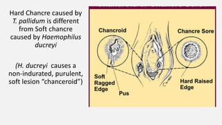
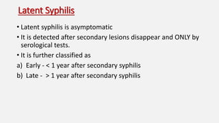
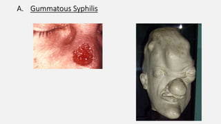
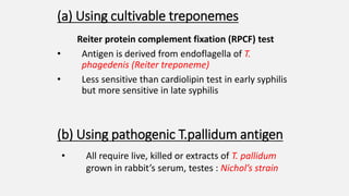
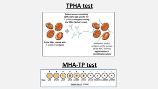
Spirochaetes
Anuradha Bhagwat
Isha Joshi
Sanika Deodhar
Shivani Deshpande
CONTENTS
• General Introduction-Spirochaetes
• Treponema
• Treponema Palladium:
• Morphology
• Cultivation
• Antigenic Structure
• Pathogenesis
• Laboratory Diagnosis
General Introduction
• Spirochaetes are elongated ,motile,flexible,bacteria twisted along the
long axis.
• Various Species of spirochaetes are:Borrelia,Treponema,Leptospira
CELL WALL
Spirochaetes have gram
negative type of cell wall.
The constituents if the cell wall
are:
1. Inner most layer –
Peptidoglycan
2. Lipoprotein layer
3. Outer Membrane-Outer
membrane protein,porins
and phospholipid
4. Outermost layer –
Lipopolysaccharide(not
present here)
ENDOFLAGELLA
• Motility of spirochaetes can be explained on the basis of presence of
endoflagella .
• Endoflagella are fine fibrils attached subterminally at each pole of the cell and
extend towards the opposite pole between outer membrane and peptidoglycan
layer of the cell wall.
• NUMBER OF ENDOFLAGELLA :
• Treponema-3 to 4
• Borrelia-15 to 20
• Leptospira-1
The spiral shape and the serpentine motility depend upon integrity of
the endoflagella .The motility is of three types:
1. Flexion and Extension
2. Corkscrew like rotatory movement
3. Translatory motion
TREPONEMA
• Treponema-(trepos meaning to turn and nema meaning thread)
• Thus these are slender spirochaetes with fine sprials and pointed
ends
• Some of them are pathogenic in humans while others occur a
commensals in mouth and genetalia.
• Cannot be cultivated in culture media and are maintained in
susceptible animals.
PATHOGENIC
TREPONEMA SPECIES
Vereneal
treponematoses :
Vereneal Syphilis-
Treponema Palladium
Non-Vereneal
treponematoses:
Yaws
Bejel
Pinta
YAWS-Caused by
T.pertenue
Causes skin sores and
diffusing growth on legs
around nose and mouth
ENDEMIC
SYPHILIS(BEJEL)-Caused
by T.endemicum
Causes mouth sores and
destrucctive lumps in
bone
PINTA-T.Carateum
Causes itchy
,thickened,disolored
patches on the skin
TREPONEMA PALLADIUM-MORPHOLOGY
• It is thin ,delicate ,elongated,flexible,twisted along the long axis and
motile.
• It is 10um long and 0.1-0.2um wide .
• They possesses 3 to 4 endoflagella attached sub-terminally ,they are
more than half length of the organism so might interdigitate in the
center .
• Transverse Section may show 3 -4 at the terminal and 4-6 at the
center.
• MOTILITY IS SEEN BY:
Flexion and extension, Corkscrew-like rotatory movement, Translatory
motion
• Best seen in wet living preparation with dark ground microscope
DRIED PREPARATION
• Here it needs to be thickened by silver impregnanation
method:Levaditi for tissue section and Fonatana’s method for staining
films
CULTIVATION
• Pathogenic treponemes cannot be cultivated in artificial media.
• Nichol’s strain is maintained in rabbit testis obtained from CSF of a
patient with neuro-syphilis.
• Non pathogenic strain is called Reiter strain shown by T.refringens and
T.phagedenis.
• They can be grown under anaerobic condition in Smith –Noguchi
medium or digest broth enriched with serum.
ANTIGENIC STRUCTURE
• The antigenic structure is poorly understood.Infection due to these
treponemes leads to production of 3 types of antibodies.
• On this basis they may be divided as speific and non-specific antigen.
SPECIFIC ANTIGEN
• Group specific antigen:Protein in nature Present in both pathogenic
as well as non-pathogenic(Reiter treponeme).Antigen used here to
detect group specificc antibodies is Reiter treponeme.
• Species –specific treponemal antigen:Polysacccharide in
nature,Antgen used here is T.palladium.
NON-SPECIFIC ANTIGEN
• A non-specific antibody appears in sera of patients that reacts with
lipid hapten extracted from beef heart .
• This hapten is called cardiolipin.
Pathogenesis
Treponemal species pathogenic to man include causative
agents of
• Venereal Syphilis - T. pallidum
• Non- Venereal treponematoses – Yaws T. pertenue
• Endemic Syphilis – T. endemicum
• Pinta – T. carateum
• T. pallidum is a strict parasite with a limited life span outside
the body
• Syphilitic infections can be transmitted via
1) Sexual intercourse
2) Materno-foetal transmission – congenital syphilis
3) Intimate person-to-person contact
4) Transfusion of infected blood
5) Accidental finger prick with an infected needle in medical
personnel
• Treponemes are present in superficial genital lesions and
pass from one partner to the other either via intact mucous
membrane or minor skin abrasions to multiply at the sight of
entry
• After an incubation period of 10 – 90 days clinical disease
sets in
Acquired Syphilis is divided into 4 stages based on period
after which lesions appear and the type of lesions
1. Primary Syphilis
2. Secondary Syphilis
3. Latent Syphilis
4. Tertiary Syphilis
Primary Syphilis
The characteristic lesion of primary syphilis is a Chancre.
It is also know as Hunterian Chancre after John Hunter
• It is 1-2cm in diameter and appears 2-4 weeks after exposure
to treponeme
• Painless papule
• Central ulceration
• Avascular
• Circumscribed
• Indurated when fully developed
• Accompanied by Lymphadenitis
• The bacteria invade regional lymph nodes ( inguinal in males
and pelvic in females) and through these enter blood
circulation
• The lymph nodes appear swollen, discrete, rubbery and non
tender
• Chancre heals in 3-6 weeks leaving a very thin scar.
• Antibody tests are +ve 1-3 weeks after appearance of
chancre
Hard Chancre caused by
T. pallidum is different
from Soft chancre
caused by Haemophilus
ducreyi
(H. ducreyi causes a
non-indurated, purulent,
soft lesion “chanceroid”)
 Histology
It is oedematous with dense infiltration of plasma cells ,
lymphocytes and macrophages
Perivascular aggregation of mononuclear cells
Proliferation of vascular endothelium
 Common sites
External genitalia – prepuce, penis, labia, etc
Cervix, perianal areas, anal canal, tongue, lips, cheeks,
nipples,etc
Chancre usually occurs singly, but immunocompromised
individuals may be develop multiple
Secondary Syphilis
• Inadequately treated patients develop secondary syphilis
• It sets in 2-6 months after the Chancre heals during which
the patient is asymptomatic
• marked constitutional symptoms include
Mucocutaneous lesions – oropharynx , vagina
Diffuse erythematous lesions - particularly on trunk and
extremities
Condyloma lata – anogenital regions, mucocutaneous
junctions
It is a highly infective stage and lesions heal in 4-5 years.
Erythematous lesions
Oropharyngeal
condyloma lata
Macules and papules
seen in Secondary
Syphilis
Latent Syphilis
• Latent syphilis is asymptomatic
• It is detected after secondary lesions disappear and ONLY by
serological tests.
• It is further classified as
a) Early - < 1 year after secondary syphilis
b) Late - > 1 year after secondary syphilis
Tertiary Syphilis
• It develops between 3 years to decades after primary
infection.
• It is a progressive stage prevalent in 3 forms
1) Gummatous Syphilis
2) Cardiovascular Syphilis
3) Neurosyphilis
A. Gummatous Syphilis
• Syphilitic gumma are solitary, rubbery , pink, small fleshy
masses containing bacteria
• They may be nodular, tumorous or ulcerative
• Lesions may show characteristic central necrosis
• 1mm-1cm in size
• Present predominantly in Brain, testes, bone, liver, etc.
B. Cardiovascular Syphilis
• More serious lesions of heart and aorta are seen in this case
Inflammatory response to spirochetes in arterial walls
Chronic inflammation of Tunica adventitia of elastic arteries
Resultant endarteritis and periarteritis of vaso vasorum
Ischaemia of t. media and t. adventitia
Weakening of wall of aorta and resultant dilatation
Aortic Aneurysm
Hepar Lobatum
Aorta
C. Neurosyphilis
• It may manifest as
1. Meningovascular Syphilis
2. Tabes dorsalis affecting the spinal cord - Tabes dorsalis is a
slow degeneration of the nerve cells and nerve fibers that
carry sensory information to the brain
3. General paresis of the insane - a severe neuropsychiatric
disorder. It is a result of cerebral atrophy caused due to
chronic meningoencephalitis
Argyll Robertson Pupil
Congenital Syphilis
It is seen in a foetus of more than 16 weeks gestation who is
exposed to maternal spirochaetemia.
Features include
• Saddle - shaped nose deformity
• Hutchinson’s teeth
• Interstitial keratitis
• Corneal opacity
• Epiphysitis
• Periostitis
Treatment
• T. pallidum is particularly sensitive to Pencillin
• In early cases, 2.4 million units of Benzathine
penicillin is adequate
For late syphilis this maybe repeated weekly for 3
weeks
• Doxycyline is administered in patients allergic to Penicillin
• Ceftriaxone is effective particularly in Neurosyphilis
 Antibiotic therapy particularly with penicillin may induce
Jarisch-Herxheimer Reaction due to release of endotoxins
following lysis of spirochaetes . Its onset is within 1-2 hours and
reaction subsides in 12-24 hours
• Fever
• Chills
• Myalgia
• Tachycardia
• Hypervetilation
• Vasodilation
• Hypotension
Laboratory diagnosis
CLINICAL DIAGNOSIS OF SYPHILLIS
Demonstration of treponemes
in exudate.
demonstration of
treponemes in tissue
Demonstration of treponemal
antigen in the lesion
Serological tests
1)Demonstration of treponema pallidum in
exudate
A) DARK GROUND MICROSCOPY:
The surface of the lesion is cleaned with
gauze dipped in warm normal saline and the
margins are gently scraped.
Gentle pressure is applied to base of lesion
till serum exudates.
The material is collected directly on
coverslip, placed on a glass slide &
examined under dark ground microscope.
T. Pallidum is recognized by slender
structure, regularity of its spirals and
pointed ends.
DISADVANTAGE: This method cannot be
used for oral lesions as the non pathogenic
treponemes can be confused with
T.pallidum.
B) DIRECT FLUORESCENT ANTIBODY STAINING FOR T.
PALLIDUM (DFA-TP)
 This method provides a more definite
diagnosis.
 The smear made on glass slide is fixed by
acetone and sent to laboratory.
 This is then stained by fluorescein labeled
pathogen specific antibody and examined
under fluorescence microscope.
 T.Pallidum appears as distinctive, sharply
outlined structure which has apple green
fluorescence.
 ADVANTAGE: This can be used to diagnose
oral lesions which is not possible by dark
ground microscopy.
3)DEMONSTRATION OF TREPONEMAL
ANTIGEN IN THE LESIONS
A) ENZYME IMMUNOASSAY
B)POLYMERASE CHAIN REACTION
4) SEROLOGICAL DIAGNOSIS
Based on serological diagnosis:
 nontreponemal/standard test for syphillis (STS)
 Treponemal tests
Nontreponemal tests:
1) Wasserman & kahn test
2) Veneral disease research laboratory (VDRL)
3) Rapid plasma reagin (RPR)
Antigen used in these tests is an alcoholic extract of beef
heart tissue (cardiolipin) to which lecithin and cholesterol
are added.
2)VDRL TEST
It is performed as a slide test on glass side which consists of
inactivated patient serum+ antigen (cardiolipin,
lecithin,cholesterol)
Mixture is rotated for 4 mins mechanically after which
flocculation can be detected under low power microscope.
This test is used as screening test in syphillis.
Positive in 70% primary and 90% secondary syphillis
Patients with late syphillis are non reactive.
ADVANTAGE: standard test for testing of CSF.
VDRL TEST
3)RAPID PLASMA REAGIN (RPR) TEST
VDRL antigen is adsorbed on finely divided carbon particles
& suspended in choline chloride.
Antigen is also stabilized by EDTA.
One drop patient serum+ 1 drop of modified antigen taken on
disposable plastic card by a disposable stick.
Card is rocked gently for 8 mins
+ve test: flocculation of -ve test: absence of
Carbon visible to naked black aggregates
eye( black aggeregates) with uniform grey
background.
If the test is positive the quantitative tests should be done
by doubling dilutions of serum (1:2, 1:4, 1:6, 1:8,…….. 1:32).
Most labs use this test as screening for syphillis.
Sensitivity and specificity: same as VDRL test but is is rapid
and easy to perform.
DISADVANTAGE: cannot be used to test CSF.
BIOLOGICAL FALSE POSITIVE REACTIONS
Antibodies against cardiolipin may be detected in absence
of T. pallidum infection giving rise to false positive rections
(BFP).
These are of 2 types:
A. ACUTE/TRANSIENT BFP REACTION:
After acute febrile infectious diseases.
B. CHRONIC BFP REACTION:
 Autoimmune diseases
 Infectious mononucleosis, eosinophilia
 Leprosy
 Malaria
 Relapsing fever
 Hepatitis
Treponemal Tests
• They detect specific antibody to T. pallidum proteins
• Used to confirm the infection and determine whether the disease is
active
• False positive results can occur and may be due to other infections
Treponemal
Tests
Using Cultivable
Treponemes
-RPCF test
Using
pathogenic T.
pallidum
antigen
Test using
live T. pallidum
-TPI test
Tests using
killed T. pallidum
-TPA test
-TPIA test
-FTA test
FTA-ABS test
IgM FTA-ABS test
Tests using
T.pallidum extract
-TPHA test
-EIA test
(a) Using cultivable treponemes
Reiter protein complement fixation (RPCF) test
• Antigen is derived from endoflagella of T.
phagedenis (Reiter treponeme)
• Less sensitive than cardiolipin test in early syphilis
but more sensitive in late syphilis
(b) Using pathogenic T.pallidum antigen
• All require live, killed or extracts of T. pallidum
grown in rabbit’s serum, testes : Nichol’s strain
• Suspension of treponemes, complement + Test serum
-----> Incubate anaerobically
• Examine under Dark-ground microscope
• In presence of antibody : Treponemes found to be
immobilized
i. Treponema pallidum immobilization (TPI) test
I. Tests using live T. pallidum
ii. Treponema pallidum immune adherence (TPIA)
test
• Suspension of
inactivated
treponemes + Test
serum,
complement, fresh
heparinised whole
blood ----->
Incubate
• In presence of antibody :
Treponemes adhere to erythrocytes
• In absence : No immune
adherence
i. Treponema pallidum agglutination (TPA) test
• Suspension of T.pallidum (inactivated by formalin) +
Test serum -----> Incubate
• Examined under Dark-ground microscope
• In presence of antibody : Treponemes found to be
agglutinated
II. Tests using killed T. pallidum
• Indirect immunofluorescence antibody test
• Diluted patient’s serum is absorbed with extract of
Reiter treponemes -----> layered on a slide to which T.
pallidum (pallidum) has been fixed
• In presence of antibody: antibody will coat the
treponeme
• Has high specificity (92%-99%) and sensitivity
• Modification: IgM FTA-ABS test: detects IgM
antibodies in congenital syphilis, distinguishing it from
IgG
iii. Fluorescent treponemal antibody- absorption (FTA-ABS) test
FTA-ABS test
i. Treponema pallidum haemagglutination (TPHA)
test
• T.pallidum antigen absorbed on RBC surface + patient
serum
• In presence of antibody : haemagglutination is seen
• May be performed in microlitre plates : Microlitre
haemagglutination T.pallidum (MHA-TP) test
• Test sensitivity : 65%
III. Tests using T. pallidum extract
TPHA test
MHA-TP test
ii. Enzyme immunoassay (EIA) test
• Ultrasonicate of T. pallidum antigen coated on tubes
or ferrous metal beads.
• Antibody in patient serum is detected by EIA.
• Test sensitivity : 90%
• Test specificity : 98%
Prophylaxis
• Source of infection : infected individual , for 3-5 yrs during early
syphilis.
• Transmission is by direct contact.
• Avoidance of sexual contact with infected individual.
• Use of antibiotic prophylaxis : penicillin
Borrelia vincentii
• Morphology : 7 -18µm long, 0.2 -
0.6µm wide, with 3-8 loose open
coils of variable size, motile
• Cultural characteristics: Gram
negative, stained with dilute carbol
fuchsin, methyl violet, Giemsa &
Leishman stains, obligate anaerobe,
cultured in digest broth enriched
with ascitic fluid.
• Pathogenesis: gives rise to ulcerative
gingivostomatitis or oropharyngitis
(vincent’s angina)
• Diagnosis: smears from ulcerative
lesions stained with dilute carbol
fuchsin
Spirochates

Spirochates

  • 1.
  • 2.
    CONTENTS • General Introduction-Spirochaetes •Treponema • Treponema Palladium: • Morphology • Cultivation • Antigenic Structure • Pathogenesis • Laboratory Diagnosis
  • 3.
    General Introduction • Spirochaetesare elongated ,motile,flexible,bacteria twisted along the long axis. • Various Species of spirochaetes are:Borrelia,Treponema,Leptospira
  • 4.
    CELL WALL Spirochaetes havegram negative type of cell wall. The constituents if the cell wall are: 1. Inner most layer – Peptidoglycan 2. Lipoprotein layer 3. Outer Membrane-Outer membrane protein,porins and phospholipid 4. Outermost layer – Lipopolysaccharide(not present here)
  • 5.
    ENDOFLAGELLA • Motility ofspirochaetes can be explained on the basis of presence of endoflagella .
  • 6.
    • Endoflagella arefine fibrils attached subterminally at each pole of the cell and extend towards the opposite pole between outer membrane and peptidoglycan layer of the cell wall.
  • 7.
    • NUMBER OFENDOFLAGELLA : • Treponema-3 to 4 • Borrelia-15 to 20 • Leptospira-1 The spiral shape and the serpentine motility depend upon integrity of the endoflagella .The motility is of three types: 1. Flexion and Extension 2. Corkscrew like rotatory movement 3. Translatory motion
  • 8.
    TREPONEMA • Treponema-(trepos meaningto turn and nema meaning thread) • Thus these are slender spirochaetes with fine sprials and pointed ends • Some of them are pathogenic in humans while others occur a commensals in mouth and genetalia. • Cannot be cultivated in culture media and are maintained in susceptible animals.
  • 9.
    PATHOGENIC TREPONEMA SPECIES Vereneal treponematoses : VerenealSyphilis- Treponema Palladium Non-Vereneal treponematoses: Yaws Bejel Pinta
  • 10.
    YAWS-Caused by T.pertenue Causes skinsores and diffusing growth on legs around nose and mouth
  • 11.
  • 12.
  • 13.
    TREPONEMA PALLADIUM-MORPHOLOGY • Itis thin ,delicate ,elongated,flexible,twisted along the long axis and motile. • It is 10um long and 0.1-0.2um wide . • They possesses 3 to 4 endoflagella attached sub-terminally ,they are more than half length of the organism so might interdigitate in the center . • Transverse Section may show 3 -4 at the terminal and 4-6 at the center.
  • 14.
    • MOTILITY ISSEEN BY: Flexion and extension, Corkscrew-like rotatory movement, Translatory motion • Best seen in wet living preparation with dark ground microscope
  • 15.
    DRIED PREPARATION • Hereit needs to be thickened by silver impregnanation method:Levaditi for tissue section and Fonatana’s method for staining films
  • 16.
    CULTIVATION • Pathogenic treponemescannot be cultivated in artificial media. • Nichol’s strain is maintained in rabbit testis obtained from CSF of a patient with neuro-syphilis. • Non pathogenic strain is called Reiter strain shown by T.refringens and T.phagedenis. • They can be grown under anaerobic condition in Smith –Noguchi medium or digest broth enriched with serum.
  • 17.
    ANTIGENIC STRUCTURE • Theantigenic structure is poorly understood.Infection due to these treponemes leads to production of 3 types of antibodies. • On this basis they may be divided as speific and non-specific antigen.
  • 18.
    SPECIFIC ANTIGEN • Groupspecific antigen:Protein in nature Present in both pathogenic as well as non-pathogenic(Reiter treponeme).Antigen used here to detect group specificc antibodies is Reiter treponeme. • Species –specific treponemal antigen:Polysacccharide in nature,Antgen used here is T.palladium.
  • 19.
    NON-SPECIFIC ANTIGEN • Anon-specific antibody appears in sera of patients that reacts with lipid hapten extracted from beef heart . • This hapten is called cardiolipin.
  • 20.
    Pathogenesis Treponemal species pathogenicto man include causative agents of • Venereal Syphilis - T. pallidum • Non- Venereal treponematoses – Yaws T. pertenue • Endemic Syphilis – T. endemicum • Pinta – T. carateum
  • 21.
    • T. pallidumis a strict parasite with a limited life span outside the body • Syphilitic infections can be transmitted via 1) Sexual intercourse 2) Materno-foetal transmission – congenital syphilis 3) Intimate person-to-person contact 4) Transfusion of infected blood 5) Accidental finger prick with an infected needle in medical personnel
  • 22.
    • Treponemes arepresent in superficial genital lesions and pass from one partner to the other either via intact mucous membrane or minor skin abrasions to multiply at the sight of entry • After an incubation period of 10 – 90 days clinical disease sets in
  • 23.
    Acquired Syphilis isdivided into 4 stages based on period after which lesions appear and the type of lesions 1. Primary Syphilis 2. Secondary Syphilis 3. Latent Syphilis 4. Tertiary Syphilis
  • 24.
    Primary Syphilis The characteristiclesion of primary syphilis is a Chancre. It is also know as Hunterian Chancre after John Hunter • It is 1-2cm in diameter and appears 2-4 weeks after exposure to treponeme • Painless papule • Central ulceration • Avascular • Circumscribed • Indurated when fully developed • Accompanied by Lymphadenitis
  • 25.
    • The bacteriainvade regional lymph nodes ( inguinal in males and pelvic in females) and through these enter blood circulation • The lymph nodes appear swollen, discrete, rubbery and non tender • Chancre heals in 3-6 weeks leaving a very thin scar. • Antibody tests are +ve 1-3 weeks after appearance of chancre
  • 26.
    Hard Chancre causedby T. pallidum is different from Soft chancre caused by Haemophilus ducreyi (H. ducreyi causes a non-indurated, purulent, soft lesion “chanceroid”)
  • 27.
     Histology It isoedematous with dense infiltration of plasma cells , lymphocytes and macrophages Perivascular aggregation of mononuclear cells Proliferation of vascular endothelium  Common sites External genitalia – prepuce, penis, labia, etc Cervix, perianal areas, anal canal, tongue, lips, cheeks, nipples,etc Chancre usually occurs singly, but immunocompromised individuals may be develop multiple
  • 29.
    Secondary Syphilis • Inadequatelytreated patients develop secondary syphilis • It sets in 2-6 months after the Chancre heals during which the patient is asymptomatic • marked constitutional symptoms include Mucocutaneous lesions – oropharynx , vagina Diffuse erythematous lesions - particularly on trunk and extremities Condyloma lata – anogenital regions, mucocutaneous junctions It is a highly infective stage and lesions heal in 4-5 years.
  • 30.
  • 31.
    Macules and papules seenin Secondary Syphilis
  • 32.
    Latent Syphilis • Latentsyphilis is asymptomatic • It is detected after secondary lesions disappear and ONLY by serological tests. • It is further classified as a) Early - < 1 year after secondary syphilis b) Late - > 1 year after secondary syphilis
  • 33.
    Tertiary Syphilis • Itdevelops between 3 years to decades after primary infection. • It is a progressive stage prevalent in 3 forms 1) Gummatous Syphilis 2) Cardiovascular Syphilis 3) Neurosyphilis
  • 34.
  • 35.
    • Syphilitic gummaare solitary, rubbery , pink, small fleshy masses containing bacteria • They may be nodular, tumorous or ulcerative • Lesions may show characteristic central necrosis • 1mm-1cm in size • Present predominantly in Brain, testes, bone, liver, etc.
  • 36.
    B. Cardiovascular Syphilis •More serious lesions of heart and aorta are seen in this case Inflammatory response to spirochetes in arterial walls Chronic inflammation of Tunica adventitia of elastic arteries Resultant endarteritis and periarteritis of vaso vasorum Ischaemia of t. media and t. adventitia Weakening of wall of aorta and resultant dilatation Aortic Aneurysm
  • 37.
  • 38.
    C. Neurosyphilis • Itmay manifest as 1. Meningovascular Syphilis 2. Tabes dorsalis affecting the spinal cord - Tabes dorsalis is a slow degeneration of the nerve cells and nerve fibers that carry sensory information to the brain 3. General paresis of the insane - a severe neuropsychiatric disorder. It is a result of cerebral atrophy caused due to chronic meningoencephalitis
  • 40.
  • 41.
    Congenital Syphilis It isseen in a foetus of more than 16 weeks gestation who is exposed to maternal spirochaetemia. Features include • Saddle - shaped nose deformity • Hutchinson’s teeth • Interstitial keratitis • Corneal opacity • Epiphysitis • Periostitis
  • 43.
    Treatment • T. pallidumis particularly sensitive to Pencillin • In early cases, 2.4 million units of Benzathine penicillin is adequate For late syphilis this maybe repeated weekly for 3 weeks • Doxycyline is administered in patients allergic to Penicillin • Ceftriaxone is effective particularly in Neurosyphilis  Antibiotic therapy particularly with penicillin may induce Jarisch-Herxheimer Reaction due to release of endotoxins following lysis of spirochaetes . Its onset is within 1-2 hours and reaction subsides in 12-24 hours
  • 44.
    • Fever • Chills •Myalgia • Tachycardia • Hypervetilation • Vasodilation • Hypotension
  • 45.
  • 46.
    CLINICAL DIAGNOSIS OFSYPHILLIS Demonstration of treponemes in exudate. demonstration of treponemes in tissue Demonstration of treponemal antigen in the lesion Serological tests
  • 47.
    1)Demonstration of treponemapallidum in exudate A) DARK GROUND MICROSCOPY: The surface of the lesion is cleaned with gauze dipped in warm normal saline and the margins are gently scraped. Gentle pressure is applied to base of lesion till serum exudates. The material is collected directly on coverslip, placed on a glass slide & examined under dark ground microscope. T. Pallidum is recognized by slender structure, regularity of its spirals and pointed ends. DISADVANTAGE: This method cannot be used for oral lesions as the non pathogenic treponemes can be confused with T.pallidum.
  • 48.
    B) DIRECT FLUORESCENTANTIBODY STAINING FOR T. PALLIDUM (DFA-TP)  This method provides a more definite diagnosis.  The smear made on glass slide is fixed by acetone and sent to laboratory.  This is then stained by fluorescein labeled pathogen specific antibody and examined under fluorescence microscope.  T.Pallidum appears as distinctive, sharply outlined structure which has apple green fluorescence.  ADVANTAGE: This can be used to diagnose oral lesions which is not possible by dark ground microscopy.
  • 49.
    3)DEMONSTRATION OF TREPONEMAL ANTIGENIN THE LESIONS A) ENZYME IMMUNOASSAY B)POLYMERASE CHAIN REACTION
  • 50.
    4) SEROLOGICAL DIAGNOSIS Basedon serological diagnosis:  nontreponemal/standard test for syphillis (STS)  Treponemal tests Nontreponemal tests: 1) Wasserman & kahn test 2) Veneral disease research laboratory (VDRL) 3) Rapid plasma reagin (RPR) Antigen used in these tests is an alcoholic extract of beef heart tissue (cardiolipin) to which lecithin and cholesterol are added.
  • 51.
    2)VDRL TEST It isperformed as a slide test on glass side which consists of inactivated patient serum+ antigen (cardiolipin, lecithin,cholesterol) Mixture is rotated for 4 mins mechanically after which flocculation can be detected under low power microscope. This test is used as screening test in syphillis. Positive in 70% primary and 90% secondary syphillis Patients with late syphillis are non reactive. ADVANTAGE: standard test for testing of CSF.
  • 52.
  • 53.
    3)RAPID PLASMA REAGIN(RPR) TEST VDRL antigen is adsorbed on finely divided carbon particles & suspended in choline chloride. Antigen is also stabilized by EDTA. One drop patient serum+ 1 drop of modified antigen taken on disposable plastic card by a disposable stick. Card is rocked gently for 8 mins +ve test: flocculation of -ve test: absence of Carbon visible to naked black aggregates eye( black aggeregates) with uniform grey background.
  • 54.
    If the testis positive the quantitative tests should be done by doubling dilutions of serum (1:2, 1:4, 1:6, 1:8,…….. 1:32). Most labs use this test as screening for syphillis. Sensitivity and specificity: same as VDRL test but is is rapid and easy to perform. DISADVANTAGE: cannot be used to test CSF.
  • 55.
    BIOLOGICAL FALSE POSITIVEREACTIONS Antibodies against cardiolipin may be detected in absence of T. pallidum infection giving rise to false positive rections (BFP). These are of 2 types: A. ACUTE/TRANSIENT BFP REACTION: After acute febrile infectious diseases. B. CHRONIC BFP REACTION:  Autoimmune diseases  Infectious mononucleosis, eosinophilia  Leprosy  Malaria  Relapsing fever  Hepatitis
  • 56.
    Treponemal Tests • Theydetect specific antibody to T. pallidum proteins • Used to confirm the infection and determine whether the disease is active • False positive results can occur and may be due to other infections
  • 57.
    Treponemal Tests Using Cultivable Treponemes -RPCF test Using pathogenicT. pallidum antigen Test using live T. pallidum -TPI test Tests using killed T. pallidum -TPA test -TPIA test -FTA test FTA-ABS test IgM FTA-ABS test Tests using T.pallidum extract -TPHA test -EIA test
  • 58.
    (a) Using cultivabletreponemes Reiter protein complement fixation (RPCF) test • Antigen is derived from endoflagella of T. phagedenis (Reiter treponeme) • Less sensitive than cardiolipin test in early syphilis but more sensitive in late syphilis (b) Using pathogenic T.pallidum antigen • All require live, killed or extracts of T. pallidum grown in rabbit’s serum, testes : Nichol’s strain
  • 59.
    • Suspension oftreponemes, complement + Test serum -----> Incubate anaerobically • Examine under Dark-ground microscope • In presence of antibody : Treponemes found to be immobilized i. Treponema pallidum immobilization (TPI) test I. Tests using live T. pallidum
  • 60.
    ii. Treponema pallidumimmune adherence (TPIA) test • Suspension of inactivated treponemes + Test serum, complement, fresh heparinised whole blood -----> Incubate • In presence of antibody : Treponemes adhere to erythrocytes • In absence : No immune adherence
  • 61.
    i. Treponema pallidumagglutination (TPA) test • Suspension of T.pallidum (inactivated by formalin) + Test serum -----> Incubate • Examined under Dark-ground microscope • In presence of antibody : Treponemes found to be agglutinated II. Tests using killed T. pallidum
  • 62.
    • Indirect immunofluorescenceantibody test • Diluted patient’s serum is absorbed with extract of Reiter treponemes -----> layered on a slide to which T. pallidum (pallidum) has been fixed • In presence of antibody: antibody will coat the treponeme • Has high specificity (92%-99%) and sensitivity • Modification: IgM FTA-ABS test: detects IgM antibodies in congenital syphilis, distinguishing it from IgG iii. Fluorescent treponemal antibody- absorption (FTA-ABS) test
  • 63.
  • 64.
    i. Treponema pallidumhaemagglutination (TPHA) test • T.pallidum antigen absorbed on RBC surface + patient serum • In presence of antibody : haemagglutination is seen • May be performed in microlitre plates : Microlitre haemagglutination T.pallidum (MHA-TP) test • Test sensitivity : 65% III. Tests using T. pallidum extract
  • 65.
  • 66.
    ii. Enzyme immunoassay(EIA) test • Ultrasonicate of T. pallidum antigen coated on tubes or ferrous metal beads. • Antibody in patient serum is detected by EIA. • Test sensitivity : 90% • Test specificity : 98%
  • 67.
    Prophylaxis • Source ofinfection : infected individual , for 3-5 yrs during early syphilis. • Transmission is by direct contact. • Avoidance of sexual contact with infected individual. • Use of antibiotic prophylaxis : penicillin
  • 68.
    Borrelia vincentii • Morphology: 7 -18µm long, 0.2 - 0.6µm wide, with 3-8 loose open coils of variable size, motile • Cultural characteristics: Gram negative, stained with dilute carbol fuchsin, methyl violet, Giemsa & Leishman stains, obligate anaerobe, cultured in digest broth enriched with ascitic fluid. • Pathogenesis: gives rise to ulcerative gingivostomatitis or oropharyngitis (vincent’s angina) • Diagnosis: smears from ulcerative lesions stained with dilute carbol fuchsin