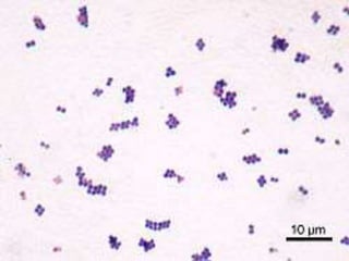
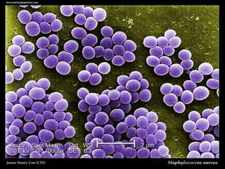
STAPHYLOCOCCUS
RATHEESH R L
• Medically important species is staphylococcus
aureus
MORPHOLOGY
• They are gram positive, ovoid, arranged in
bunches, non- motile, non- sporing and
anaerobic type.
• They measure 0.8 to 0.9 micrometer in
diameter.
CULTURAL CHARACTERISTICS
Staphylococcus grows usually in simple or
ordinary culture media under aerobic or
anaerobic conditions at optimum temperature
of 37 C and pH 7.5
NUTRIENT AGAR MEDIUM
• After over night incubation, it mostly produces
pigmented, golden yellow, round, smooth,
raised colonies with 2-4mm diameter
BLOOD AGAR MEDIUM
• The organism produces beta hemolysis in this
medium containing. rabbit Or sheep blood.
LIQUID MEDIUM
• In nutrient broth the organism produces
uniform turbidity
MacConkey's agar medium
• The organism produces pink colonies which
are of pinhead size indicating that they are
lactose fermenting
PATHOGENECITY
• The pathogenecity of organism is mainly due
to coagulase produced which inhibiting
phagocytosis
Cutaneous infection
• The organism causes superficial infections
like,pestules, abscesses, carbuncles, styes,
impetigo, furunctes (boils)
Deep infection
These include acute osteomyelitis, tonsilitis,
pharyngitis, pneumonitis,meningitis
Skin exfoliation/exfoliative disease
• The toxin (epidermolytic toxin or TSST-l)
produced by the organism causes striping of
superficial layers of skin that causes a clinical
condition known as Ritter's disease or scalded
skin syndrome (SSS) characterized by painful
rashes.
Toxic food poisoning
• When preformed toxins of the organism
growing In contaminated food (especially fish,
meat, milk & milk products) are ingested, the
toxins act on mucosal surface of GI tract
causing Staphylococcal food poisoning within
2-5 hours of ingestion characterized by
nausea, vomiting and diarrhea.
Toxic Shock Syndrome
• It is usually caused by the enterotoxin
(enterotoxin F) produced by organism in gut
characterized by fever, hypotension, vomiting,
confusion, headache, diarrhea, & rashes.
LABORATORY DIAGNOSIS
• Haematological investigations
Total leucocyte count (TLC):
Shows leucocytosis (count > 10.000
cells/cubic mm)
Differential leucocyte count (DLC) :
Shows increase in neutrophil count (more
than 80%
Bacteriological investigation
• Specimen: Depending upon the nature of
lesions specimen Is collected like pus from
suppurative lesion, CSF from meningitis, blood
from septicemia, midstream urine from UTI,
sputum from respiratory infection &
suspected food, feces or vomits from food
poisoning cases.
1. Microscopy:
Smear is made from the above
specimen on a clean slide & subjected to Gram
staining. Examination of Gram stained smear
shows Gram-positive cocci in clusters.
2. Culture:
• The above specimen is inoculated on
Nutrient agar medium, Blood Agar medium or
Mannitol agar medium (selective media) & the
plates are incubated at 37 C for 24 hours
• On nutrient agar colonies will appear as
golden yellow or white which are confirmed
by coagulase test
• On blood agar plates the organism shows beta
hemolysis and on mannitol agar colonies will
be pink due to fermentation of lactose by the
organism.
TREATMENT
• Benzyl penicillin, methicillin and cloxacillin are
effective.
• At present vancomycin is most effect drug
against staphylococci.

1. staphylococcus

  • 1.
  • 2.
    • Medically importantspecies is staphylococcus aureus
  • 4.
    MORPHOLOGY • They aregram positive, ovoid, arranged in bunches, non- motile, non- sporing and anaerobic type. • They measure 0.8 to 0.9 micrometer in diameter.
  • 6.
    CULTURAL CHARACTERISTICS Staphylococcus growsusually in simple or ordinary culture media under aerobic or anaerobic conditions at optimum temperature of 37 C and pH 7.5
  • 7.
    NUTRIENT AGAR MEDIUM •After over night incubation, it mostly produces pigmented, golden yellow, round, smooth, raised colonies with 2-4mm diameter
  • 8.
    BLOOD AGAR MEDIUM •The organism produces beta hemolysis in this medium containing. rabbit Or sheep blood.
  • 9.
    LIQUID MEDIUM • Innutrient broth the organism produces uniform turbidity
  • 10.
    MacConkey's agar medium •The organism produces pink colonies which are of pinhead size indicating that they are lactose fermenting
  • 11.
    PATHOGENECITY • The pathogenecityof organism is mainly due to coagulase produced which inhibiting phagocytosis
  • 12.
    Cutaneous infection • Theorganism causes superficial infections like,pestules, abscesses, carbuncles, styes, impetigo, furunctes (boils)
  • 13.
    Deep infection These includeacute osteomyelitis, tonsilitis, pharyngitis, pneumonitis,meningitis
  • 14.
    Skin exfoliation/exfoliative disease •The toxin (epidermolytic toxin or TSST-l) produced by the organism causes striping of superficial layers of skin that causes a clinical condition known as Ritter's disease or scalded skin syndrome (SSS) characterized by painful rashes.
  • 15.
    Toxic food poisoning •When preformed toxins of the organism growing In contaminated food (especially fish, meat, milk & milk products) are ingested, the toxins act on mucosal surface of GI tract causing Staphylococcal food poisoning within 2-5 hours of ingestion characterized by nausea, vomiting and diarrhea.
  • 16.
    Toxic Shock Syndrome •It is usually caused by the enterotoxin (enterotoxin F) produced by organism in gut characterized by fever, hypotension, vomiting, confusion, headache, diarrhea, & rashes.
  • 17.
    LABORATORY DIAGNOSIS • Haematologicalinvestigations Total leucocyte count (TLC): Shows leucocytosis (count > 10.000 cells/cubic mm) Differential leucocyte count (DLC) : Shows increase in neutrophil count (more than 80%
  • 18.
    Bacteriological investigation • Specimen:Depending upon the nature of lesions specimen Is collected like pus from suppurative lesion, CSF from meningitis, blood from septicemia, midstream urine from UTI, sputum from respiratory infection & suspected food, feces or vomits from food poisoning cases.
  • 19.
    1. Microscopy: Smear ismade from the above specimen on a clean slide & subjected to Gram staining. Examination of Gram stained smear shows Gram-positive cocci in clusters.
  • 20.
    2. Culture: • Theabove specimen is inoculated on Nutrient agar medium, Blood Agar medium or Mannitol agar medium (selective media) & the plates are incubated at 37 C for 24 hours • On nutrient agar colonies will appear as golden yellow or white which are confirmed by coagulase test
  • 21.
    • On bloodagar plates the organism shows beta hemolysis and on mannitol agar colonies will be pink due to fermentation of lactose by the organism.
  • 22.
    TREATMENT • Benzyl penicillin,methicillin and cloxacillin are effective. • At present vancomycin is most effect drug against staphylococci.