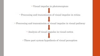
• Visual impulsein photoreceptors
• Processing and transmission of visual impulse in retina
• Processing and transmission of visual impulse in visual pathway
• Analysis of visual impulse in visual cortex
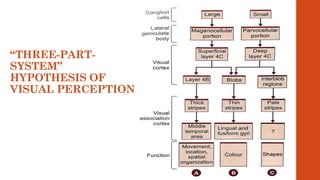
• Three part system hypothesis of visual perception
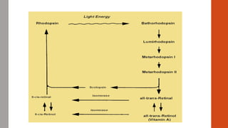
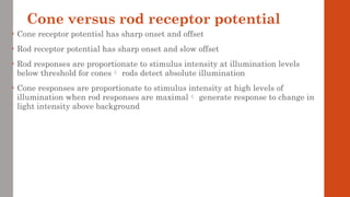
Cone versus rodreceptor potential
• Cone receptor potentisl has sharp onset and offset
• Rod receptor potential has sharp onset and slow offset
• Rod responses are proportionate to stimulus intensity at illumination levels
below threshold for cones rods detect absolute illumination
• Cone responses are proportionate to stimulus intensity at high levels of
illumination when rod responses are maximal generate response to change in
light intensity above background
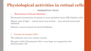
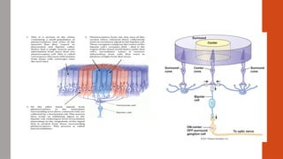
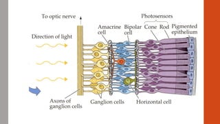
Physiological activities inretinal cells
• HORIZONTAL CELLS
1. Phenomenon of lateral inhibition-
Horizontal transmission of signals in outer plexiform layer (PR->bipolar cells)
Minute spot of light central most area excited area around (surround)
inhibited
Enhance visual contrast by lateral inhibition
2. Concept of receptive field-
The influence area of a sensory neuron
Receptive field of horizontal cells is very large as compared to the
photoreceptor cell
11.
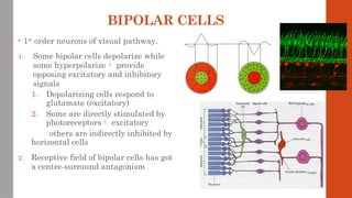
BIPOLAR CELLS
• 1st
orderneurons of visual pathway.
1. Some bipolar cells depolarize while
some hyperpolarize provide
opposing excitatory and inhibitory
signals
1. Depolarizing cells respond to
glutamate (excitatory)
2. Some are directly stimulated by
photoreceptors excitatory
others are indirectly inhibited by
horizontal cells
2. Receptive field of bipolar cells has got
a centre-surround antagonism
14.
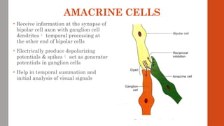
AMACRINE CELLS
• Receiveinformation at the synapse of
bipolar cell axon with ganglion cell
dendrites temporal processing at
the other end of bipolar cells
• Electrically produce depolarizing
potentials & spikes act as generator
potentials in ganglion cells
• Help in temporal summation and
initial analysis of visual signals
15.
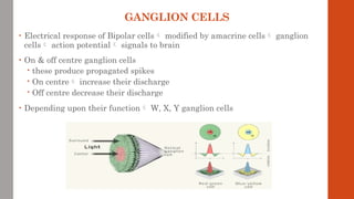
GANGLION CELLS
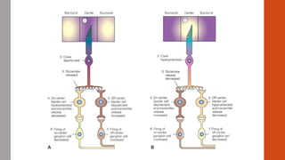
• Electricalresponse of Bipolar cells modified by amacrine cells ganglion
cells action potential signals to brain
• On & off centre ganglion cells
these produce propagated spikes
On centre increase their discharge
Off centre decrease their discharge
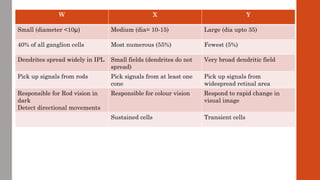
• Depending upon their function W, X, Y ganglion cells
17.
W X Y
Small(diameter <10μ) Medium (dia= 10-15) Large (dia upto 35)
40% of all ganglion cells Most numerous (55%) Fewest (5%)
Dendrites spread widely in IPL Small fields (dendrites do not
spread)
Very broad dendritic field
Pick up signals from rods Pick signals from at least one
cone
Pick up signals from
widespread retinal area
Responsible for Rod vision in
dark
Detect directional movements
Responsible for colour vision Respond to rapid change in
visual image
Sustained cells Transient cells
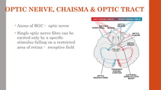
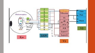
OPTIC NERVE, CHAISMA& OPTIC TRACT
• Axons of RGC optic nevre
• Single optic nerve fibre can be
excited only by a specific
stimulus falling on a restricted
area of retina receptive field
20.
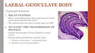
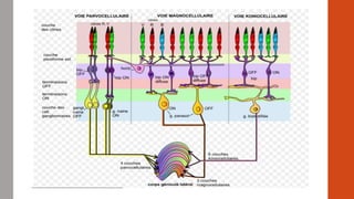
LAERAL GENICULATE BODY
•2 principal functions
1. RELAY STATION-
Relay visual information from optic tract to visual
cortex (geniculocalcarine tract)
The signals from two eyes are kept apart in LGB
2. TO “GATE” THE TRANSMISSION OF
SIGNALS-
Control the passage of visual signals to visual
cortex
Receive gating (inhibitory) controls from-
1. Primary visual cortex corticofugal fibres
2. Reticular area of mesencephalon
21.
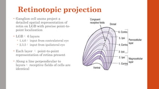
Retinotopic projection
• Ganglioncell axons project a
detailed spatial representation of
retin on LGB with precise point-to-
point localization
• LGB 6 layers
1,4,6 input from contralateral eye
2,3,5 input from ipsilateral eye
• Each layer point-to-point
representation of retina present
• Along a line perpendicular to
layers receptive fields of cells are
identical
22.
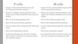
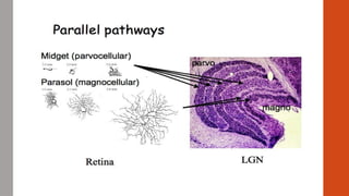
P cells
• projectto the parvocellular layers of
the lateral geniculate nucleus.
• known as midget retinal ganglion cells
small sizes of their dendritic trees and cell
bodies.
• 80% of all retinal ganglion cells
• part of the parvocellular pathway.
• receive inputs from relatively few rods and
cones.
• have slow conduction velocity
• respond to changes in color but respond
only weakly to changes in contrast unless
the change is great
M cells
• project to the magnocellular layers of the
lateral geniculate nucleus.
• known as parasol retinal ganglion cells
large sizes of their dendritic trees and cell
bodies.
• 10% of all retinal ganglion cells
• part of the magnocellular pathway.
• receive inputs from relatively many rods and
cones.
• have fast conduction velocity
• can respond to low-contrast stimuli, but are
not very sensitive to changes in color
25.
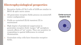
Electrophysiological properties
1. Receptivefields of P & I cells of LGB are similar to
RGCs & optic nerve axons
2. All geniculate receptive fields process on-center/off-
center configuration
3. Fields as sustained (X) & transient (Y) is
maintained
4. High degree of peripheral suppression in
geniculate receptive field. Larger “off” periphery
cancels effects of “on” centre. They are sensitive in
responding to spatial differences in retinal
illumination.
5. Geniculate relay cells have binocular receptive
fields.
26.
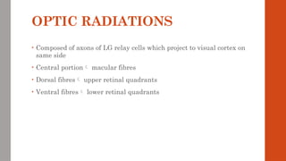
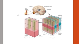
OPTIC RADIATIONS
• Composedof axons of LG relay cells which project to visual cortex on
same side
• Central portion macular fibres
• Dorsal fibres upper retinal quadrants
• Ventral fibres lower retinal quadrants
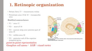
1. Retinopic organization
•Striate Area 17 visuosensory retina
• Peristriate area 18 & 19 visuopsychic
area
Modified nomenclature
• V1 area 17
• V2 most of 18
• V3 narrow strip over anterior part of
area 18
• V4 within area 19
• V5 posterior end of the superior
temporal gyrus
point-to-point representation
Ganglion cell axons LGBvisual cortex
29.
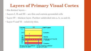
Layers of PrimaryVisual Cortex
• Six distinct layers –
• Layers I, II and III – are thin and contain pyramidal cells
• Layer IV – thickest layer. Further subdivided into a, b, ca and cb.
• Layer V and VI – relatively thin.
30.
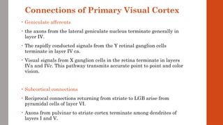
Connections of PrimaryVisual Cortex
• Geniculate afferents
• the axons from the lateral geniculate nucleus terminate generally in
layer IV.
• The rapidly conducted signals from the Y retinal ganglion cells
terminate in layer IV ca.
• Visual signals from X ganglion cells in the retina terminate in layers
IVa and IVc. This pathway transmits accurate point to point and color
vision.
• Subcortical connections
• Reciprocal connections returning from striate to LGB arise from
pyramidal cells of layer VI.
• Axons from pulvinar to striate cortex terminate among dendrites of
layers I and V.
31.
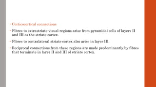
• Corticocortical connections
•Fibres to extrastriate visual regions arise from pyramidal cells of layers II
and III os the striate cortex.
• Fibres to contralateral striate cortex also arise in layer III.
• Reciprocal connections from these regions are made predominantly by fibres
that terminate in layer II and III of striate cortex.
32.
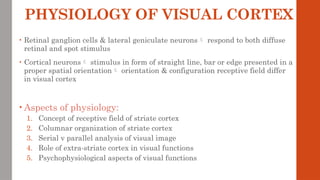
PHYSIOLOGY OF VISUALCORTEX
• Retinal ganglion cells & lateral geniculate neurons respond to both diffuse
retinal and spot stimulus
• Cortical neurons stimulus in form of straight line, bar or edge presented in a
proper spatial orientation orientation & configuration receptive field differ
in visual cortex
• Aspects of physiology:
1. Concept of receptive field of striate cortex
2. Columnar organization of striate cortex
3. Serial v parallel analysis of visual image
4. Role of extra-striate cortex in visual functions
5. Psychophysiological aspects of visual functions
33.
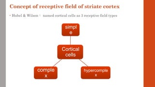
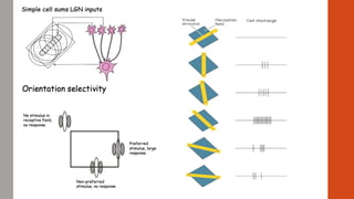
Concept of receptivefield of striate cortex
• Hubel & Wilson named cortical cells as 3 receptive field types
Cortical
cells
simpl
e
hypercomple
x
comple
x
34.
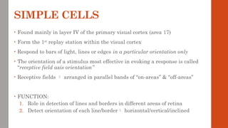
SIMPLE CELLS
• Foundmainly in layer IV of the primary visual cortex (area 17)
• Form the 1st
replay station within the visual cortex
• Respond to bars of light, lines or edges in a particular orientation only
• The orientation of a stimulus most effective in evoking a response is called
“receptive field axis orientation”
• Receptive fields arranged in parallel bands of “on-areas” & “off-areas”
• FUNCTION:
1. Role in detection of lines and borders in different areas of retina
2. Detect orientation of each line/border horizontal/vertical/inclined
36.
COMPLEX CELLS
• Foundin cortical layers above and below layer IV of areas 17, 18, 19
• Require preferred orientation of linear stimulus but are less dependent upon
the location of a stimulus in the visual field
• Respond maximally when stimulus is moved laterally without change in
orientation
• On and off areas cannot be mapped in their receptive fields
• Receive input from both eyes called binocular
• 4 types of receptive field a/c preferred stimulus
1. Activated by a slit-nonuniform field
2. Activated by a slit-uniform field
3. Activated by an edge
4. Activated by a dark bar
37.
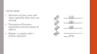
• FUNCTION-
1. Detectionof lines, bars and
edges specially when they are
moving
2. Perception of features,
orientation and movement of
objects
3. Simple + complex cells =
feature detectors
39.
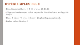
HYPERCOMPLEX CELLS
• Foundin cortical layers II & III of areas 17, 18, 19
• All properties of complex cells + require the line stimulus to be of specific
length
• Hubel & wiesel = 6 types (4 lower + 2 higher) hypercomplex cells
• Dreher = class I & class II
41.
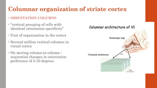
Columnar organization ofstriate cortex
• ORIENTATION COLUMNS
• “vertical grouping of cells with
identical orientation specificity”
• Unit of organization in the cortex
• Several million vertical columns in
visual cortex
• On moving column-to-column
sequential changes in orientation
preference of 5-10 degress
42.
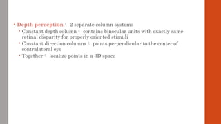
• Depth perception2 separate column systems
Constant depth column contains binocular units with exactly same
retinal disparity for properly oriented stimuli
Constant direction columns points perpendicular to the center of
contralateral eye
Together localize points in a 3D space
43.
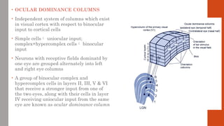
• OCULAR DOMINANCECOLUMNS
• Independent system of columns which exist
in visual cortex with respect to binocular
input to cortical cells
• Simple cells uniocular input;
complex+hypercomplex cells binocular
input
• Neurons with receptive fields dominatd by
one eye are grouped alternately into left
and right eye columns
• A group of binocular complex and
hypercomplex cells in layers II, III, V & VI
that receive a stronger input from one of
the two eyes, along with their cells in layer
IV receiving uniocular input from the same
eye are known as ocular dominance column
44.
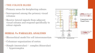
• THE COLOURBLOBS
• Primary areas for deciphering colours
• Interspersed among the primary visual
columns
• Receive lateral signals from adjacent
visual column and respond specifically to
colour signals
SERIAL Vs PARALLEL ANALYSIS
• Hierarchical model for cell interconnection
• Columnar organization of cortex
• Simple (monocular) complex (binocular)
hypercomplex
46.
EXTRASTRIATE CORTEX
• Neuronsof straite cortex (area 17 or VI)
extrastraite cortex [area 18 (V2), area 19
(V2), V3 V4 MT] strait cortex
• Pontifical cells receive information from
the feature detectors (simple & complex
cells)
• Specialized extrastriate areas
1. Colour processing area V4 (rhesus
monkeytrial)
2. Movement processing area MT. cells
show strong preference for stimuli
moving in a particular direction
3. Stereoscopic depth perception area
V2 & V3
47.
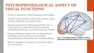
PSYCHOPHYSIOLOGICAL ASPECT OF
VISUALFUNCTIONS
• Vision is related to verbal language and reading
• Visual cortex connects with tactile sensory motor
auditoy, olfactory and speech areas
• Angular gyrus (area 40)of parietal lobe acts as
visual memory centre for words by forming
associations between visual and auditory centres
• Corpus callosum connets the two hemispheres
and help perceieve the several qualities
simultaneously and synthesize a unified picture
• Brain’s response to stimuli is in the form of an
over all picture


![EXTRASTRIATE CORTEX
• Neurons of straite cortex (area 17 or VI)
extrastraite cortex [area 18 (V2), area 19
(V2), V3 V4 MT] strait cortex
• Pontifical cells receive information from
the feature detectors (simple & complex
cells)
• Specialized extrastriate areas
1. Colour processing area V4 (rhesus
monkeytrial)
2. Movement processing area MT. cells
show strong preference for stimuli
moving in a particular direction
3. Stereoscopic depth perception area
V2 & V3](https://image.slidesharecdn.com/physiologyofvision3-200522070350-250704044845-6ec42ecd/85/physiologyofvision3-200522070350-ppt-pptx-46-320.jpg)


![Midterm NOTES [CH6] - Vision.PDF](https://cdn.slidesharecdn.com/ss_thumbnails/midtermnotesch6-vision-221030101221-b6edbebe-thumbnail.jpg?width=640&height=640&fit=bounds)


