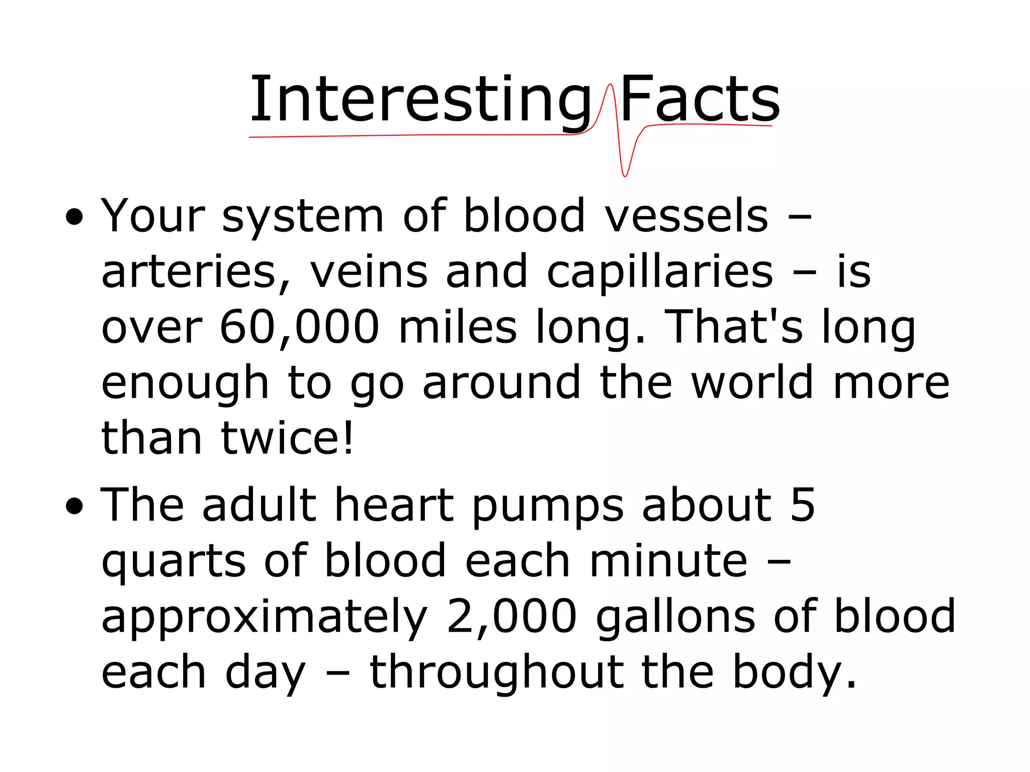
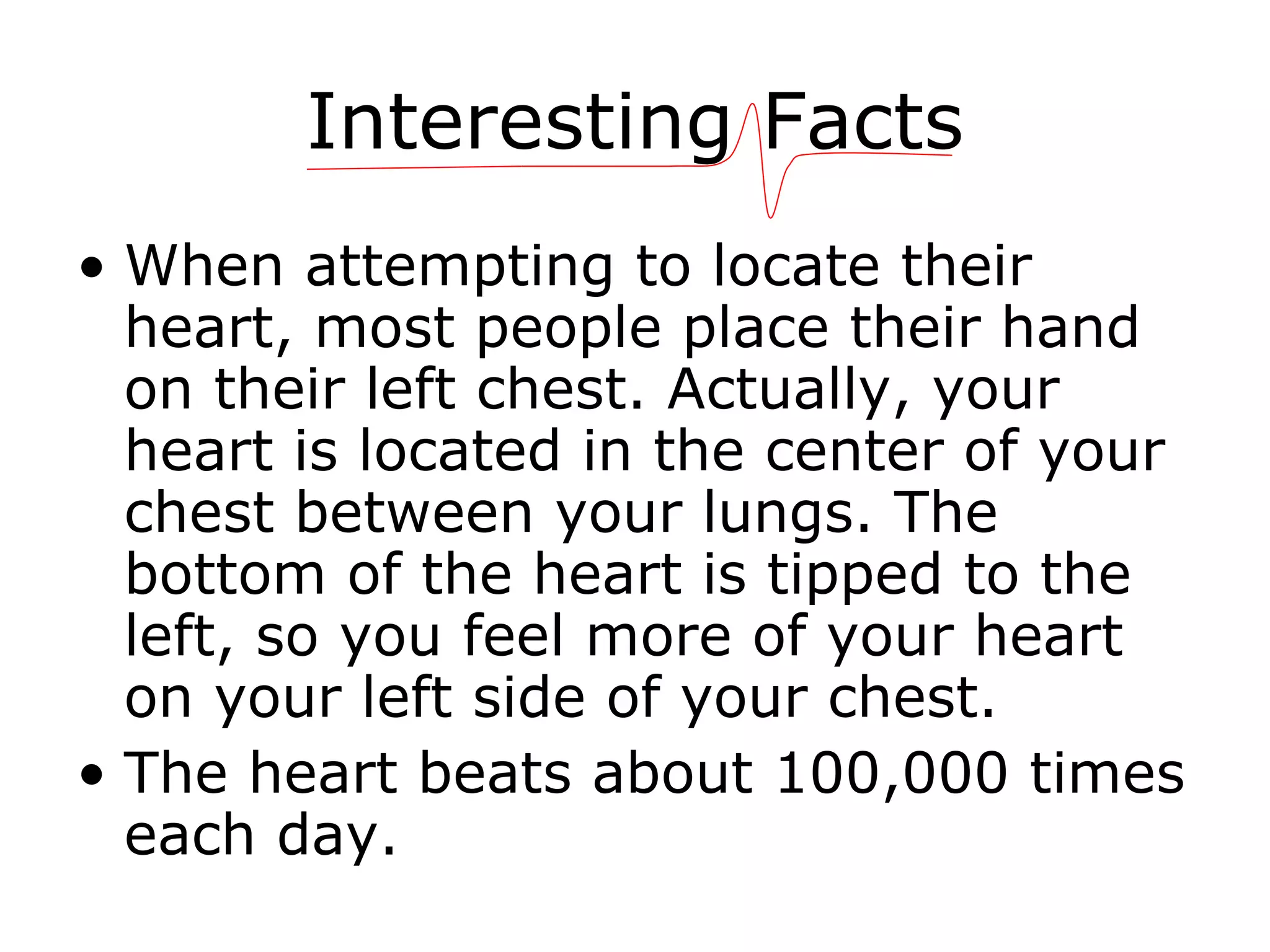
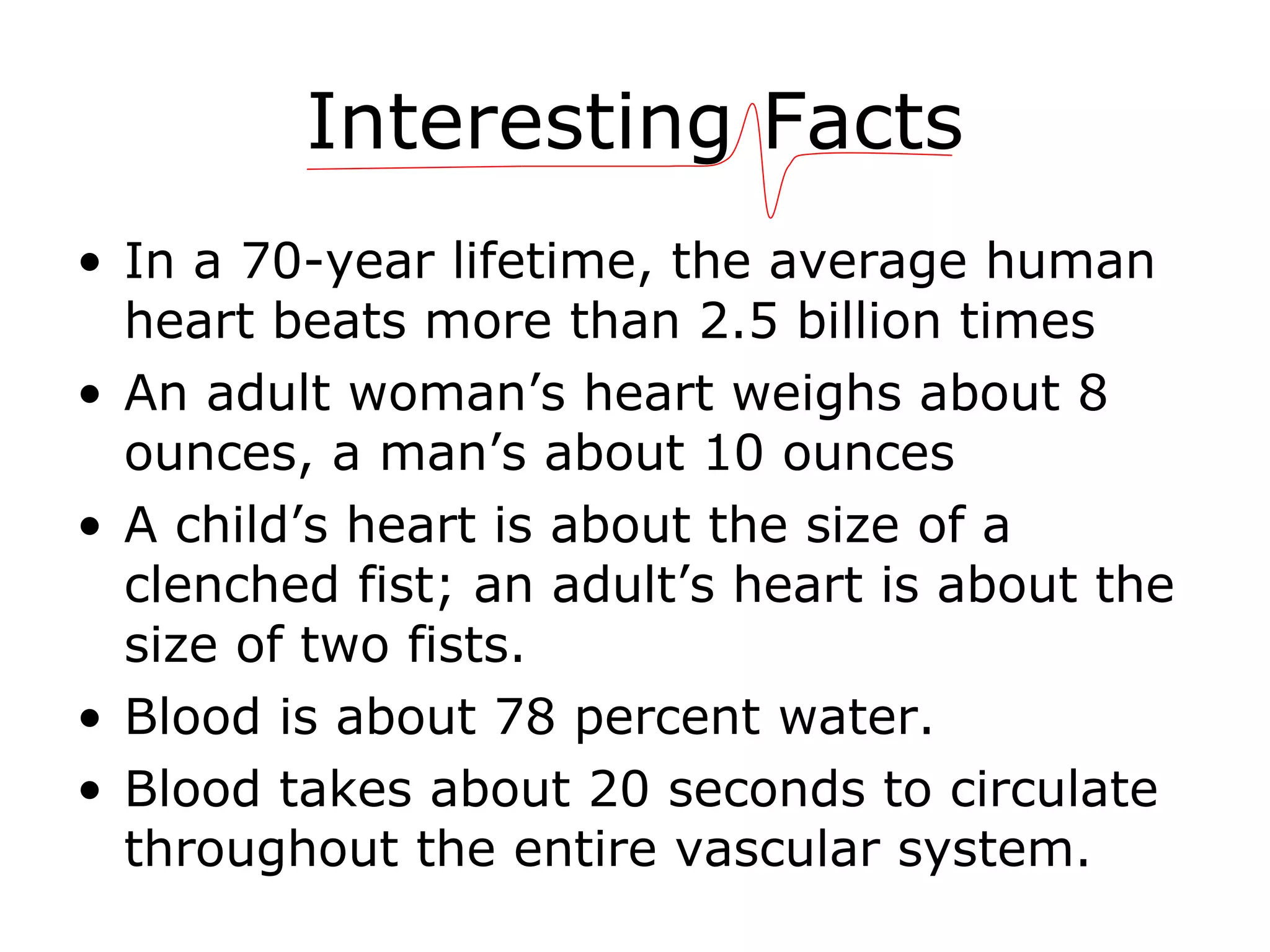
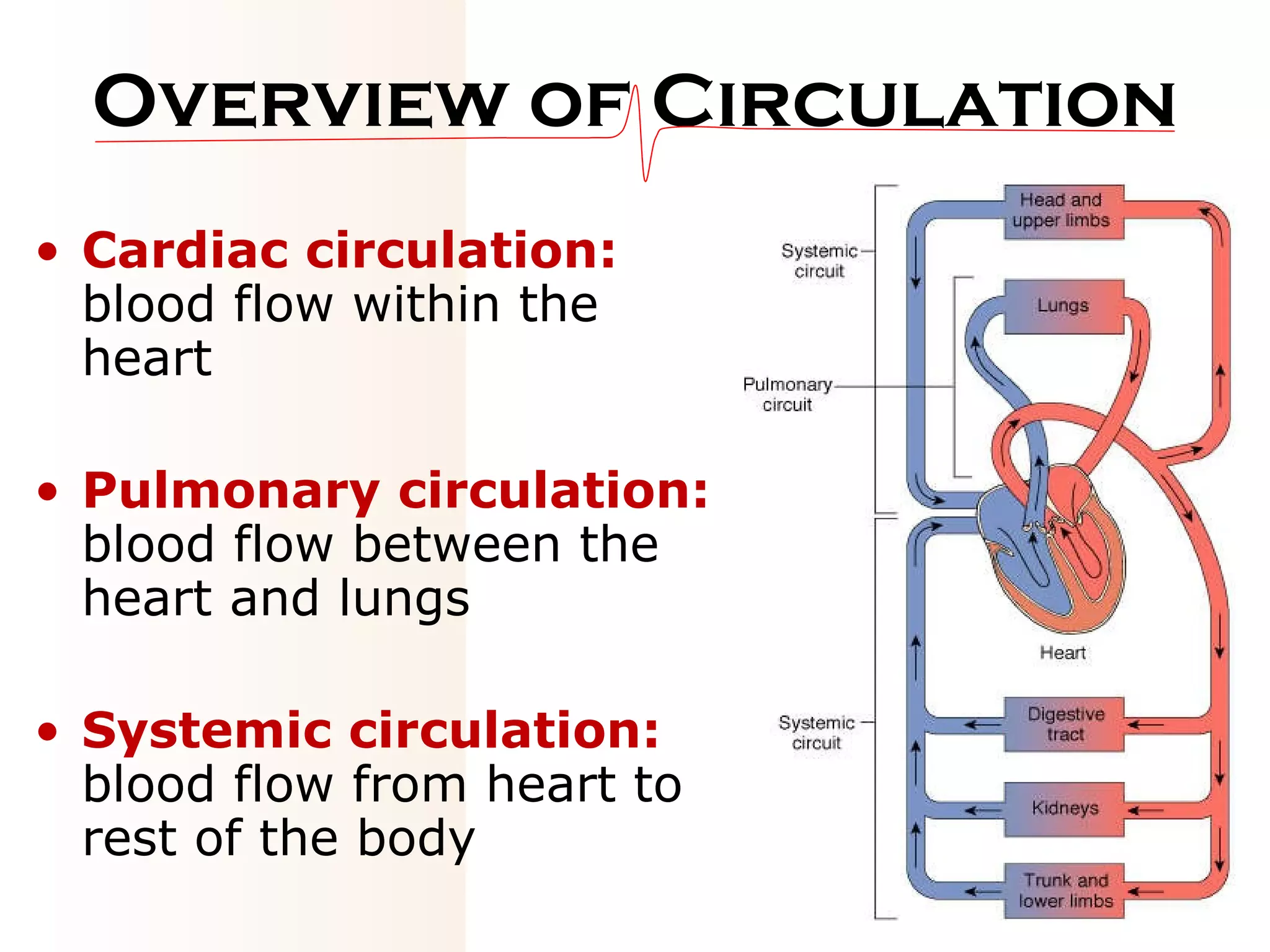
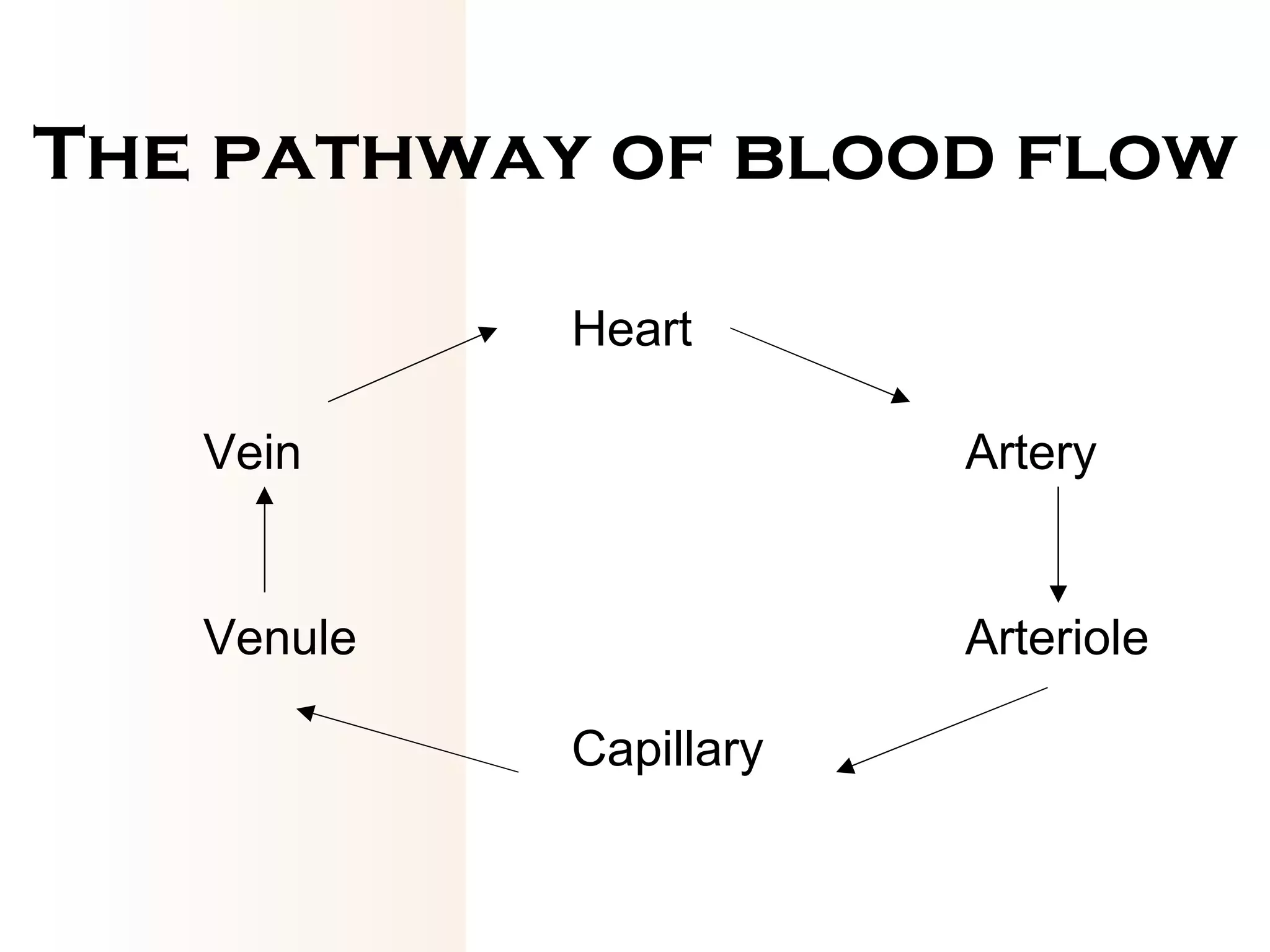
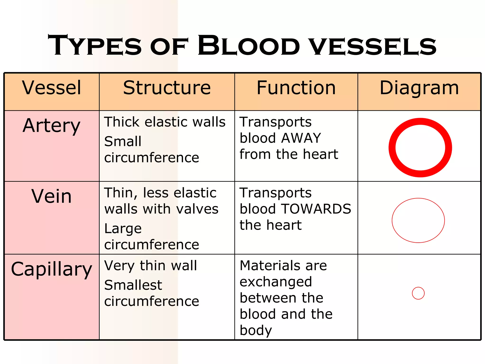
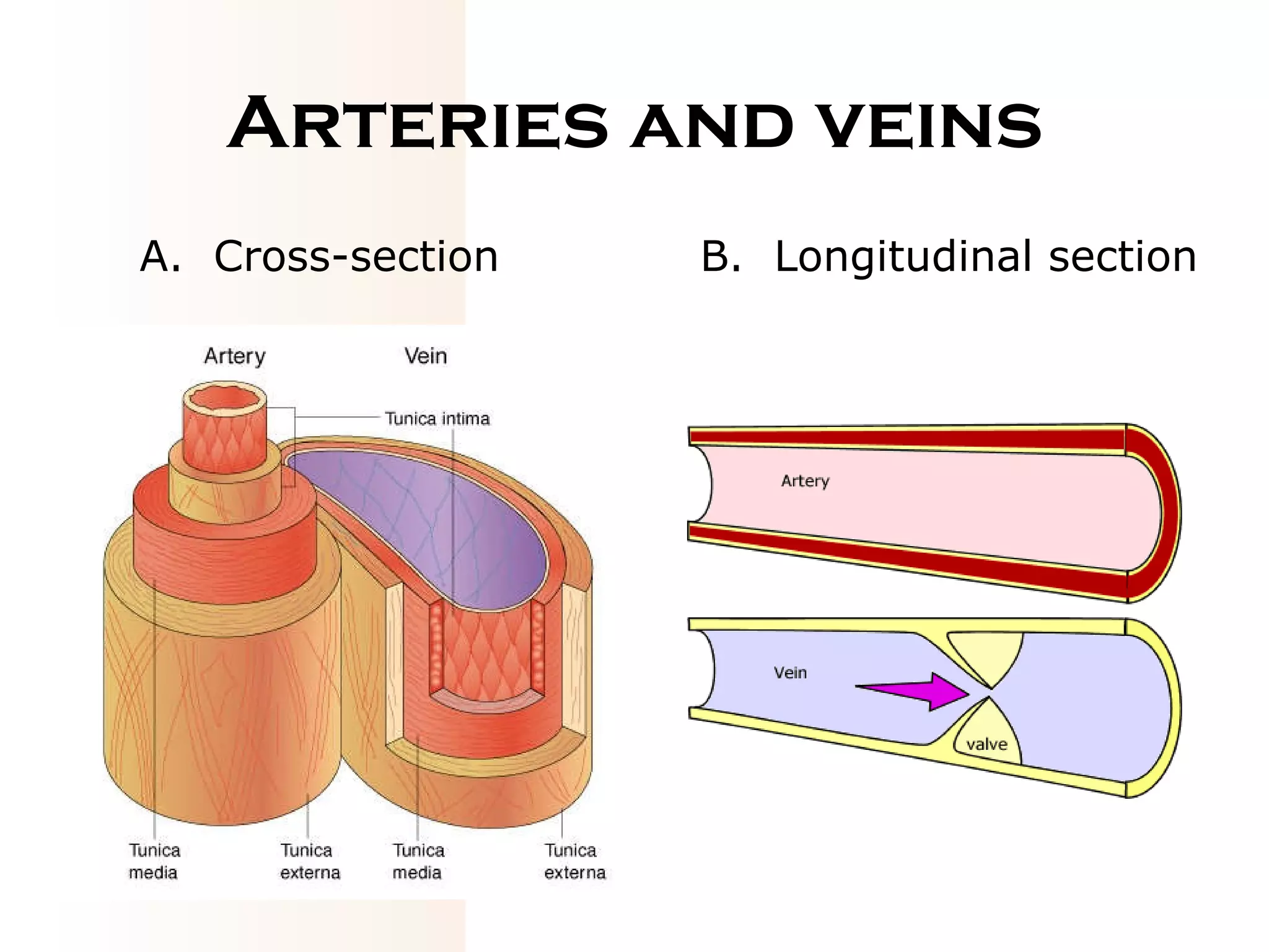
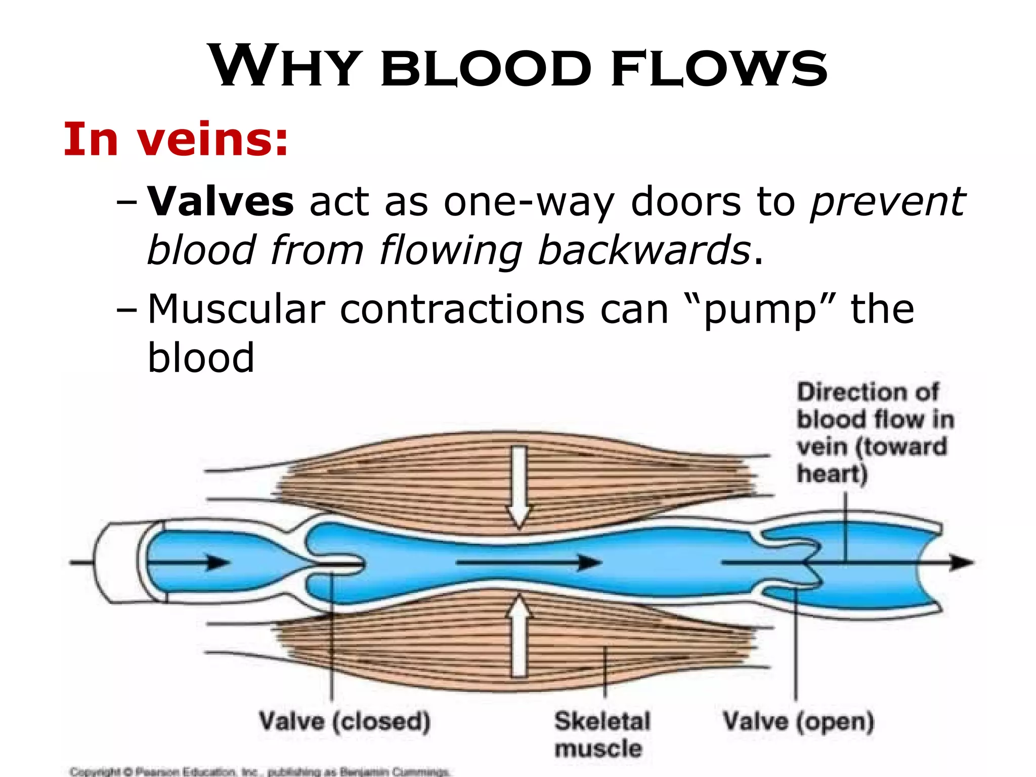
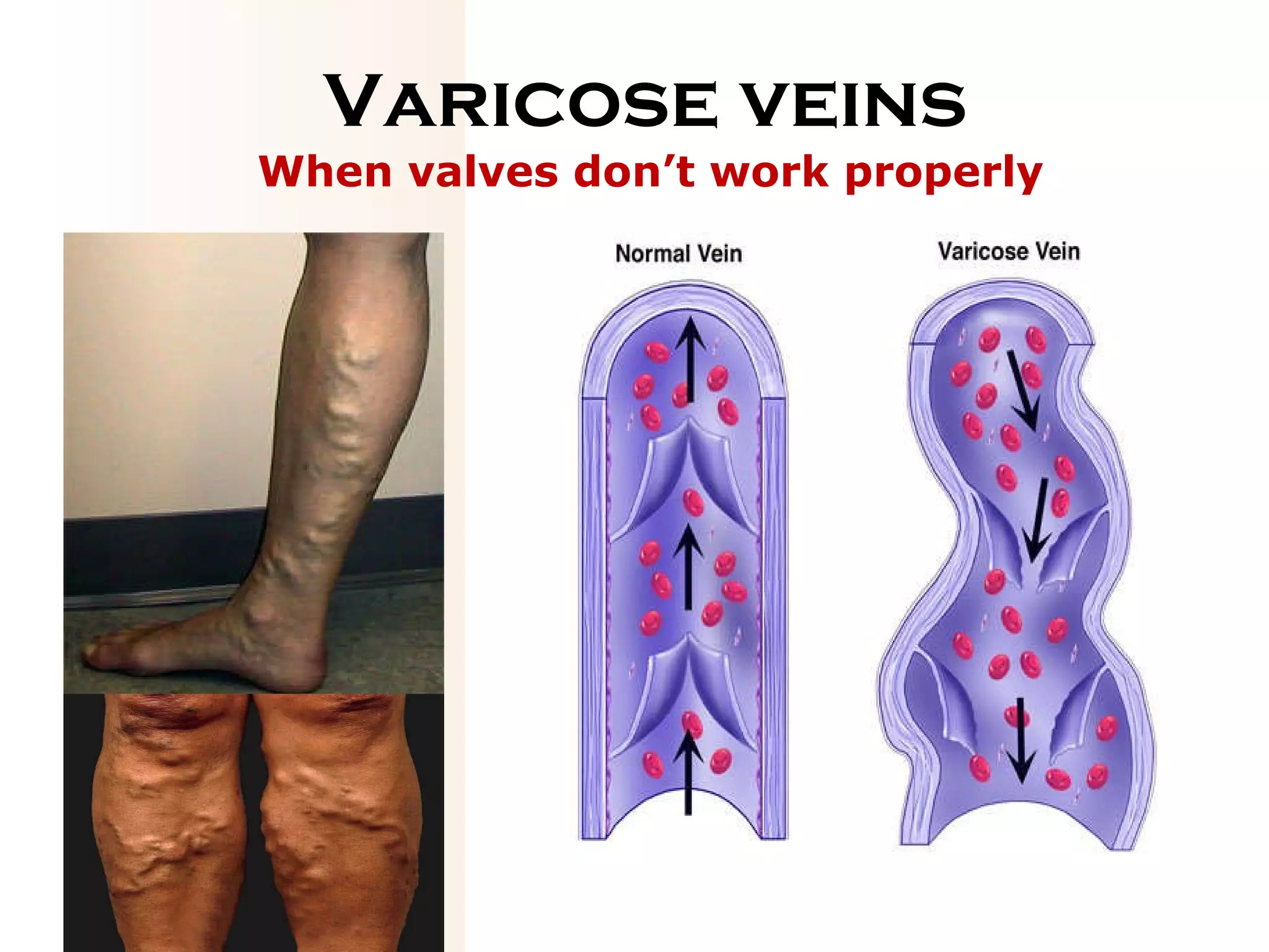
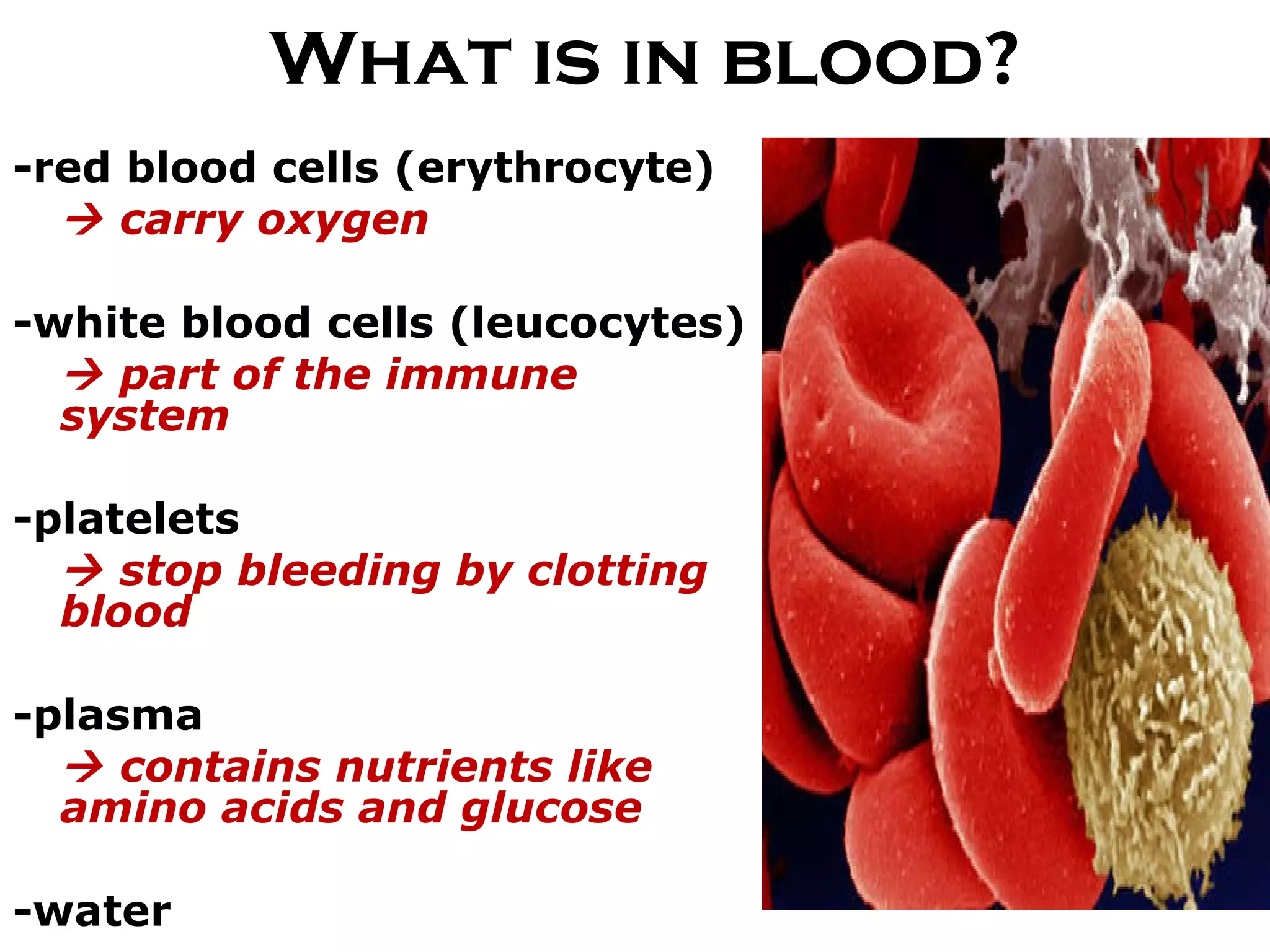
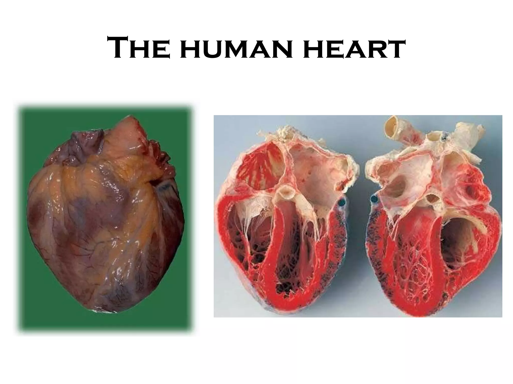
The Circulatory System SBI3U
Interesting Facts Your system of blood vessels – arteries, veins and capillaries – is over 60,000 miles long. That's long enough to go around the world more than twice!  The adult heart pumps about 5 quarts of blood each minute – approximately 2,000 gallons of blood each day – throughout the body.
Interesting Facts When attempting to locate their heart, most people place their hand on their left chest. Actually, your heart is located in the center of your chest between your lungs. The bottom of the heart is tipped to the left, so you feel more of your heart on your left side of your chest.  The heart beats about 100,000 times each day.
Interesting Facts In a 70-year lifetime, the average human heart beats more than 2.5 billion times  An adult woman’s heart weighs about 8 ounces, a man’s about 10 ounces  A child’s heart is about the size of a clenched fist; an adult’s heart is about the size of two fists.  Blood is about 78 percent water.  Blood takes about 20 seconds to circulate throughout the entire vascular system.
Overview of Circulation Cardiac circulation:  blood flow within the heart Pulmonary circulation:  blood flow between the heart and lungs Systemic circulation:  blood flow from heart to rest of the body
The pathway of blood flow Heart  Vein Artery Venule Arteriole Capillary
Types of Blood vessels Vessel Structure Function Diagram Artery Thick elastic walls Small circumference Transports blood AWAY from the heart Vein Thin, less elastic walls with valves Large circumference Transports blood TOWARDS the heart Capillary Very thin wall Smallest circumference Materials are exchanged between the blood and the body
Arteries and veins A.  Cross-section B.  Longitudinal section
Capillaries Walls of blood vessels are made up of cells called  endothelial cells . The  thin  layer of endothelial cells allows diffusion to occur more rapidly.
Capillaries Red blood cells travel through the capillary  in single file. Blood velocity is the  slowest  in capillaries
Why blood flows In arteries: Elastic walls  expand and contract. As they contract, this helps to pump the blood around the body. 3 structural layers.  Outer layer  - covering of connective tissue. Middle layer (thickest layer)  - alternating circular bands of elastic fibres and smooth muscle Inner layer  - only a single cell thick, contains smooth endothelial cells that serve to reduce friction as blood courses through the artery.
Why blood flows In veins:  Valves  act as one-way doors to  prevent blood from flowing backwards .  Muscular contractions can “pump” the blood
Varicose veins When valves don’t work properly
What is in blood? -red blood cells (erythrocyte)     carry oxygen -white blood cells (leucocytes)    part of the immune system -platelets     stop bleeding by clotting blood -plasma    contains nutrients like amino acids and glucose -water
The human heart
Direction of blood flow Oxygenated blood: Lung      pulmonary vein      left atrium     bicuspid valve     left ventricle     aortic semilunar valve     aorta      rest of body Deoxygenated blood: Vena cava     tricuspid valve     right atrium right ventricle   pulmonary semilunar valve     pulmonary artery     lungs
Cardiac Circulation
Cardiac Circulation
Comparing atria and ventricles Atria Ventricles Location Upper half of heart Lower half of heart Thick-ness Thinner walls (pumps blood to ventricles below) Thicker walls (pumps blood to lungs or to rest of the body)
The heartbeat The heartbeat is controlled by the  sinoatrial   node  (also known as the  pacemaker ). The SA node sends  electrical signals  that cause the heart muscle to contract. The heart makes a  ‘lup-dup ’ sound as blood flows through and the valves close.
The SA node and the AV node SA node  = sinoatrial node AV node  = atrioventricular node
Electrocardiographs (ECGs) The electrical signals of the heart can be measured using a machine called an  electrocardiograph  ( ECG ).
ECG analysis P:  atria contract (ventricles relax) QRS:  ventricles contract (atria relax) T:  both atria and ventricles relax
Cardiovascular disease AORTIC ANEURYSM
Cardiovascular disease ATHEROSCLEROSIS
Treatments CORONARY BYPASS
Treatments ANGIOPLASTY & STENT
Mammalian Heart
Pericardium
Tricuspid Valve
Bicuspid Valve
Chordae Tendinae “the heart strings”
Prosthetic Valves

05 circulation

  • 1.
  • 2.
    Interesting Facts Yoursystem of blood vessels – arteries, veins and capillaries – is over 60,000 miles long. That's long enough to go around the world more than twice! The adult heart pumps about 5 quarts of blood each minute – approximately 2,000 gallons of blood each day – throughout the body.
  • 3.
    Interesting Facts Whenattempting to locate their heart, most people place their hand on their left chest. Actually, your heart is located in the center of your chest between your lungs. The bottom of the heart is tipped to the left, so you feel more of your heart on your left side of your chest. The heart beats about 100,000 times each day.
  • 4.
    Interesting Facts Ina 70-year lifetime, the average human heart beats more than 2.5 billion times An adult woman’s heart weighs about 8 ounces, a man’s about 10 ounces A child’s heart is about the size of a clenched fist; an adult’s heart is about the size of two fists. Blood is about 78 percent water. Blood takes about 20 seconds to circulate throughout the entire vascular system.
  • 5.
    Overview of CirculationCardiac circulation: blood flow within the heart Pulmonary circulation: blood flow between the heart and lungs Systemic circulation: blood flow from heart to rest of the body
  • 6.
    The pathway ofblood flow Heart Vein Artery Venule Arteriole Capillary
  • 7.
    Types of Bloodvessels Vessel Structure Function Diagram Artery Thick elastic walls Small circumference Transports blood AWAY from the heart Vein Thin, less elastic walls with valves Large circumference Transports blood TOWARDS the heart Capillary Very thin wall Smallest circumference Materials are exchanged between the blood and the body
  • 8.
    Arteries and veinsA. Cross-section B. Longitudinal section
  • 9.
    Capillaries Walls ofblood vessels are made up of cells called endothelial cells . The thin layer of endothelial cells allows diffusion to occur more rapidly.
  • 10.
    Capillaries Red bloodcells travel through the capillary in single file. Blood velocity is the slowest in capillaries
  • 11.
    Why blood flowsIn arteries: Elastic walls expand and contract. As they contract, this helps to pump the blood around the body. 3 structural layers. Outer layer - covering of connective tissue. Middle layer (thickest layer) - alternating circular bands of elastic fibres and smooth muscle Inner layer - only a single cell thick, contains smooth endothelial cells that serve to reduce friction as blood courses through the artery.
  • 12.
    Why blood flowsIn veins: Valves act as one-way doors to prevent blood from flowing backwards . Muscular contractions can “pump” the blood
  • 13.
    Varicose veins Whenvalves don’t work properly
  • 14.
    What is inblood? -red blood cells (erythrocyte)  carry oxygen -white blood cells (leucocytes)  part of the immune system -platelets  stop bleeding by clotting blood -plasma  contains nutrients like amino acids and glucose -water
  • 15.
  • 16.
    Direction of bloodflow Oxygenated blood: Lung  pulmonary vein  left atrium  bicuspid valve  left ventricle  aortic semilunar valve  aorta  rest of body Deoxygenated blood: Vena cava  tricuspid valve  right atrium right ventricle  pulmonary semilunar valve  pulmonary artery  lungs
  • 17.
  • 18.
  • 19.
    Comparing atria andventricles Atria Ventricles Location Upper half of heart Lower half of heart Thick-ness Thinner walls (pumps blood to ventricles below) Thicker walls (pumps blood to lungs or to rest of the body)
  • 20.
    The heartbeat Theheartbeat is controlled by the sinoatrial node (also known as the pacemaker ). The SA node sends electrical signals that cause the heart muscle to contract. The heart makes a ‘lup-dup ’ sound as blood flows through and the valves close.
  • 21.
    The SA nodeand the AV node SA node = sinoatrial node AV node = atrioventricular node
  • 22.
    Electrocardiographs (ECGs) Theelectrical signals of the heart can be measured using a machine called an electrocardiograph ( ECG ).
  • 23.
    ECG analysis P: atria contract (ventricles relax) QRS: ventricles contract (atria relax) T: both atria and ventricles relax
  • 24.
  • 25.
  • 26.
  • 27.
  • 28.
  • 29.
  • 30.
  • 31.
  • 32.
    Chordae Tendinae “theheart strings”
  • 33.

Editor's Notes

  • #11 When capillaries unite to form venules, and then veins, the area decreases and the blood flow speeds up