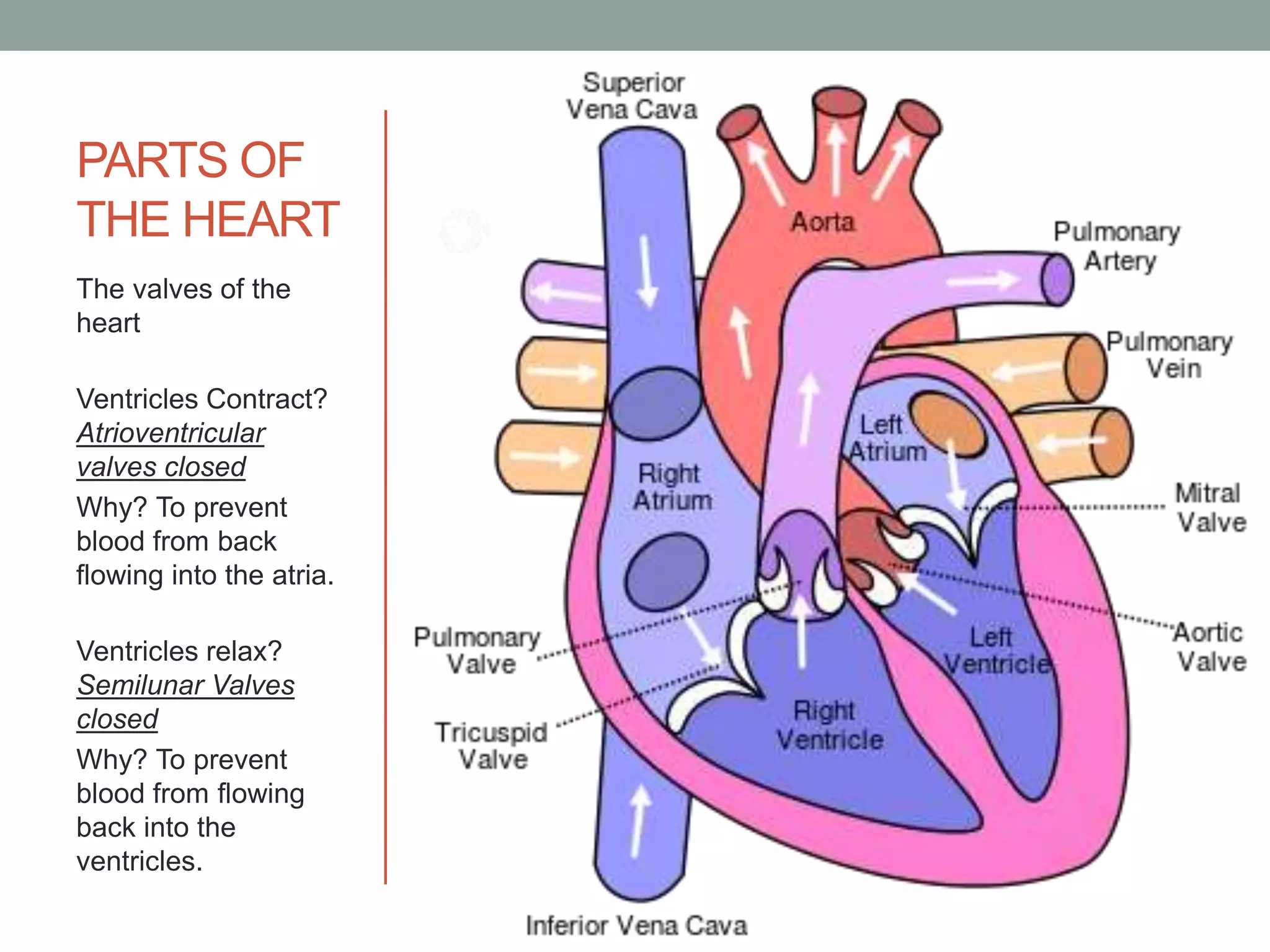
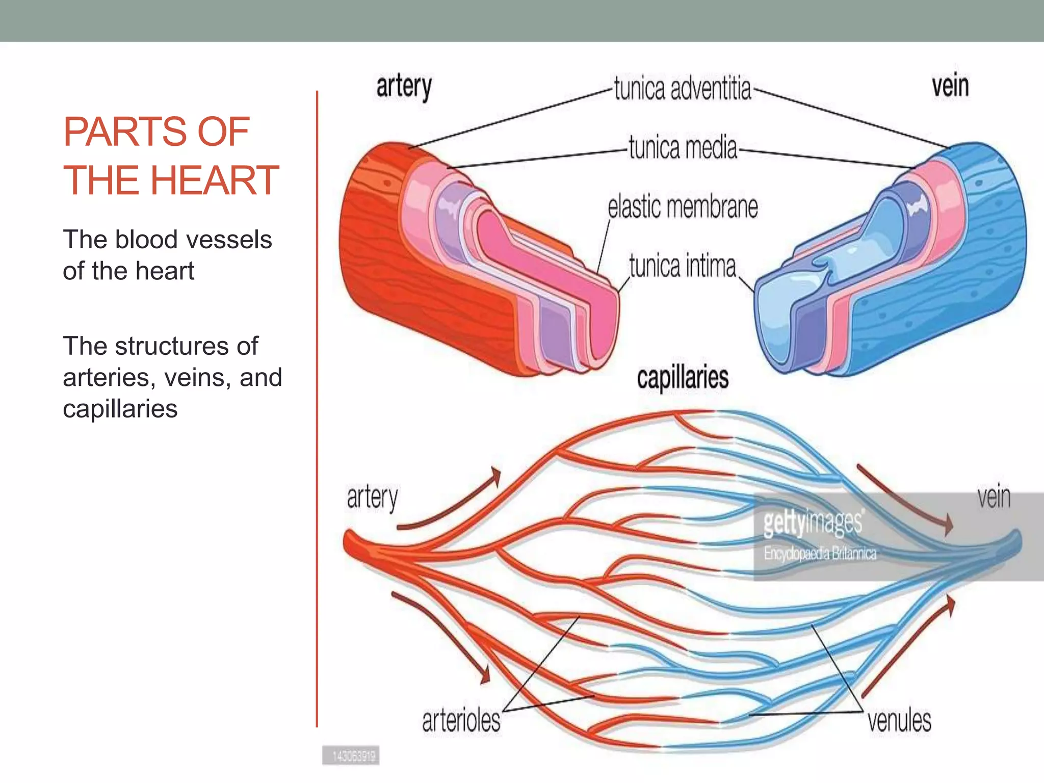
THE ORGANS OF THE
CARDIOVASCULAR SYSTEM
CIRCULATORY SYSTEM
• Responsible in transporting materials throughout
the entire body
• Transports nutrients, water, and oxygen to the
billions of body cells composing the multicellular
body, heals and fights infections, and at the same
time, removes wastes from them
• Functions as an intricate highway that travels
throughout the entire body supplying the body
cells with the materials they need to survive
TWO DIVISIONS OF CIRCULATORY
SYSTEM
CARDIOVASCULAR
• Transports blood
LYMPHATIC
• Transports the fluid
called lymph
CARDIOVASCULAR SYSTEM
• The organ system that transports nutrients
(digestive products), gases, hormones, and other
materials to and from the cells of the body
• Functions in fighting diseases and helps stabilize
body temperature and pH to maintain
homeostasis
• Heart, Blood, Blood Vessels
HEART
• A muscular organ that pumps blood to the
different parts of the body.
• Middle of the chest cavity with its tip or apex
slightly tilted towards the left.
• Just a big as one’s own clenched fist.
• Pericardium – a sac that encloses it.
THREE LAYERS OF TISSUES FORM
THE WALLS OF THE HEART
• Epicardium - outer layer; the inner part of the
pericardium that is closely adhered to the heart
• Myocardium - middle layer; composed of cardiac
muscle tissue
• Endocardium - inner layer; thin layer of tissue that
lines the muscles of the heart
• Pericardial Cavity – filled with pericardial fluid;
between the outer pericardium and the
epicardium.
PARTS OF
THE HEART
The tissue layers
of the heart
CHAMBERS OF THE HEART
• Right Atrium
• Left Atrium
• Right Ventricle
• Left Ventricle
• Septum – a muscle that divides the heart into
four chambers
PARTS OF
THE HEART
The chambers of
the heart
The heart
chambers differ in
thickness (due to
variations in the
amount of
myocardium
present)
• Atria – receiving chambers of the heart; thin-
walled
• Right atrium receives oxygen-poor blood from the
vena cava, a large vein
• Left atrium receives blood from the pulmonary
veins
• Ventricles – pumping chambers that force blood
out of the heart; thick-walled
• Right ventricle pumps blood to the lungs for
oxygenation
• Left ventricle pumps oxygen-rich blood to all parts
of the body
VALVES OF THE HEART
• Valves – flaps of muscles that prevent blood from
back flowing; two sets of valves work to ensure
that blood will move only in one direction
• Atrioventricular valves (cuspid valves) located
between at the atria and ventricles
• Semilunar valves located at the bases of the
large vessels leaving the ventricles
•  right atrioventricular valve: tricuspid valve; left
atrioventricular valve : bicuspid/mitral valve
•  Right ventricle: pulmonary semilunar valve:
Pulmonary artery
•  Left ventricle: aortic semilunar valve: Aorta
PARTS OF
THE HEART
The valves of the
heart
Ventricles Contract?
Atrioventricular
valves closed
Why? To prevent
blood from back
flowing into the atria.
Ventricles relax?
Semilunar Valves
closed
Why? To prevent
blood from flowing
back into the
ventricles.
BLOOD VESSELS IN THE HEART
• They are connected to the heart.
• Vena cava – largest vein connected to the heart;
conveys oxygen-poor blood coming from all parts
of the body back to the right atrium.
• Aorta – largest artery; conveys oxygen-rich blood
pumped by the left ventricle to all parts of the
body.
• Pulmonary artery conveys blood pumped by the
right ventricle to the lungs for oxygenation
(branches are connected to the lungs)
• Pulmonary veins convey oxygen-rich blood back
to the left atrium of the heart
• Heart needs blood to sustain itself.
(DIFFERENT) BLOOD VESSELS
• Network of channels that convey blood to all parts
of the body.
• As the blood travels throughout the body, it
remains confined within these blood vessels.
• Closed circulation
• Arteries, veins, capillaries
ARTERIES
• Conveys oxygen-rich blood away from the heart.
• Most of them transport oxygenated blood from
the left ventricles to the body tissues.
• From aorta, arteries branch repeatedly into
smaller and smaller arteries until the branching
results in the smallest arteries (arterioles, who
plays a key role in regulating blood flow into the
tissue capillaries).
ARTERIES
• Elastic blood vessels
• Blood passing through the arteries: high pressure
exerted by the pumping action of the heart
• Structure: adapted to carry on its function
efficiently
VEINS
• Convey blood coming from all parts of the body
toward the heart
• From capillaries of the different tissues and
organs in the body, blood enters the smallest
veins (venules)
• From venules, blood flows through larger and
larger veins until the blood ends into the largest
vein: vena cava.
VEINS
• Blood passing through the veins has reduced
oxygen has already been used for metabolic
activities of the cells
• Only blood passing through the veins w/ high
oxygen content: blood in the pulmonary veins
(oxygenated in the lungs)
PARTS ARTERIES VEINS
Tunica
Externa/Tunica
Adventitia
Connective tissue with
elastic and collagenous
fibers that are tough and
strong
Same as the arteries
Tunica Media Smooth muscle layer that
supports the entire vessel
& changes the diameter of
the vessel to regulate
blood flow and to handle
blood pressure of the
blood flowing with it
Thin layer that has wider
lumen and is equipped
with valves that prevent
the back flowing of blood;
it is easily collapse when
cut (fewer elastic
muscles)
Tunica Intima Connective tissue lined
with simple squamous
epithelium; in direct
contact with the blood flow
Same as the arteries
CAPILLARIES
• Smallest and most numerous blood vessels
• Form the connection between arterioles &
venules
• Exchange of materials – gases, nutrients, and
waste products of metabolism between the blood
and tissue cells
• One-cell thick: materials easily move in and out of
these blood vessels
• Movement of substances: diffusion, filtration,
osmosis
PARTS OF
THE HEART
The blood vessels
of the heart
The structures of
arteries, veins, and
capillaries
PATHWAYS OF BLOOD
CIRCULATION
• Pulmonary Pathway/Circulation
• Systemic Pathway/Circulation
PATHWAY OF CIRCULATION
Pulmonary Circulation (deoxygenated) & systemic
circulation (oxygenated)
right atrium  right ventricle  pulmonary
arteries  arterioles  capillary beds of
lungs  venules pulmonary veins  left
atrium  left ventricle  aorta  systemic
arteries  arterioles  capillary beds of
body tissues  venules  systemic veins
 vena cava
Organs of Cardiovascular System

Organs of Cardiovascular System

  • 1.
    THE ORGANS OFTHE CARDIOVASCULAR SYSTEM
  • 2.
    CIRCULATORY SYSTEM • Responsiblein transporting materials throughout the entire body • Transports nutrients, water, and oxygen to the billions of body cells composing the multicellular body, heals and fights infections, and at the same time, removes wastes from them • Functions as an intricate highway that travels throughout the entire body supplying the body cells with the materials they need to survive
  • 3.
    TWO DIVISIONS OFCIRCULATORY SYSTEM CARDIOVASCULAR • Transports blood LYMPHATIC • Transports the fluid called lymph
  • 4.
    CARDIOVASCULAR SYSTEM • Theorgan system that transports nutrients (digestive products), gases, hormones, and other materials to and from the cells of the body • Functions in fighting diseases and helps stabilize body temperature and pH to maintain homeostasis • Heart, Blood, Blood Vessels
  • 5.
    HEART • A muscularorgan that pumps blood to the different parts of the body. • Middle of the chest cavity with its tip or apex slightly tilted towards the left. • Just a big as one’s own clenched fist. • Pericardium – a sac that encloses it.
  • 7.
    THREE LAYERS OFTISSUES FORM THE WALLS OF THE HEART • Epicardium - outer layer; the inner part of the pericardium that is closely adhered to the heart • Myocardium - middle layer; composed of cardiac muscle tissue • Endocardium - inner layer; thin layer of tissue that lines the muscles of the heart • Pericardial Cavity – filled with pericardial fluid; between the outer pericardium and the epicardium.
  • 8.
    PARTS OF THE HEART Thetissue layers of the heart
  • 9.
    CHAMBERS OF THEHEART • Right Atrium • Left Atrium • Right Ventricle • Left Ventricle • Septum – a muscle that divides the heart into four chambers
  • 10.
    PARTS OF THE HEART Thechambers of the heart The heart chambers differ in thickness (due to variations in the amount of myocardium present)
  • 11.
    • Atria –receiving chambers of the heart; thin- walled • Right atrium receives oxygen-poor blood from the vena cava, a large vein • Left atrium receives blood from the pulmonary veins
  • 12.
    • Ventricles –pumping chambers that force blood out of the heart; thick-walled • Right ventricle pumps blood to the lungs for oxygenation • Left ventricle pumps oxygen-rich blood to all parts of the body
  • 13.
    VALVES OF THEHEART • Valves – flaps of muscles that prevent blood from back flowing; two sets of valves work to ensure that blood will move only in one direction • Atrioventricular valves (cuspid valves) located between at the atria and ventricles • Semilunar valves located at the bases of the large vessels leaving the ventricles
  • 14.
    •  rightatrioventricular valve: tricuspid valve; left atrioventricular valve : bicuspid/mitral valve •  Right ventricle: pulmonary semilunar valve: Pulmonary artery •  Left ventricle: aortic semilunar valve: Aorta
  • 15.
    PARTS OF THE HEART Thevalves of the heart Ventricles Contract? Atrioventricular valves closed Why? To prevent blood from back flowing into the atria. Ventricles relax? Semilunar Valves closed Why? To prevent blood from flowing back into the ventricles.
  • 16.
    BLOOD VESSELS INTHE HEART • They are connected to the heart. • Vena cava – largest vein connected to the heart; conveys oxygen-poor blood coming from all parts of the body back to the right atrium. • Aorta – largest artery; conveys oxygen-rich blood pumped by the left ventricle to all parts of the body.
  • 17.
    • Pulmonary arteryconveys blood pumped by the right ventricle to the lungs for oxygenation (branches are connected to the lungs) • Pulmonary veins convey oxygen-rich blood back to the left atrium of the heart • Heart needs blood to sustain itself.
  • 18.
    (DIFFERENT) BLOOD VESSELS •Network of channels that convey blood to all parts of the body. • As the blood travels throughout the body, it remains confined within these blood vessels. • Closed circulation • Arteries, veins, capillaries
  • 19.
    ARTERIES • Conveys oxygen-richblood away from the heart. • Most of them transport oxygenated blood from the left ventricles to the body tissues. • From aorta, arteries branch repeatedly into smaller and smaller arteries until the branching results in the smallest arteries (arterioles, who plays a key role in regulating blood flow into the tissue capillaries).
  • 20.
    ARTERIES • Elastic bloodvessels • Blood passing through the arteries: high pressure exerted by the pumping action of the heart • Structure: adapted to carry on its function efficiently
  • 21.
    VEINS • Convey bloodcoming from all parts of the body toward the heart • From capillaries of the different tissues and organs in the body, blood enters the smallest veins (venules) • From venules, blood flows through larger and larger veins until the blood ends into the largest vein: vena cava.
  • 22.
    VEINS • Blood passingthrough the veins has reduced oxygen has already been used for metabolic activities of the cells • Only blood passing through the veins w/ high oxygen content: blood in the pulmonary veins (oxygenated in the lungs)
  • 23.
    PARTS ARTERIES VEINS Tunica Externa/Tunica Adventitia Connectivetissue with elastic and collagenous fibers that are tough and strong Same as the arteries Tunica Media Smooth muscle layer that supports the entire vessel & changes the diameter of the vessel to regulate blood flow and to handle blood pressure of the blood flowing with it Thin layer that has wider lumen and is equipped with valves that prevent the back flowing of blood; it is easily collapse when cut (fewer elastic muscles) Tunica Intima Connective tissue lined with simple squamous epithelium; in direct contact with the blood flow Same as the arteries
  • 24.
    CAPILLARIES • Smallest andmost numerous blood vessels • Form the connection between arterioles & venules • Exchange of materials – gases, nutrients, and waste products of metabolism between the blood and tissue cells • One-cell thick: materials easily move in and out of these blood vessels • Movement of substances: diffusion, filtration, osmosis
  • 25.
    PARTS OF THE HEART Theblood vessels of the heart The structures of arteries, veins, and capillaries
  • 26.
    PATHWAYS OF BLOOD CIRCULATION •Pulmonary Pathway/Circulation • Systemic Pathway/Circulation
  • 27.
    PATHWAY OF CIRCULATION PulmonaryCirculation (deoxygenated) & systemic circulation (oxygenated) right atrium  right ventricle  pulmonary arteries  arterioles  capillary beds of lungs  venules pulmonary veins  left atrium  left ventricle  aorta  systemic arteries  arterioles  capillary beds of body tissues  venules  systemic veins  vena cava