Downloaded 229 times

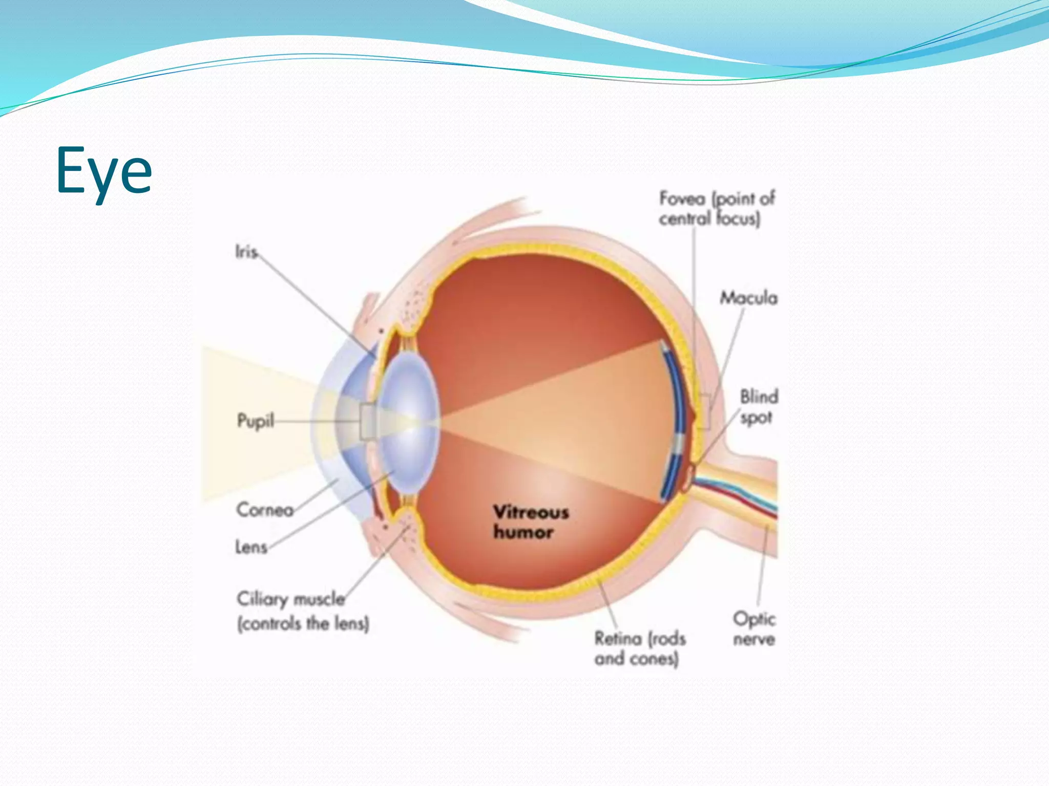

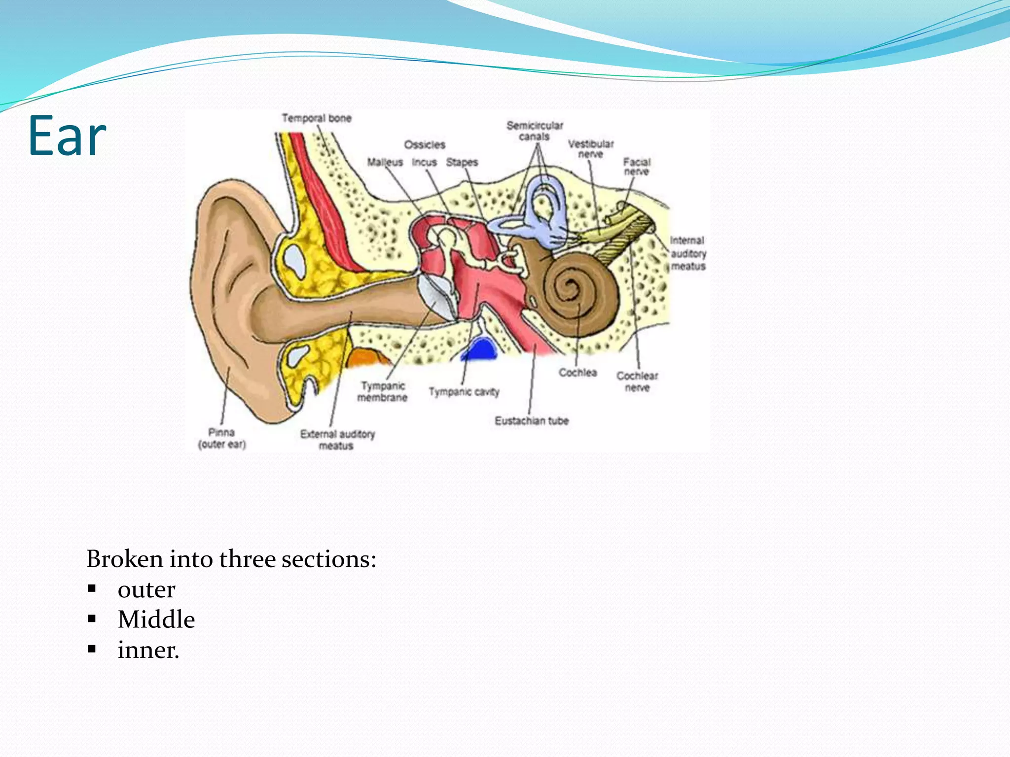
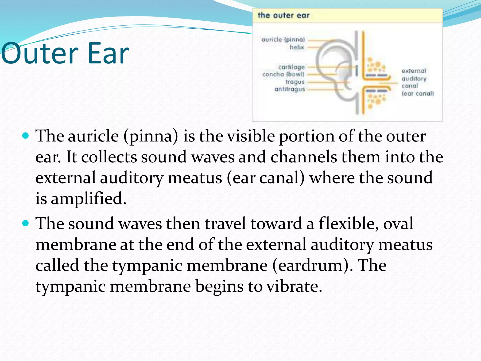
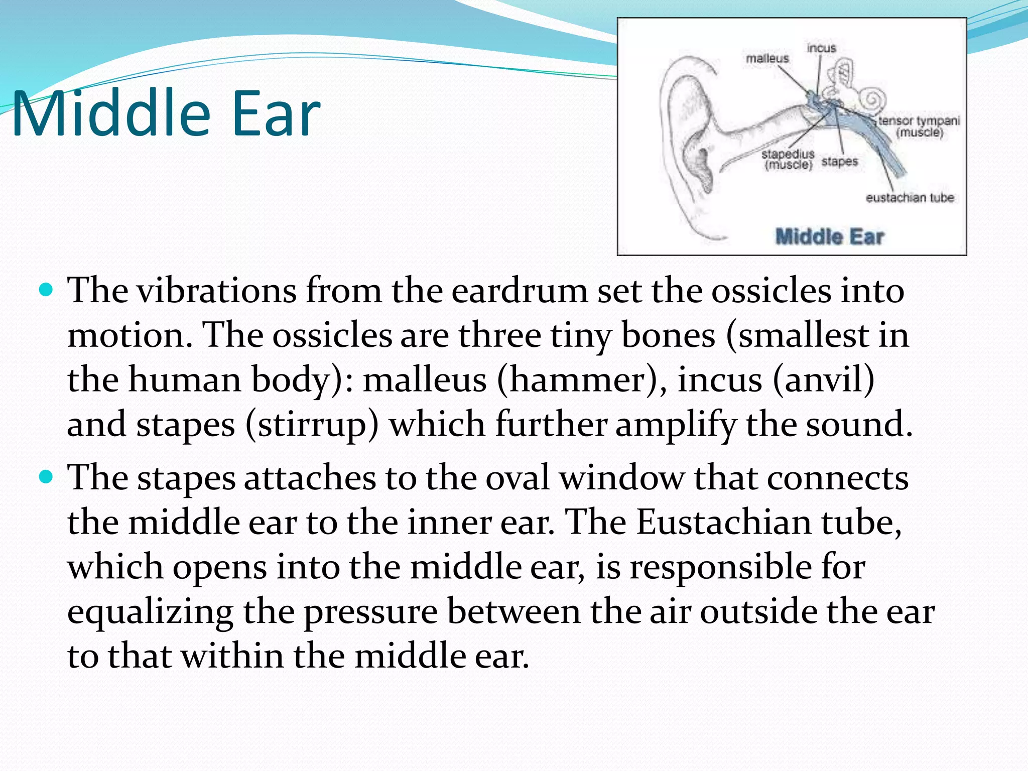
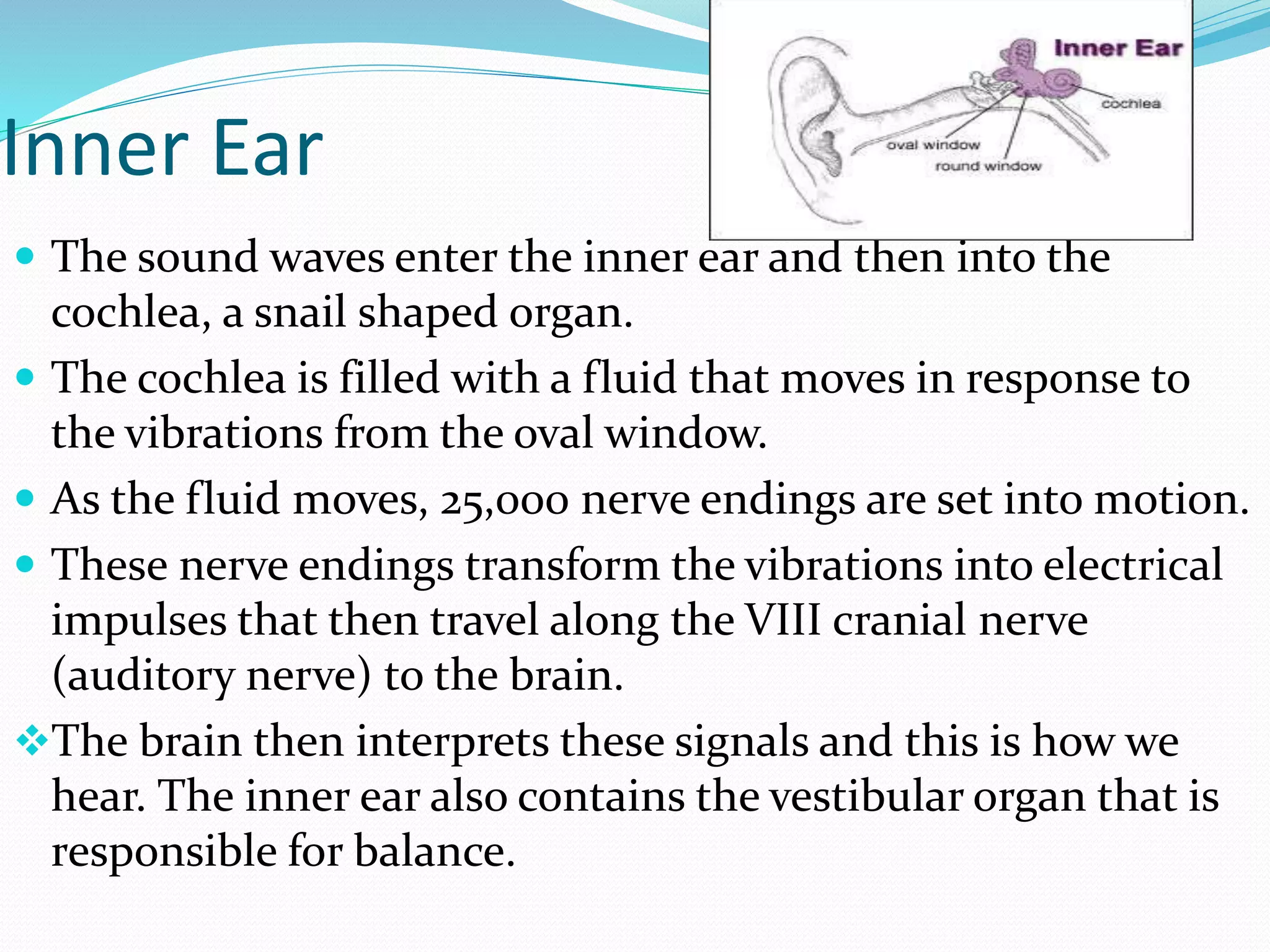


The document summarizes the structure and function of the eye and ear. It describes the three layers that make up the eye - outer fibrous tunic, middle vascular tunic, and inner retina. It explains how light enters the eye and is focused on the retina, where rods and cones detect light and color. It also outlines the three sections of the ear - outer, middle, and inner ear - and how sound waves are collected, amplified, and transmitted through the ossicles and cochlea to be interpreted as sound by the brain.


