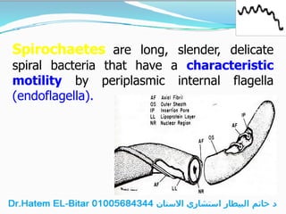
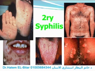
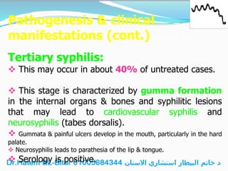
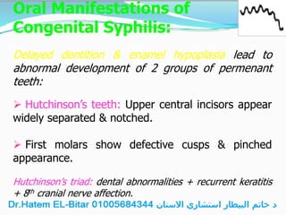
Spirochaetes are long, slender, delicate
spiral bacteria that have a characteristic
motility by periplasmic internal flagella
(endoflagella).
Periplasmic Flagella
Diagram
The family Spirochaetaceae
includes 3 important genera:
Treponema
Borrelia
Leptospira
TREPONEMA
The genus Treponema includes an important human
pathogen:
T. pallidum which causes venereal & congenital
syphilis.
Morphology:
T. pallidum is a slender spirochaete 0.2 μm in diameter &
approximate length of 10 μm.
The organism has regular coils with pointed ends.
The organism shows characteristic rapid corckscrew
(forward and backward) movement.
Unstained organisms (wet preparations) are not visible by
standard bright-field microscopy (because of the small cell
diameter), but can be seen by dark-field or phase contrast
microscopy.
T. pallidum is usually stained by silver stains (e.g. Fontana
stain).
Dark Field Microscopy Fontana Stain
Section of Liver stained with the Warthin-Starry
technique to demonstrate spirochaetes.
Isolation procedures
(Cultivability):
T. pallidum cannot be cultivated in vitro.
However, T. pallidum can be propagated by inoculation of
positive specimens into rabbit testicles.
Susceptibility to physical & chemical
agents:
Any contact with air, antiseptics or sunlight will kill
the microbe.
Its fastidious nature may account for its obligate
parasitism & rapid death outside the host.
Modes of transmission:
1. Sexual intercourse.
2. Trans-placental leading to congenital syphilis.
3. Fresh blood transfusion.
4. Occupational exposure of health care workers
through skin abrasions.
Pathogenesis & clinical
manifestations of Syphilis
Primary syphilis:
 The incubation period is 10-60 days (average 21 days).
 This is usually manifested as a single hard, painless
ulcer called chancre.
 It usually appears on the penis, labia, cervix, anorectal
region or around the mouth.
 The regional lymph nodes also become enlarged.
 The chancre heals within 4-6 weeks, even without
treatment.
1ry
Syphilis
Pathogenesis & clinical
manifestations (cont.)
Secondary syphilis:
This stage has four cardinal features:
(1) Generalized skin rash.
(2) Mucous patches in mouth & throat & snail-track
ulcers on the tongue & palate.
(3) Chondyloma lata (moist papules) form around
the genitals or anus.
(4) Generalized lymphadenopathy (including cervical
lymph nodes).
The symptoms usually last 3-6 months & disappear
spontaneously.
2ry
Syphilis
Pathogenesis & clinical
manifestations (cont.)
The Latent (hidden) stage:
There are neither symptoms nor lesions during this stage
& the serology is positive.
This stage can range from a few months to a lifetime.
Pathogenesis & clinical
manifestations (cont.)
Tertiary syphilis:
 This may occur in about 40% of untreated cases.
 This stage is characterized by gumma formation
in the internal organs & bones and syphilitic lesions
that may lead to cardiovascular syphilis and
neurosyphilis (tabes dorsalis).
 Gummata & painful ulcers develop in the mouth, particularly in the hard
palate.
 Neurosyphilis leads to parathesia of the lip & tongue.
 Serology is positive.
Tertiary Syphilis
Oral Manifestations of
Congenital Syphilis:
Delayed dentition & enamel hypoplasia lead to
abnormal development of 2 groups of permenant
teeth:
 Hutchinson’s teeth: Upper central incisors appear
widely separated & notched.
 First molars show defective cusps & pinched
appearance.
Hutchinson’s triad: dental abnormalities + recurrent keratitis
+ 8th cranial nerve affection.
Congenital
Syphilis
Laboratory Diagnosis
of Venereal Syphilis
Each stage of syphilis has a particular testing requirement as
follows:
1. Primary stage:
 Serous fluids from chancre contain numerous treponemas.
Therefore, dark-field microscopy or direct IF or PCR of
serous fluids from chancre is the method of choice for
diagnosis.
 Antibodies usually do not appear until 1-4 weeks after
the chancre has formed. Therefore, serologic tests for syphilis
are not reactive except late.
Laboratory Diagnosis
of Venereal Syphilis
2. Secondary stage:
 All serologic tests for syphilis are reactive.
 Also treponemas can be directly detected in the
mucous patches & chondyloma lata.
3. Tertiary syphilis is diagnosed only by
serologic tests.
Serologic Tests of
Syphilis
They include:
a. Non-treponemal tests: non-specific, for
screening, cheap.
b. Treponemal tests: specific, for confirmation,
expensive.
Non-Treponemal Tests
 The antigen is composed of an alcoholic solution containing
measured amounts of cardiolipin, cholesterol & lecithin to
produce standard reactivity.
 They include the following flocculation tests:
1. The venereal disease research laboratory (VDRL):
seen microscopically.
2. The rapid plasma reagin (RPR) card test: seen with
the naked eye.
RPR
-ve C +ve C
+ve
Non-Treponemal Tests
(cont.)
 These tests measure antibodies to lipoidal material released
from damaged host cells as well as from treponemas.
 These cross-reacting (heterophil) antibodies are also
produced in other conditions like autoimmune diseases,
pregnancy, leprosy, immunization & viral infections.
 Thus, false reactivity is expected during diagnosis of
syphilis. Therefore, reactivity for syphilis must be confirmed
by one of the specific treponemal tests.
Non-Treponemal Tests
(cont.)
 These tests are inexpensive, rapid and simple
to perform Therefore, they are used for
screening.
 They are also used for follow up of treatment,
since reactivity declines or disappears within 6-18
months of effective therapy.
Treponemal Tests
 The antigen is T. pallidum.
 They include:
 The fluorescent treponemal antibody absorption
(FTA-ABS) test.
 T. pallidum haemagglutination assay (TPHA).
 T. pallidum immobilization (TPI) test.
 The EIA & the Western blot.
These tests detect antibodies specific for cellular
components of the organism.
FTA-ABS
TPHA
Treponemal Tests
 They are not screening tests because they are expensive
& difficult.
 However, because they are specific, they are used in
confirming or ruling out reactive non-treponemal test
result.
 They remain reactive for life can’t be used
for follow up.
 Serologic testing for syphilis must start with the non-
treponemal tests.
 Reactive results only are then confirmed by the specific
treponemal tests.
Diagnosis of
Congenital Syphilis
It is done by detection of treponemal
IgM antibodies by EIA or Western blot.
Treatment,
Prevention & Control
 Penicillin is the drug of choice for treatment of
syphilis.
Tetracycline, erythromycin & chloramphenicol
can be used as alternative antibiotics for patients
allergic to penicillin.
 Syphilitic pregnant mothers
should be adequately treated to
prevent congenital syphilis.
FUSOSPIROCHAETAL DISEASES
• Oral spirochaetes in the genus Treponema (as T.
denticola, B. vincentii, T. pectinovarum & T.
socranskii) are found at the gingival margin.
• Under certain circumstances, particularly injury to
mucous membranes, nutritional deficiency or
concomitant infection (e.g. with herpes simplex
virus), normal oral spirochaetes together with
fusobacteria (anaerobic fusiform bacilli) multiply &
in number causing fusospirochaetal diseases.
FUSOSPIROCHAETAL DISEASES
1. Trench mouth :
It is a condition of acute necrotizing ulcerative gingivitis
(ANUG).
2. Vincent’s angina:
Fusospirochaetal infection of the pharynx with
pseudomembrane formation. Ulcers extend to the pharynx &
tonsils wiyh massive tissue destruction (similar to diphtheria
& follicular tonsillitis).
Laboratory Diagnosis
Gram stained smear from the pseudo-
membrane shows large number of Gram
negative cigar-shaped fusiform bacilli &
spirochaetes in association with pus cells &
other organisms.
Treatment
1. Penicillin combined with metronidazole.
2. Correction of the underlying condition.
Actinomyces
• They are branching filaments.
• They include:
– Aerobic: Nocardia & Streptomyces.
– Anaerobic: Actinomyces.
Actinomycosis
• It’s a chronic suppurative disease caused by A.
israelii.
• A. israelii is a normal flora in the mouth,
tonsils, GIT & vagina.
Pathogenesis
• Usually endogenous infection, following local
trauma.
• Disease is characterized by chronic
inflammatory granulomatous lesions that
discharge pus (containing sulfur granules)
from multiple sinuses.
Clinical Forms (based on site)
• Cervicofacial actinomycosis.
• Thoracic actinomycosis.
• Abdominal actinomycosis.
• Pelvic actinomycosis.
Lab. Diagnosis
• Sulphur granules are crushed then:
– Gram stain: Gram-positive filamentous
branching bacilli & coccoid forms.
– Culture anaerobically for at least 2 weeks
colonies with molar tooth appearance.
Treatment
• Surgical drainage of pus.
• Prolonged antibiotic course (e.g. penicillin,
clindamycin, erythromycin).
Candida
• Unicellular yeasts.
• Gram stain positive.
• Appear as spherical to oval budding yeast
cells or pseudohyphae (chains of elongated
budding cells).
Habitat & Transmission
• C. albicans is a normal flora of the oral
cavity, GIT, female genital tract &
sometimes the skin.
• Usually endogenous infection, but mother-
to-baby infection may occur.
P.F. to Oral Candidiasis
• Chronic local irritants as ill-fitting appliances.
• Disturbed oral ecology by antibiotics,
corticosteroids & xerostomia.
• Malignant & chronic disorders (e.g.
malignancy, diabetes).
• Radiation to the head & neck.
• Extremes of age.
• Heavy smoking.
Classification of Oral Candidiasis
• Primary (e.g., denture-induced stomatitis, angular
stomatitis/chelitis):
– Pseudomembranous (oral thrush)
– Erythematous (acute & chronic atrophic)
– Hyperplastic (leukoplakia)
• Secondary:
– Due to defect in CMI causing systemic candidiasis.
– Oral manifestations usually present as hyperplastic lesions
(leukoplakia).
Oral Thrush (acute psudomembraneous)
Sources of infection:
In infants:
• Birth canal of the
mother
• Feeding bottles
• Hands of attendants
In adults:
• Systemic antibiotics
• Diabetes & malignancy
• Corticosteroids
• Radiation therapy
Acute Atrophic Candidiasis
• Depapillated red areas on the dorsum of
tongue or palate & buccal mucosa.
Chronic Atrophic Candidiasis
• Also called denture stomatitis & denture
sore throat.
• Usually affect elderly people wearing ill-
fitting denture.
• Erythema & edema on palate.
Angular Cheilitis
• Occur in odontulous patients with long-term
wearing of denture.
• Saliva moistens the commissures & macerated
skin are infected with candida.
Chronic hyperplastic Candidiasis
(Candida Leukoplakia)
• Usually asymptomatic.
• On the inside of cheeks.
• Appear as dense opaque hard rough plaques.
• Higher incidence of cancer (9-40% of cases).
Progressive Verrucous Leukoplakia
(PVL)
• PVL is a single disease entity that
demonstrates a unique spectrum of
clinical and histopathologic
expression that may terminate in
squamous cell carcinoma
• 60-75% of PVL patients
harbored Candida albicans in
the superficial layers of their
biopsies.
Diagnosis
Swab (from the lesions) or biopsy
(from candida leukoplakia):
• Gram stain: oval yeast cells.
• Culture: on Sabauraud dextrose
agar (identified by morphology
& germ tube test).
Treatment
• Correct the P.F.
• Topical agents (e.g. gentian violet or nystatin).
• Antifungal drugs (e.g. fluconazole).
Thank You

د حاتم البيطار استشاري وجراح الفم والاسنان 01005684344 اتصل للحجز بالعيادة محاضر طبي Dr.Hatem EL-Bitar

  • 2.
    Spirochaetes are long,slender, delicate spiral bacteria that have a characteristic motility by periplasmic internal flagella (endoflagella). Periplasmic Flagella Diagram
  • 3.
    The family Spirochaetaceae includes3 important genera: Treponema Borrelia Leptospira
  • 4.
    TREPONEMA The genus Treponemaincludes an important human pathogen: T. pallidum which causes venereal & congenital syphilis.
  • 5.
    Morphology: T. pallidum isa slender spirochaete 0.2 μm in diameter & approximate length of 10 μm. The organism has regular coils with pointed ends. The organism shows characteristic rapid corckscrew (forward and backward) movement. Unstained organisms (wet preparations) are not visible by standard bright-field microscopy (because of the small cell diameter), but can be seen by dark-field or phase contrast microscopy. T. pallidum is usually stained by silver stains (e.g. Fontana stain).
  • 7.
    Dark Field MicroscopyFontana Stain
  • 8.
    Section of Liverstained with the Warthin-Starry technique to demonstrate spirochaetes.
  • 9.
    Isolation procedures (Cultivability): T. pallidumcannot be cultivated in vitro. However, T. pallidum can be propagated by inoculation of positive specimens into rabbit testicles. Susceptibility to physical & chemical agents: Any contact with air, antiseptics or sunlight will kill the microbe. Its fastidious nature may account for its obligate parasitism & rapid death outside the host.
  • 10.
    Modes of transmission: 1.Sexual intercourse. 2. Trans-placental leading to congenital syphilis. 3. Fresh blood transfusion. 4. Occupational exposure of health care workers through skin abrasions.
  • 12.
    Pathogenesis & clinical manifestationsof Syphilis Primary syphilis:  The incubation period is 10-60 days (average 21 days).  This is usually manifested as a single hard, painless ulcer called chancre.  It usually appears on the penis, labia, cervix, anorectal region or around the mouth.  The regional lymph nodes also become enlarged.  The chancre heals within 4-6 weeks, even without treatment.
  • 13.
  • 14.
    Pathogenesis & clinical manifestations(cont.) Secondary syphilis: This stage has four cardinal features: (1) Generalized skin rash. (2) Mucous patches in mouth & throat & snail-track ulcers on the tongue & palate. (3) Chondyloma lata (moist papules) form around the genitals or anus. (4) Generalized lymphadenopathy (including cervical lymph nodes). The symptoms usually last 3-6 months & disappear spontaneously.
  • 15.
  • 16.
    Pathogenesis & clinical manifestations(cont.) The Latent (hidden) stage: There are neither symptoms nor lesions during this stage & the serology is positive. This stage can range from a few months to a lifetime.
  • 17.
    Pathogenesis & clinical manifestations(cont.) Tertiary syphilis:  This may occur in about 40% of untreated cases.  This stage is characterized by gumma formation in the internal organs & bones and syphilitic lesions that may lead to cardiovascular syphilis and neurosyphilis (tabes dorsalis).  Gummata & painful ulcers develop in the mouth, particularly in the hard palate.  Neurosyphilis leads to parathesia of the lip & tongue.  Serology is positive.
  • 18.
  • 19.
    Oral Manifestations of CongenitalSyphilis: Delayed dentition & enamel hypoplasia lead to abnormal development of 2 groups of permenant teeth:  Hutchinson’s teeth: Upper central incisors appear widely separated & notched.  First molars show defective cusps & pinched appearance. Hutchinson’s triad: dental abnormalities + recurrent keratitis + 8th cranial nerve affection.
  • 20.
  • 21.
    Laboratory Diagnosis of VenerealSyphilis Each stage of syphilis has a particular testing requirement as follows: 1. Primary stage:  Serous fluids from chancre contain numerous treponemas. Therefore, dark-field microscopy or direct IF or PCR of serous fluids from chancre is the method of choice for diagnosis.  Antibodies usually do not appear until 1-4 weeks after the chancre has formed. Therefore, serologic tests for syphilis are not reactive except late.
  • 22.
    Laboratory Diagnosis of VenerealSyphilis 2. Secondary stage:  All serologic tests for syphilis are reactive.  Also treponemas can be directly detected in the mucous patches & chondyloma lata. 3. Tertiary syphilis is diagnosed only by serologic tests.
  • 23.
    Serologic Tests of Syphilis Theyinclude: a. Non-treponemal tests: non-specific, for screening, cheap. b. Treponemal tests: specific, for confirmation, expensive.
  • 24.
    Non-Treponemal Tests  Theantigen is composed of an alcoholic solution containing measured amounts of cardiolipin, cholesterol & lecithin to produce standard reactivity.  They include the following flocculation tests: 1. The venereal disease research laboratory (VDRL): seen microscopically. 2. The rapid plasma reagin (RPR) card test: seen with the naked eye. RPR -ve C +ve C +ve
  • 25.
    Non-Treponemal Tests (cont.)  Thesetests measure antibodies to lipoidal material released from damaged host cells as well as from treponemas.  These cross-reacting (heterophil) antibodies are also produced in other conditions like autoimmune diseases, pregnancy, leprosy, immunization & viral infections.  Thus, false reactivity is expected during diagnosis of syphilis. Therefore, reactivity for syphilis must be confirmed by one of the specific treponemal tests.
  • 26.
    Non-Treponemal Tests (cont.)  Thesetests are inexpensive, rapid and simple to perform Therefore, they are used for screening.  They are also used for follow up of treatment, since reactivity declines or disappears within 6-18 months of effective therapy.
  • 27.
    Treponemal Tests  Theantigen is T. pallidum.  They include:  The fluorescent treponemal antibody absorption (FTA-ABS) test.  T. pallidum haemagglutination assay (TPHA).  T. pallidum immobilization (TPI) test.  The EIA & the Western blot. These tests detect antibodies specific for cellular components of the organism. FTA-ABS TPHA
  • 28.
    Treponemal Tests  Theyare not screening tests because they are expensive & difficult.  However, because they are specific, they are used in confirming or ruling out reactive non-treponemal test result.  They remain reactive for life can’t be used for follow up.  Serologic testing for syphilis must start with the non- treponemal tests.  Reactive results only are then confirmed by the specific treponemal tests.
  • 29.
    Diagnosis of Congenital Syphilis Itis done by detection of treponemal IgM antibodies by EIA or Western blot.
  • 30.
    Treatment, Prevention & Control Penicillin is the drug of choice for treatment of syphilis. Tetracycline, erythromycin & chloramphenicol can be used as alternative antibiotics for patients allergic to penicillin.  Syphilitic pregnant mothers should be adequately treated to prevent congenital syphilis.
  • 31.
    FUSOSPIROCHAETAL DISEASES • Oralspirochaetes in the genus Treponema (as T. denticola, B. vincentii, T. pectinovarum & T. socranskii) are found at the gingival margin. • Under certain circumstances, particularly injury to mucous membranes, nutritional deficiency or concomitant infection (e.g. with herpes simplex virus), normal oral spirochaetes together with fusobacteria (anaerobic fusiform bacilli) multiply & in number causing fusospirochaetal diseases.
  • 32.
    FUSOSPIROCHAETAL DISEASES 1. Trenchmouth : It is a condition of acute necrotizing ulcerative gingivitis (ANUG). 2. Vincent’s angina: Fusospirochaetal infection of the pharynx with pseudomembrane formation. Ulcers extend to the pharynx & tonsils wiyh massive tissue destruction (similar to diphtheria & follicular tonsillitis).
  • 33.
    Laboratory Diagnosis Gram stainedsmear from the pseudo- membrane shows large number of Gram negative cigar-shaped fusiform bacilli & spirochaetes in association with pus cells & other organisms.
  • 34.
    Treatment 1. Penicillin combinedwith metronidazole. 2. Correction of the underlying condition.
  • 35.
    Actinomyces • They arebranching filaments. • They include: – Aerobic: Nocardia & Streptomyces. – Anaerobic: Actinomyces.
  • 36.
    Actinomycosis • It’s achronic suppurative disease caused by A. israelii. • A. israelii is a normal flora in the mouth, tonsils, GIT & vagina.
  • 37.
    Pathogenesis • Usually endogenousinfection, following local trauma. • Disease is characterized by chronic inflammatory granulomatous lesions that discharge pus (containing sulfur granules) from multiple sinuses.
  • 38.
    Clinical Forms (basedon site) • Cervicofacial actinomycosis. • Thoracic actinomycosis. • Abdominal actinomycosis. • Pelvic actinomycosis.
  • 39.
    Lab. Diagnosis • Sulphurgranules are crushed then: – Gram stain: Gram-positive filamentous branching bacilli & coccoid forms. – Culture anaerobically for at least 2 weeks colonies with molar tooth appearance.
  • 40.
    Treatment • Surgical drainageof pus. • Prolonged antibiotic course (e.g. penicillin, clindamycin, erythromycin).
  • 41.
    Candida • Unicellular yeasts. •Gram stain positive. • Appear as spherical to oval budding yeast cells or pseudohyphae (chains of elongated budding cells).
  • 42.
    Habitat & Transmission •C. albicans is a normal flora of the oral cavity, GIT, female genital tract & sometimes the skin. • Usually endogenous infection, but mother- to-baby infection may occur.
  • 43.
    P.F. to OralCandidiasis • Chronic local irritants as ill-fitting appliances. • Disturbed oral ecology by antibiotics, corticosteroids & xerostomia. • Malignant & chronic disorders (e.g. malignancy, diabetes). • Radiation to the head & neck. • Extremes of age. • Heavy smoking.
  • 44.
    Classification of OralCandidiasis • Primary (e.g., denture-induced stomatitis, angular stomatitis/chelitis): – Pseudomembranous (oral thrush) – Erythematous (acute & chronic atrophic) – Hyperplastic (leukoplakia) • Secondary: – Due to defect in CMI causing systemic candidiasis. – Oral manifestations usually present as hyperplastic lesions (leukoplakia).
  • 45.
    Oral Thrush (acutepsudomembraneous) Sources of infection: In infants: • Birth canal of the mother • Feeding bottles • Hands of attendants In adults: • Systemic antibiotics • Diabetes & malignancy • Corticosteroids • Radiation therapy
  • 46.
    Acute Atrophic Candidiasis •Depapillated red areas on the dorsum of tongue or palate & buccal mucosa.
  • 47.
    Chronic Atrophic Candidiasis •Also called denture stomatitis & denture sore throat. • Usually affect elderly people wearing ill- fitting denture. • Erythema & edema on palate.
  • 48.
    Angular Cheilitis • Occurin odontulous patients with long-term wearing of denture. • Saliva moistens the commissures & macerated skin are infected with candida.
  • 49.
    Chronic hyperplastic Candidiasis (CandidaLeukoplakia) • Usually asymptomatic. • On the inside of cheeks. • Appear as dense opaque hard rough plaques. • Higher incidence of cancer (9-40% of cases).
  • 50.
    Progressive Verrucous Leukoplakia (PVL) •PVL is a single disease entity that demonstrates a unique spectrum of clinical and histopathologic expression that may terminate in squamous cell carcinoma • 60-75% of PVL patients harbored Candida albicans in the superficial layers of their biopsies.
  • 51.
    Diagnosis Swab (from thelesions) or biopsy (from candida leukoplakia): • Gram stain: oval yeast cells. • Culture: on Sabauraud dextrose agar (identified by morphology & germ tube test).
  • 52.
    Treatment • Correct theP.F. • Topical agents (e.g. gentian violet or nystatin). • Antifungal drugs (e.g. fluconazole).
  • 53.