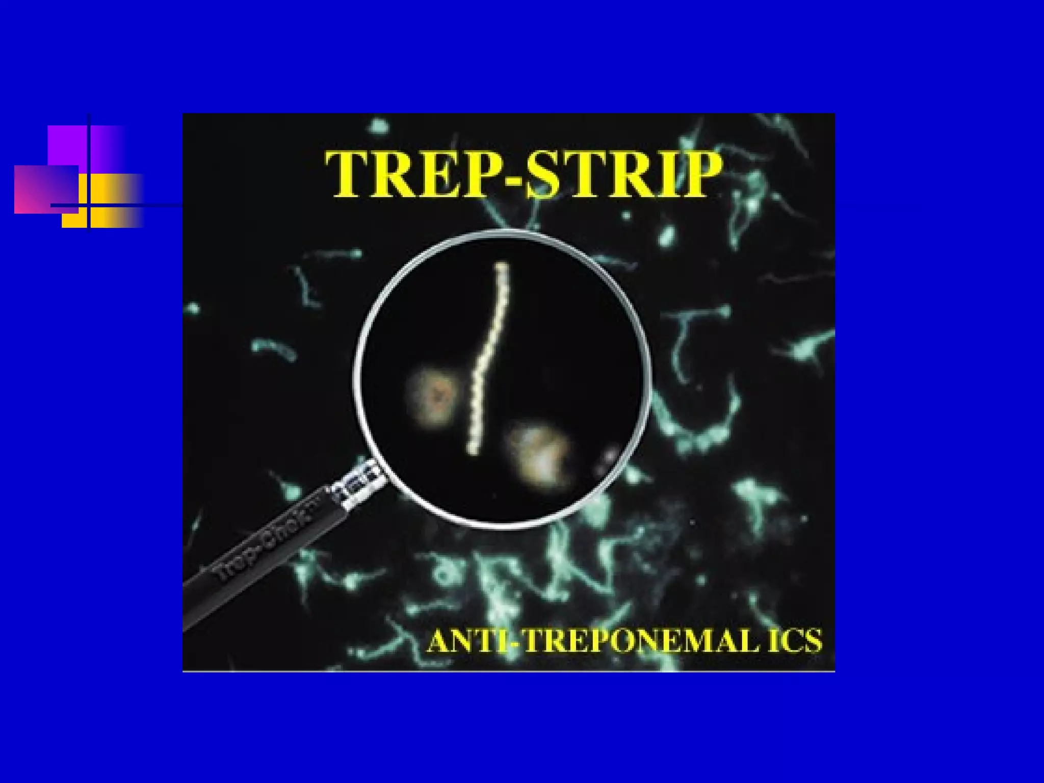
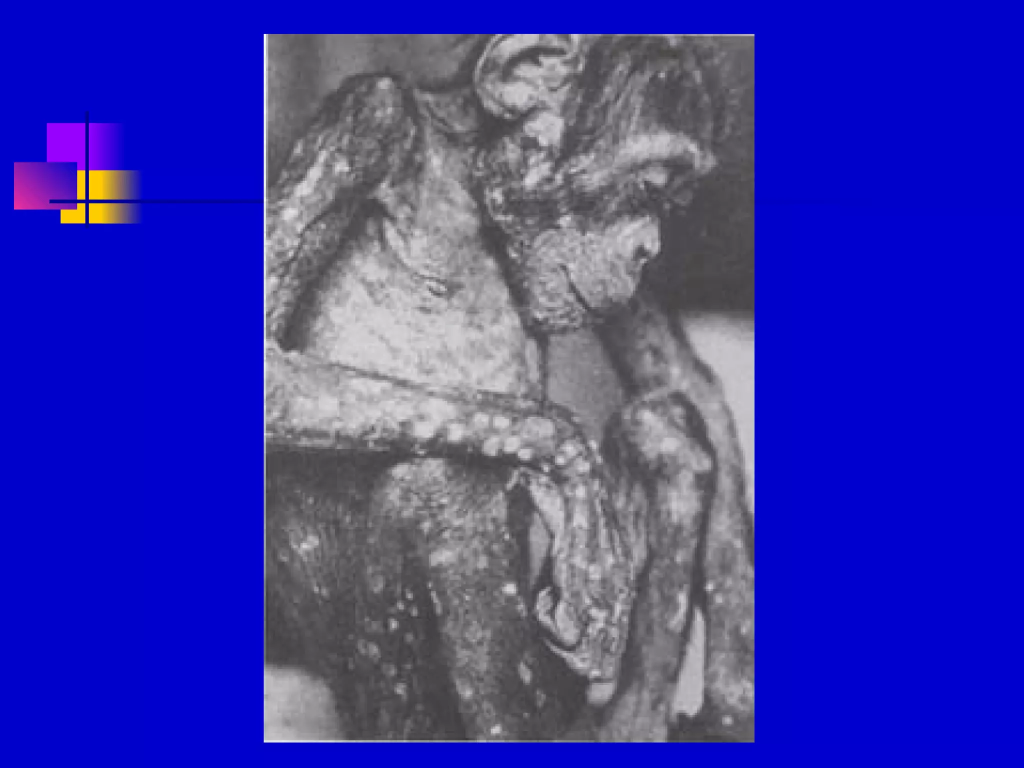
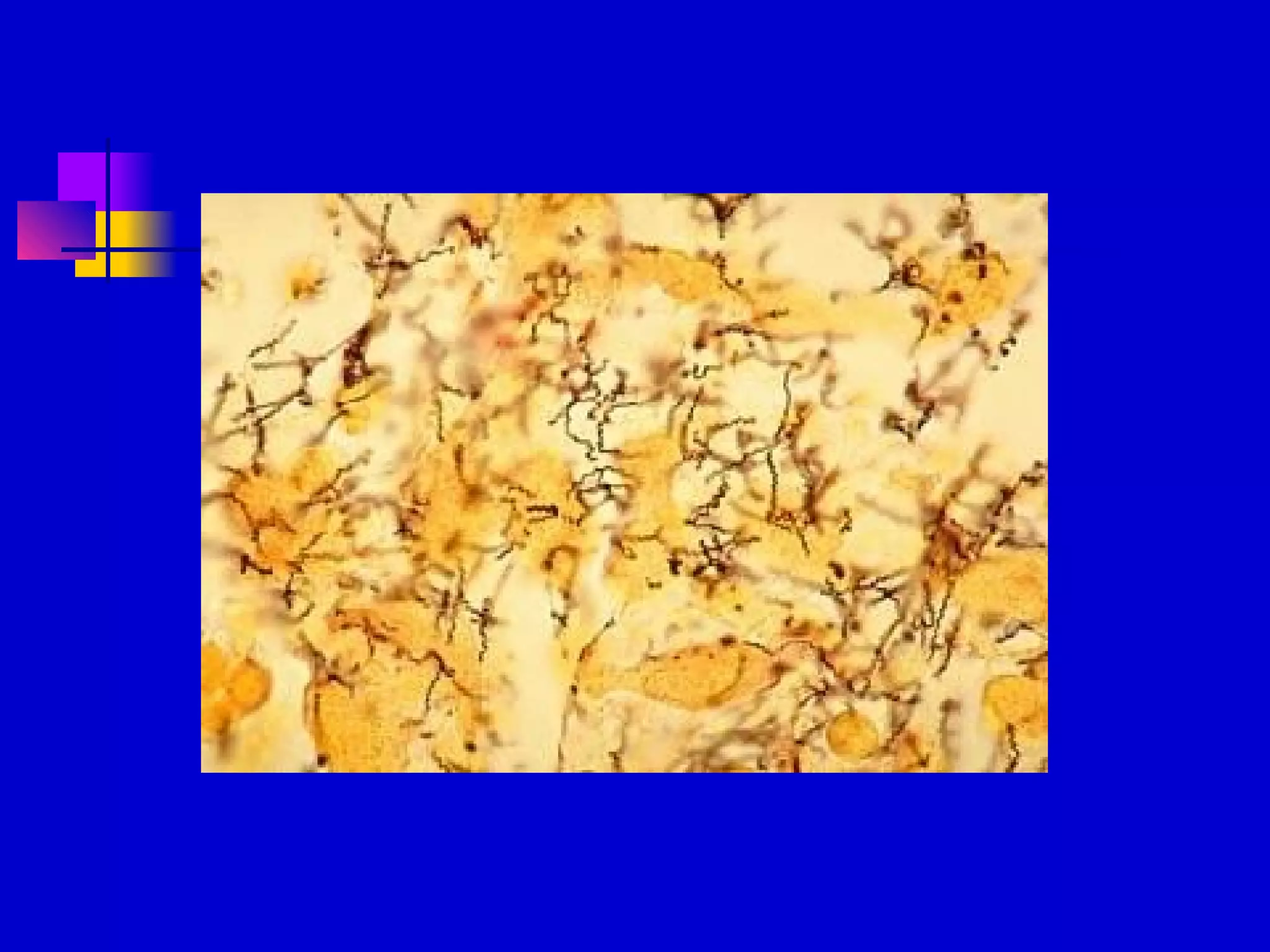
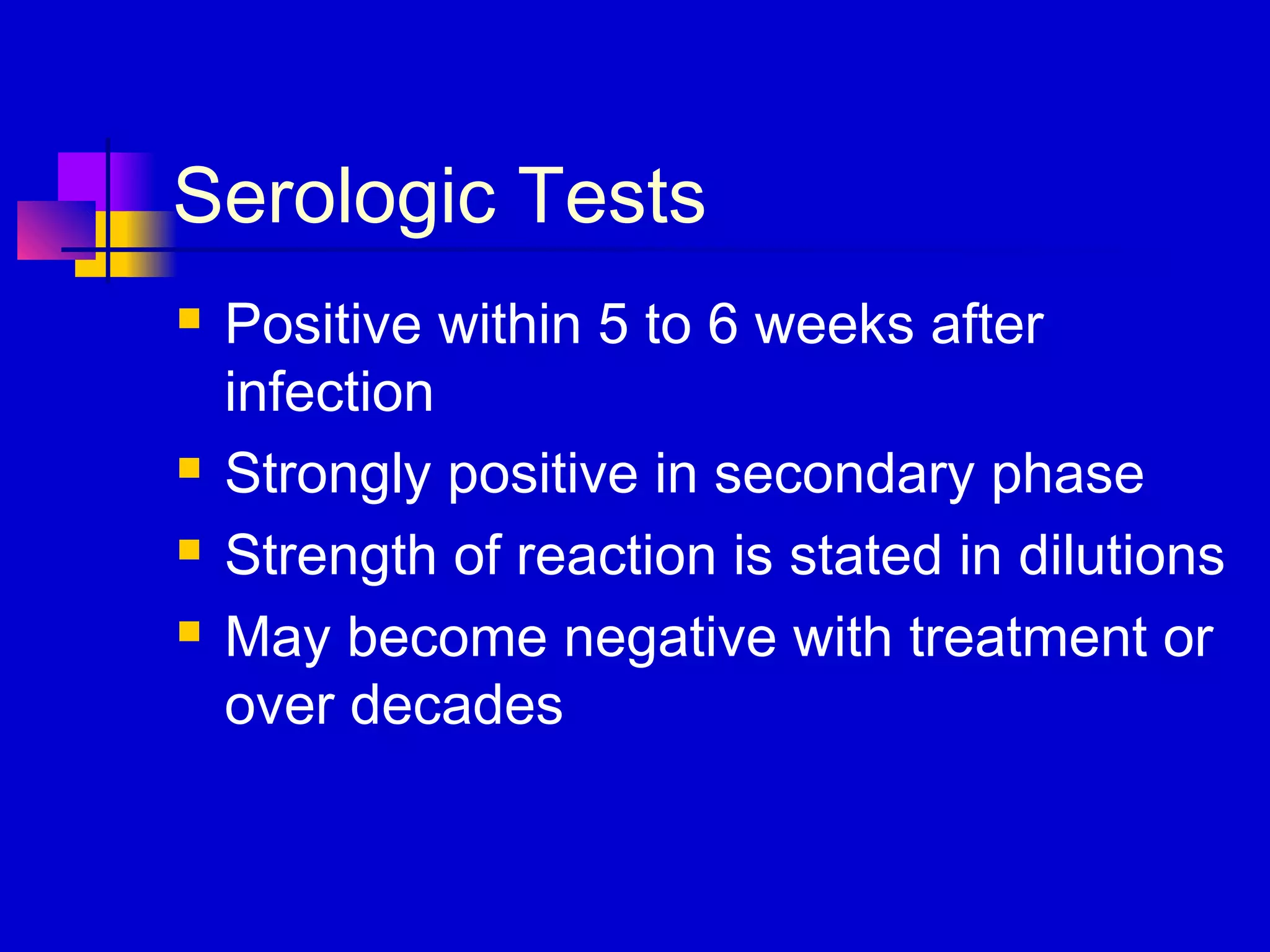
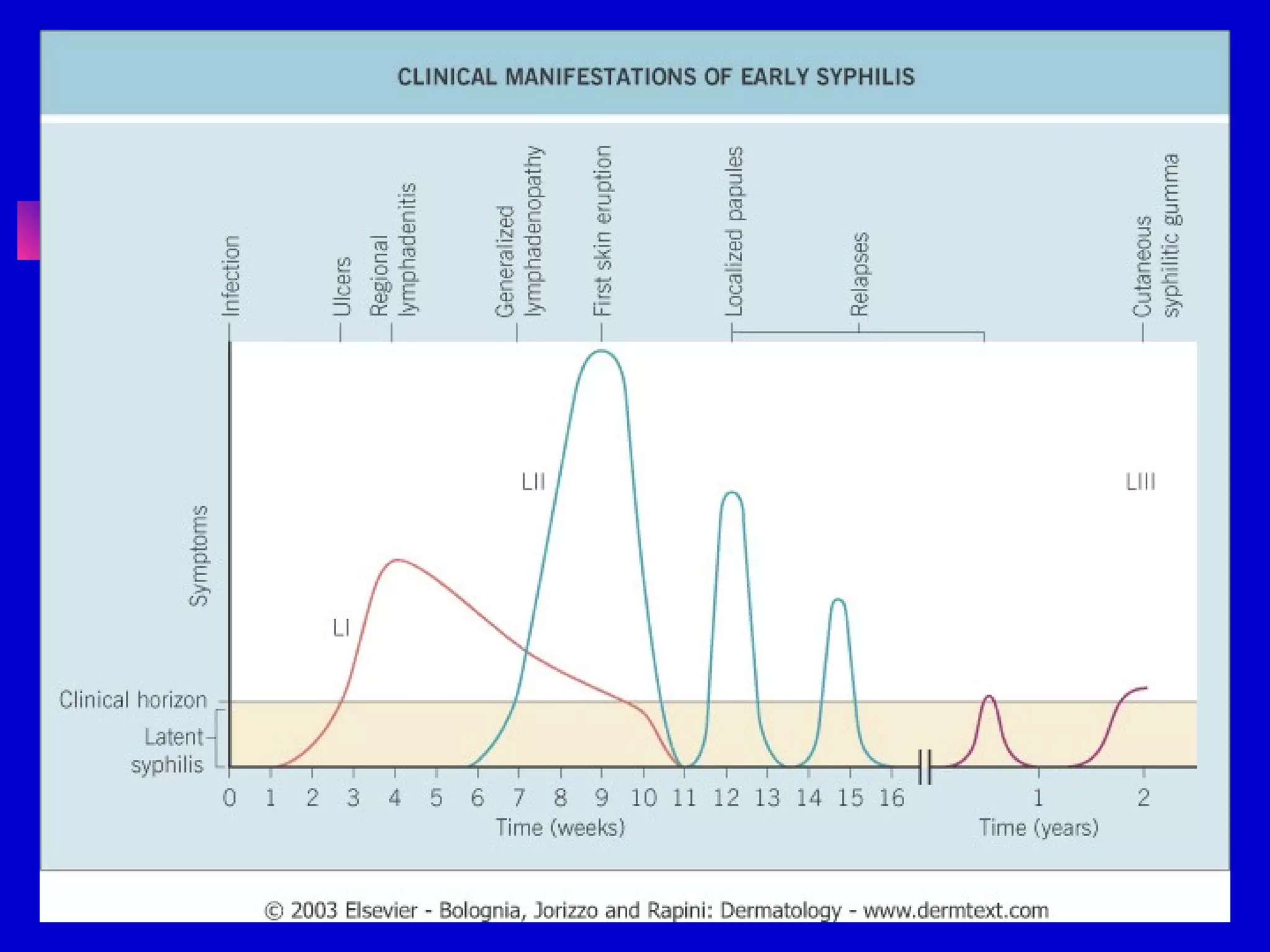
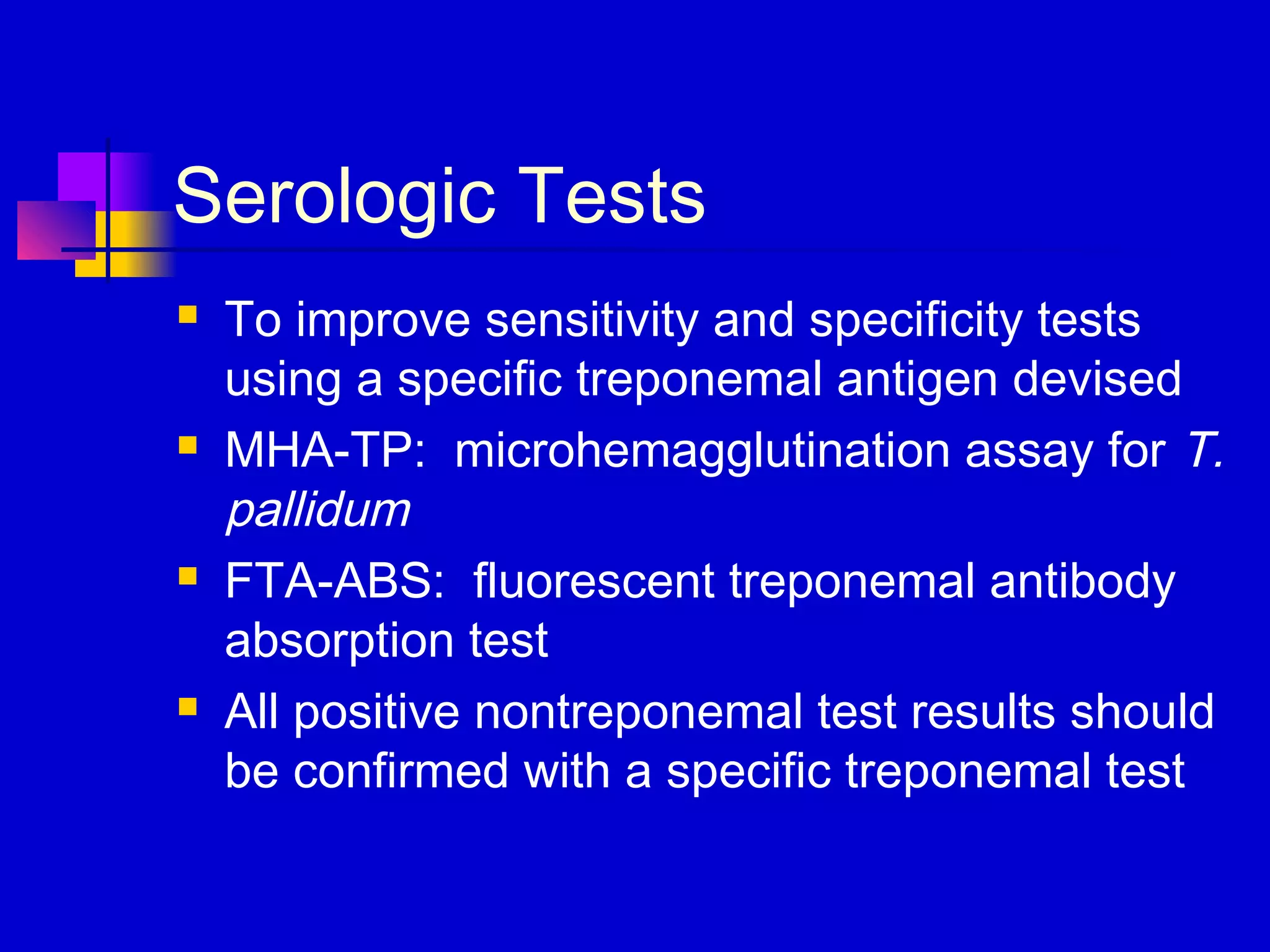
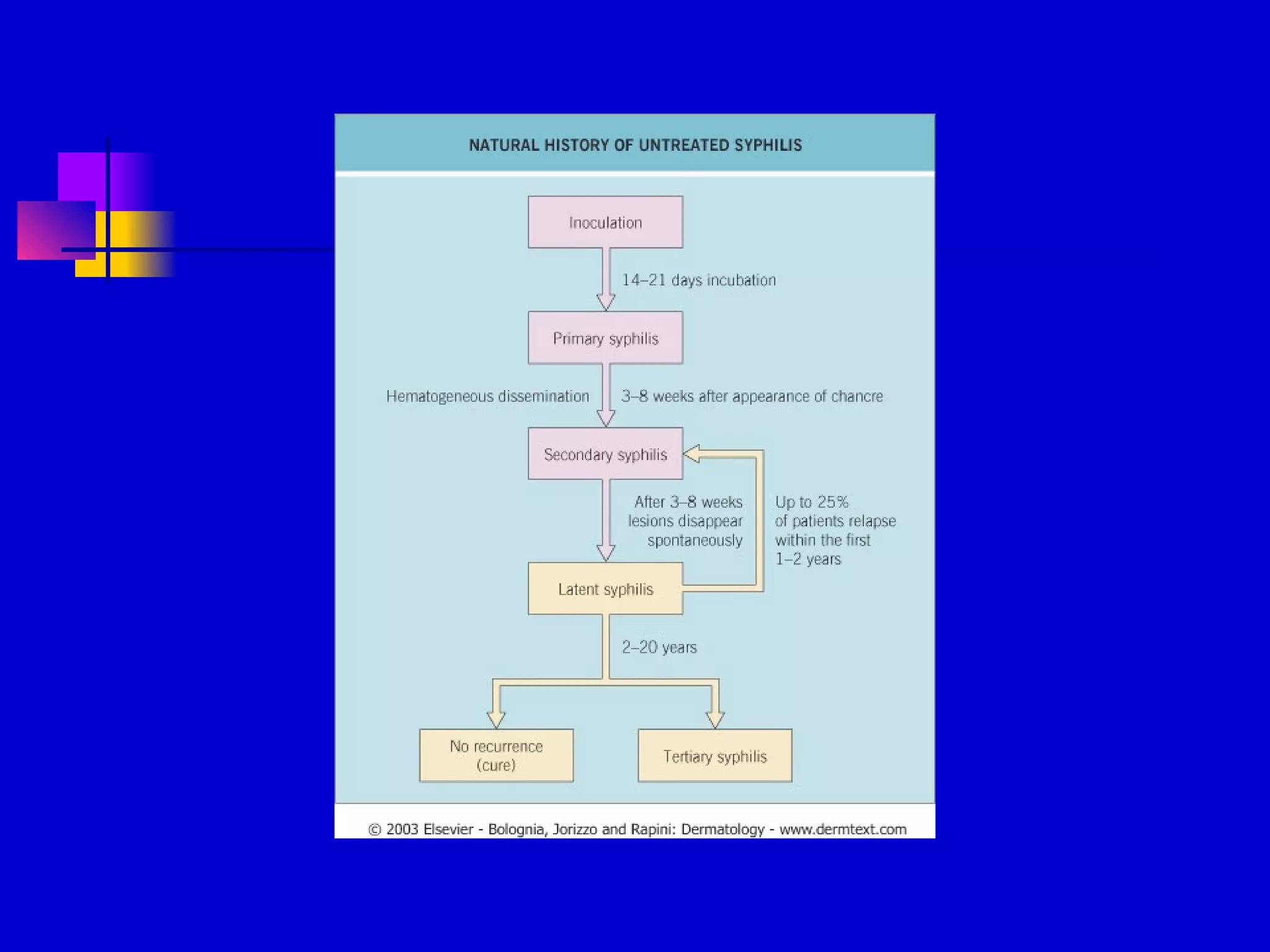
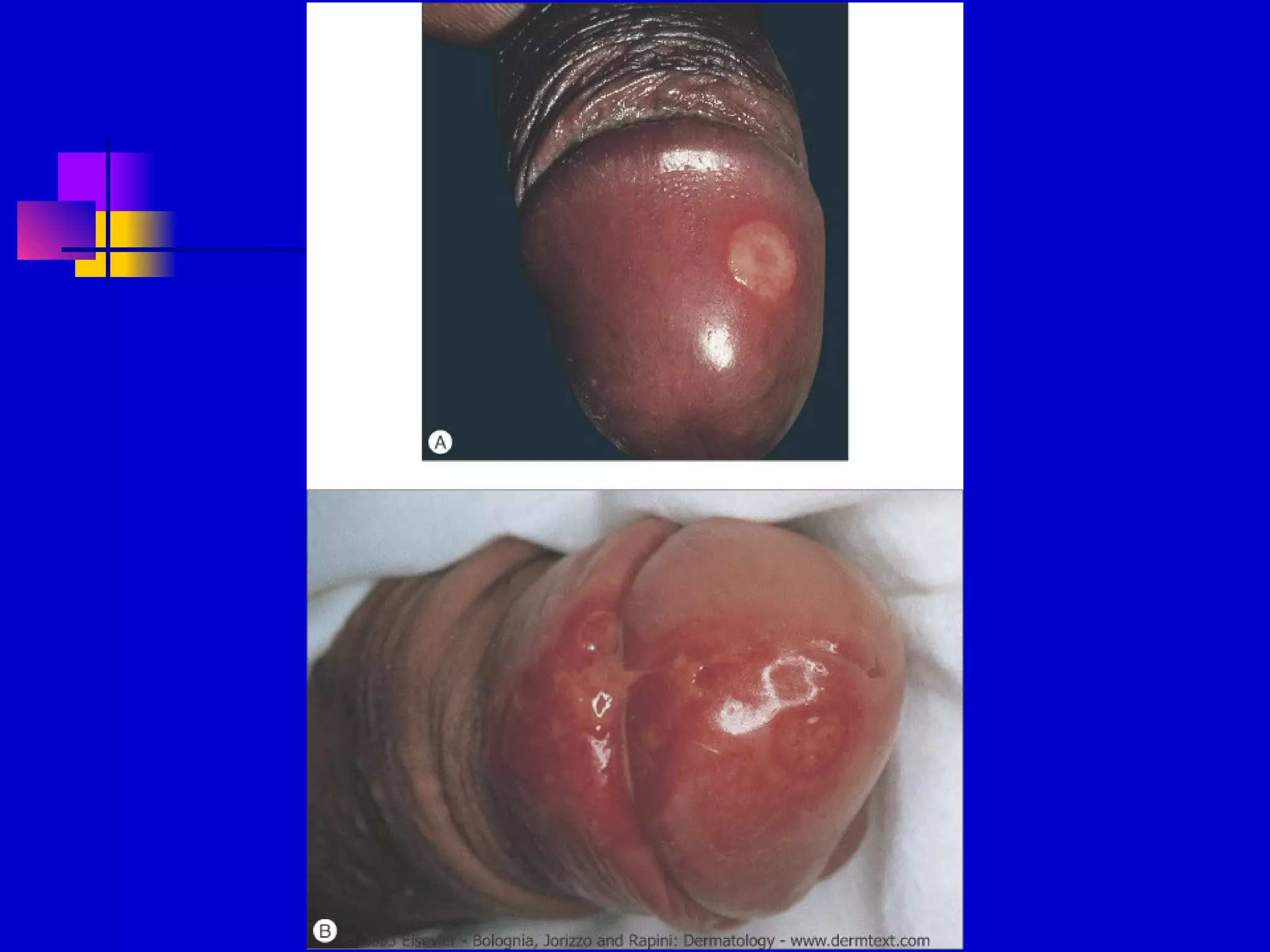
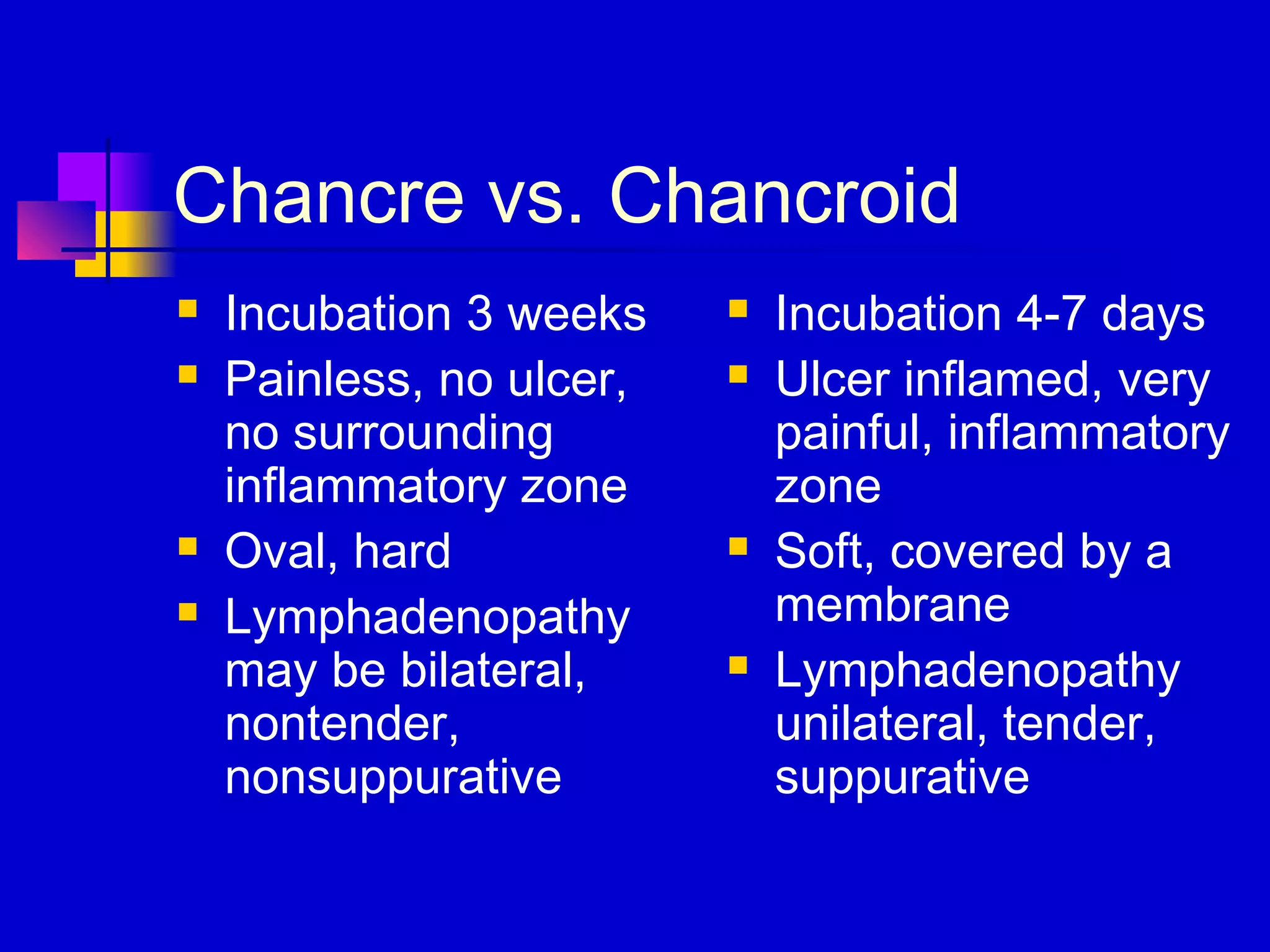
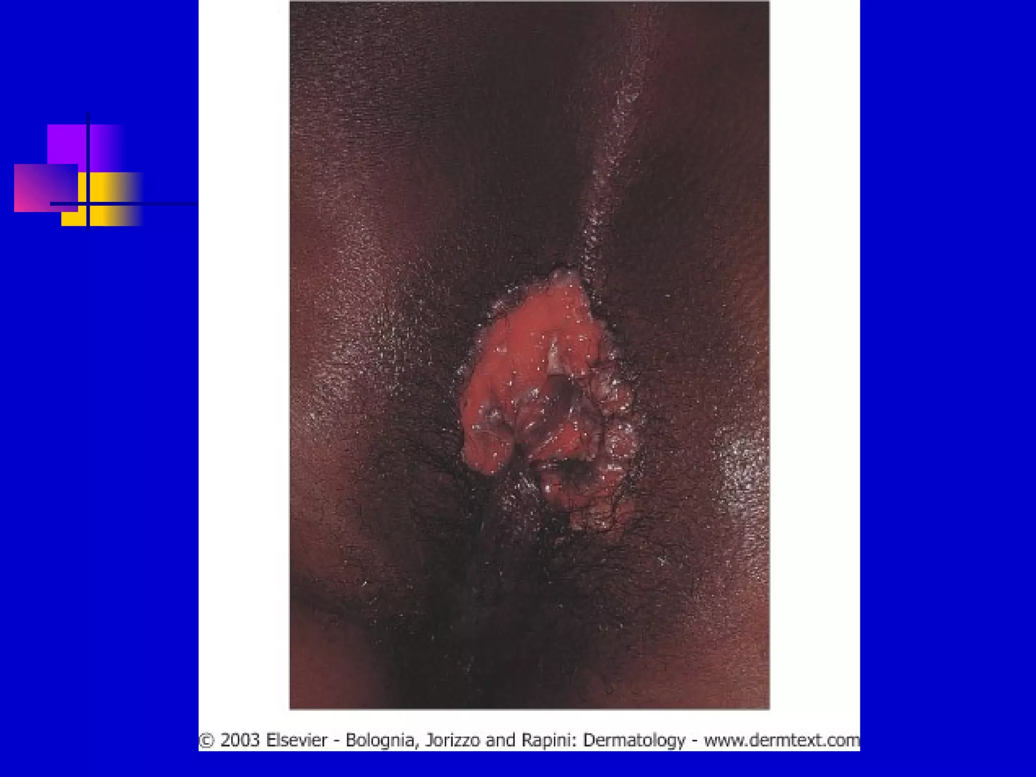
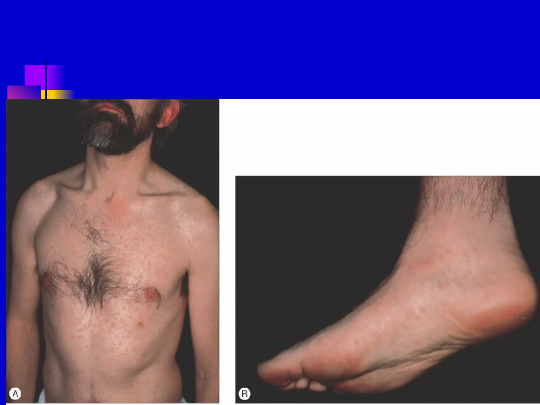
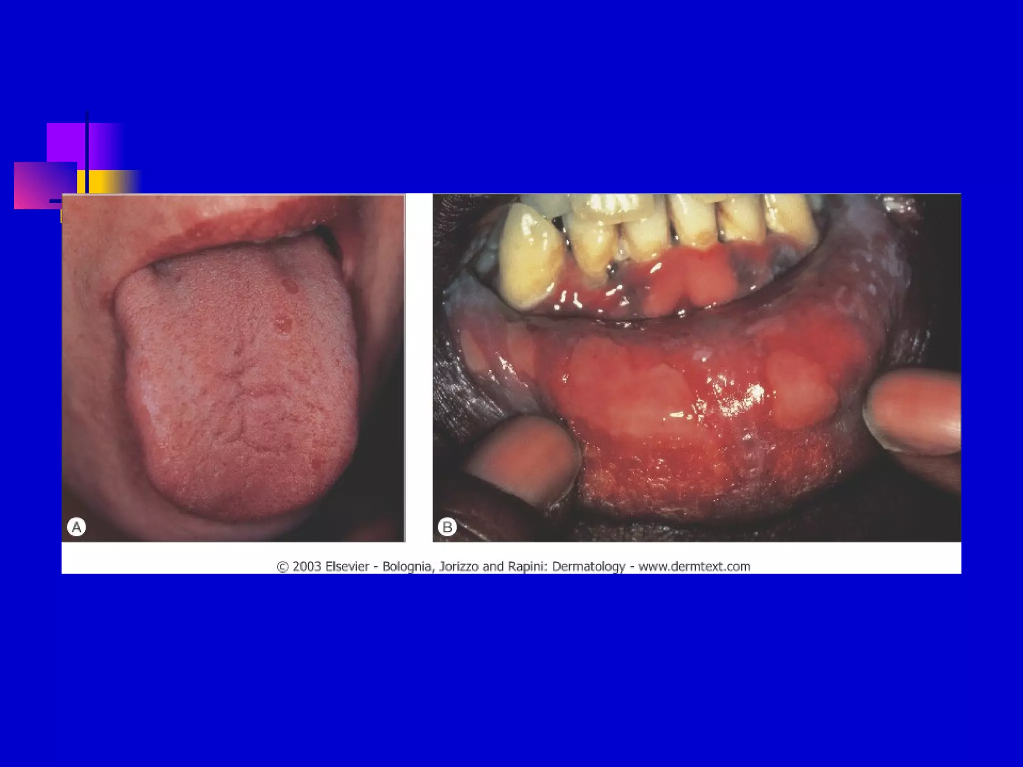
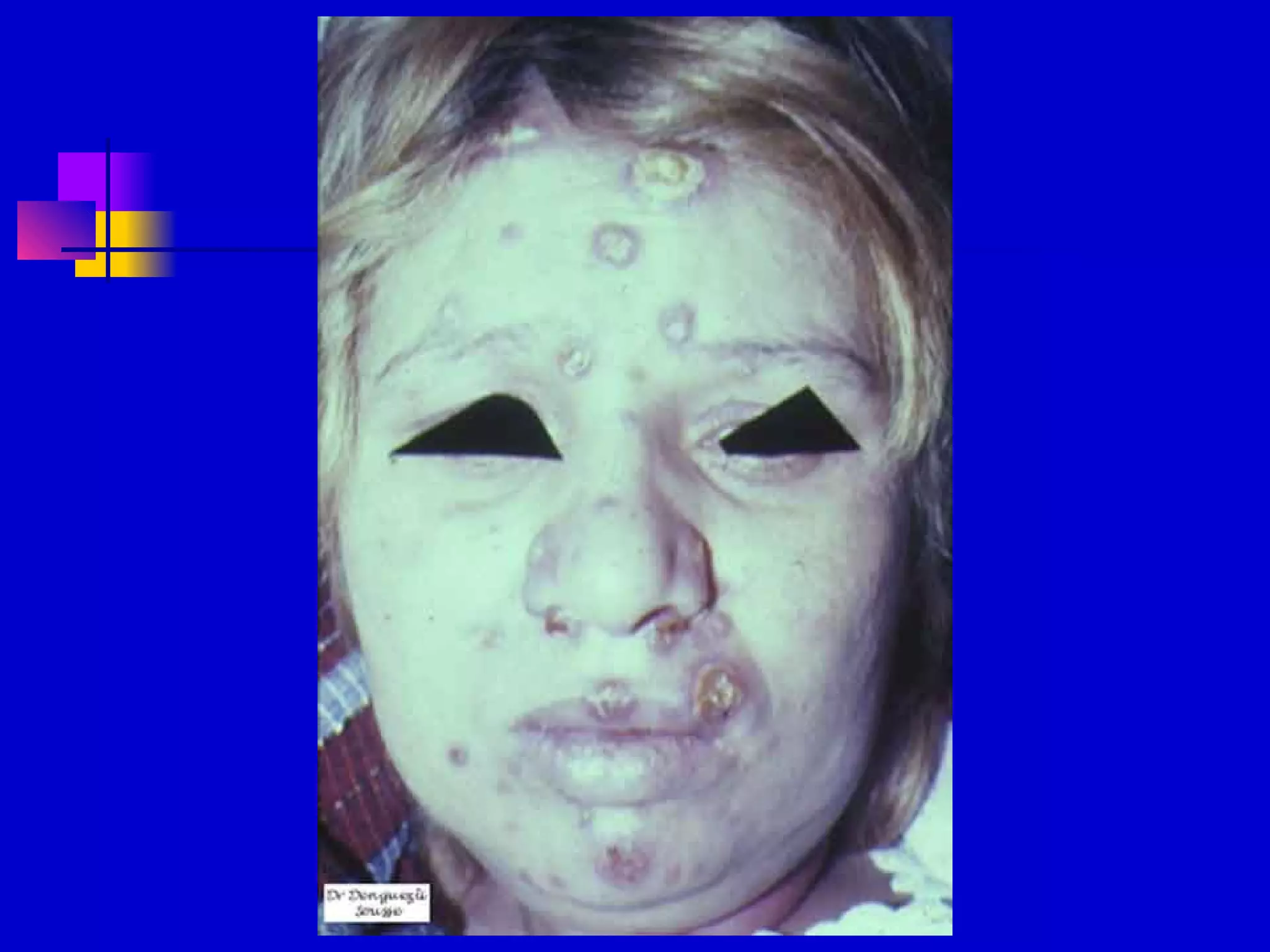
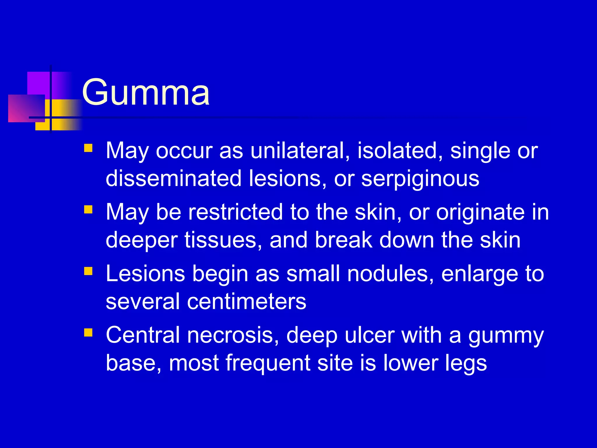
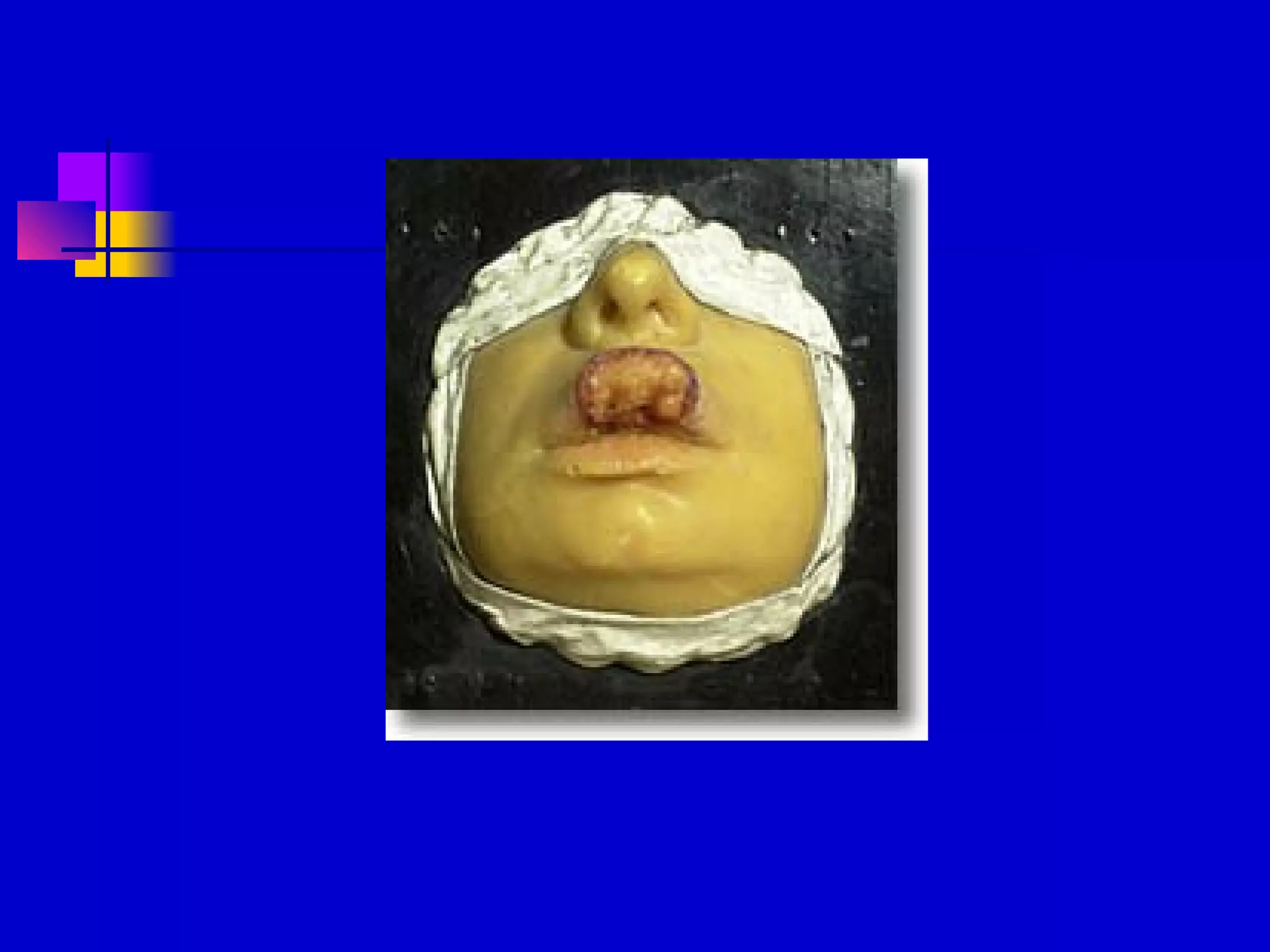
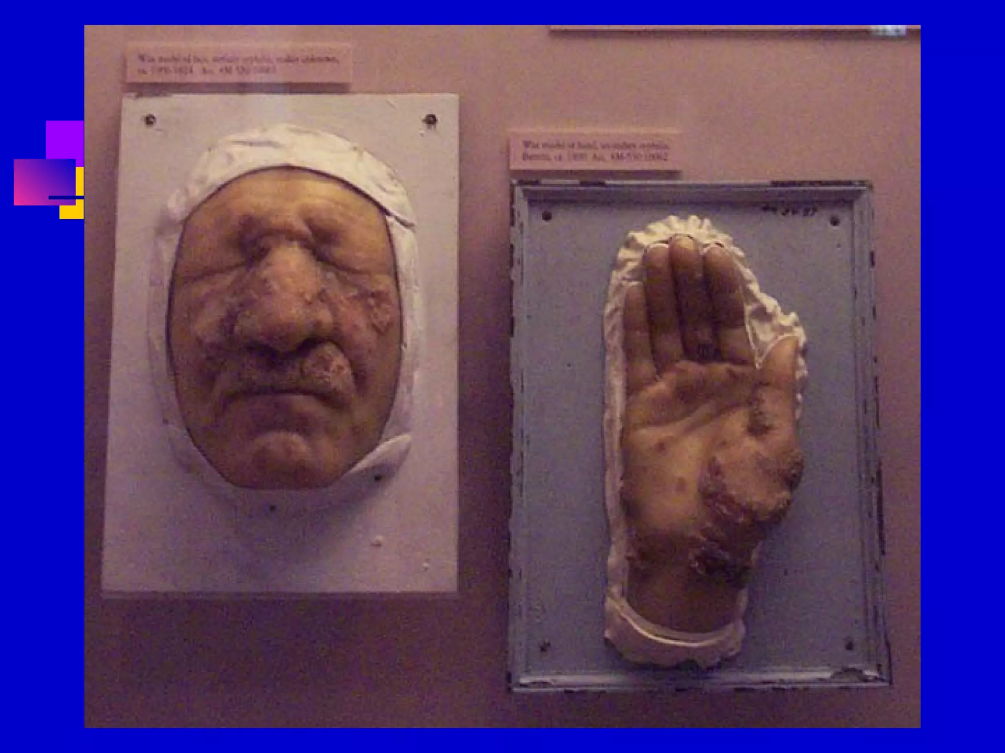
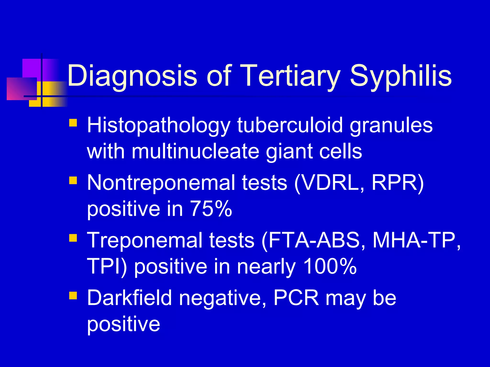
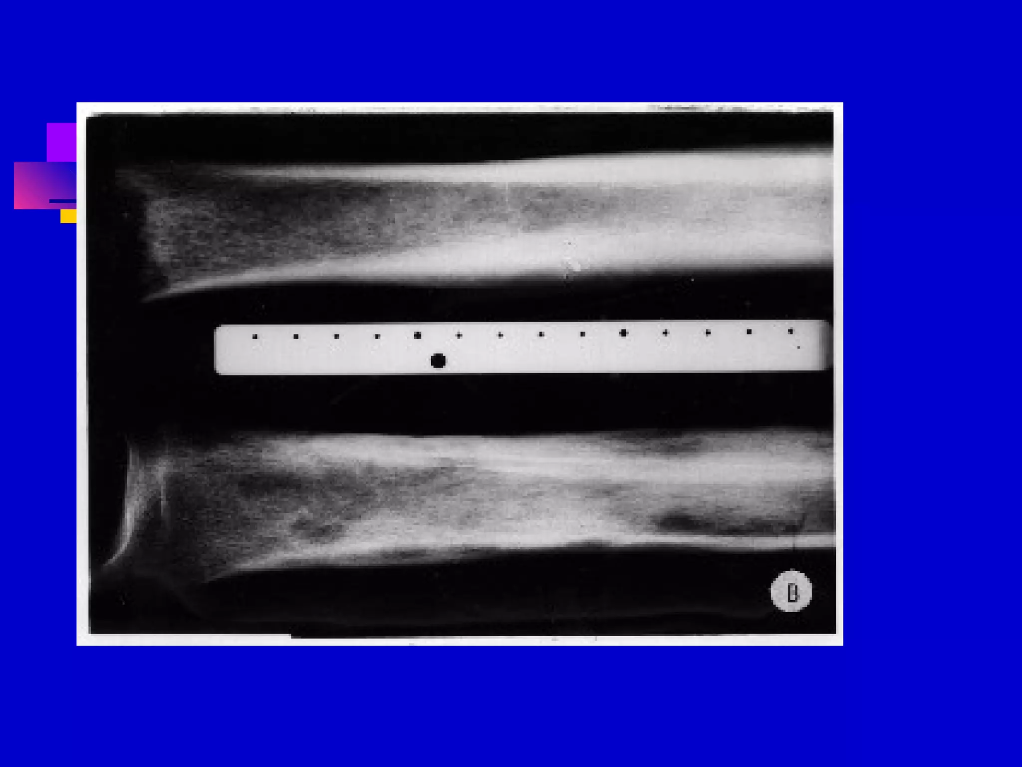
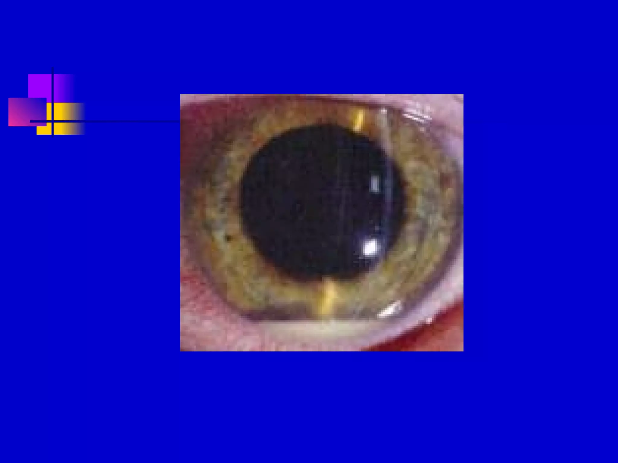
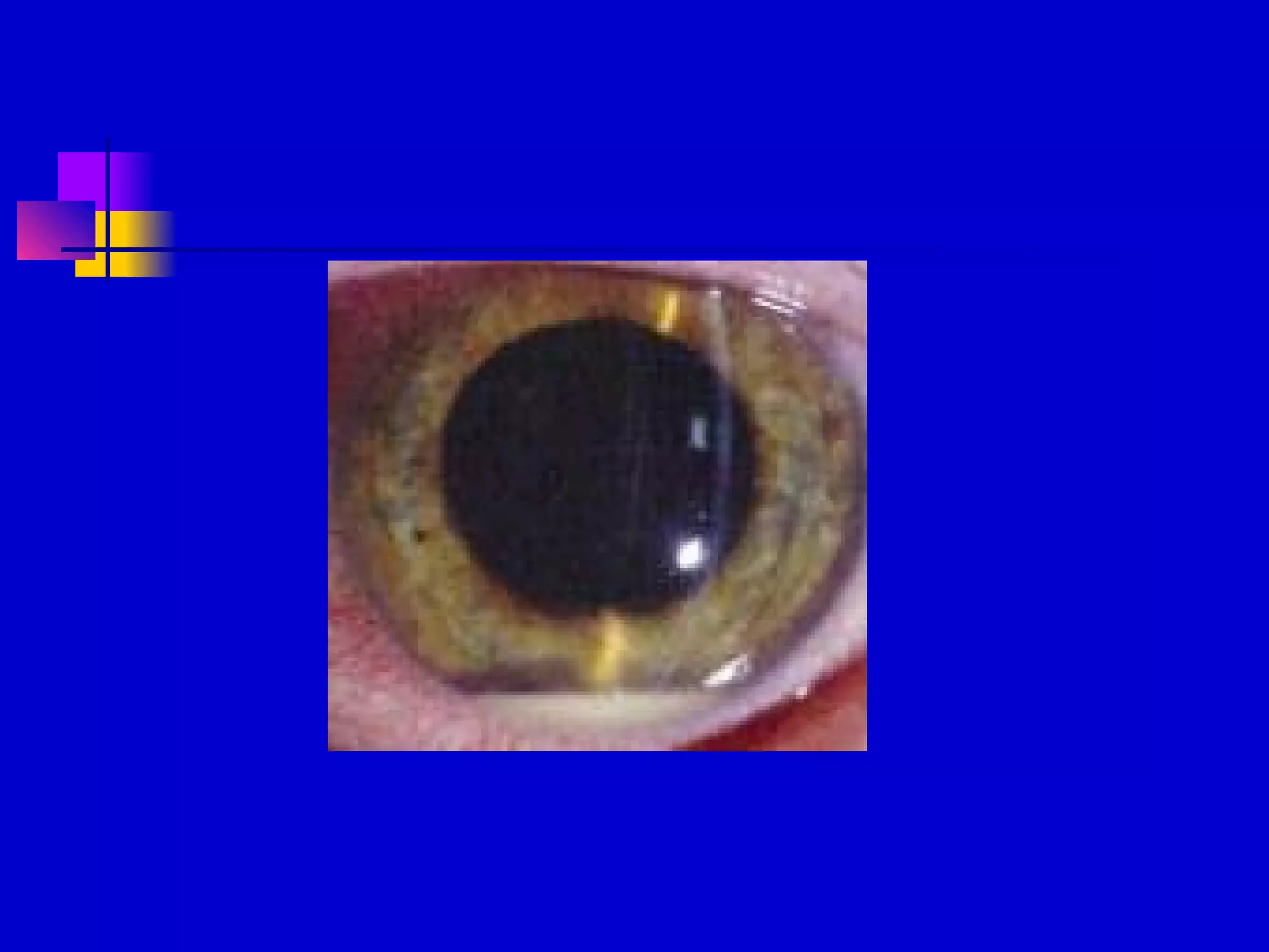
Syphilis
David M. Bracciano, D.O.
Syphilis
 Aka lues
 Contagious, sexually transmitted
disease
 Spirochete Treponema pallidum
 Enters through skin or mucous
membrane where primary
manifestations are seen
Treponema pallidum
 Spiral spirochete that is mobile
 # of spirals varies from 4 to 14
 Length 5 to 20 microns
 Can be seen on fresh primary or
secondary lesions by darkfield
microscopy or fluorescent antibody
techniques
Treponema pallidum
 Motility has three movements
 Projection and rotation in the direction
of the long axis
 Bending or twisting from side to side
 Pathogenic in apes, humans, and
rabbits
Syphilis epidemiology
 Major health problem throughout world
 U.S. 2.6 cases per 100,000 in 1999
 Lowest level ever recorded
 Concentrated in 28 counties SE U.S.
 Mainly gay men and crack cocaine
users
Syphilis epidemiology
 Enhances risk of transmission of HIV
 HIV testing recommended in all patients
with syphilis
 Reportable disease, contact tracing
Serologic Tests
 Reveal patients immune status not
whether they are currently infected
 Use lipoidal antigens rather than T.
pallidum or components of it; non-
treponemal antigen tests
 RPR; rapid plasma reagin
 VDRL; Venereal Disease Research
Laboratory
Serologic Tests
 Positive within 5 to 6 weeks after
infection
 Strongly positive in secondary phase
 Strength of reaction is stated in dilutions
 May become negative with treatment or
over decades
Serologic Tests
 To improve sensitivity and specificity tests
using a specific treponemal antigen devised
 MHA-TP: microhemagglutination assay for T.
pallidum
 FTA-ABS: fluorescent treponemal antibody
absorption test
 All positive nontreponemal test results should
be confirmed with a specific treponemal test
Serologic Tests
 Treponemal tests become positive
early, useful in confirming primary
syphilis
 Remain positive for life, useful in
diagnosing late disease
 Treatment results in loss of positivity in
13-24% of patients
Biologic False-Positive Test
Results
 Positive STS in persons with no history
or clinical evidence of syphilis
 Acute BFP: those that revert to
negative in less than 6 months
 Chronic BFP: persist > 6 months
BFP Test Results in Syphilis
 Acute BFP
 Vaccinations
 Infections
 pregnancy
 Chronic BFP
 Connective tissue
disease (SLE)
 Liver disease
 Blood transfusions
 IVDA
Cutaneous Syphilis
 Chancre is usually the first cutaneous
lesion
 18 to 21 days after infection
 Round indurated papule with eroded
surface that exudes a serous fluid
 Cartilage-like consistency
 Usually painless (Hunterian chancre is
“classic” heals without scarring)
Chancre
 Inguinal adenopathy 1-2 weeks after
chancre
 Generally occur singly, may be multiple
 Diameter mm to cm
Chancres
 Women genital chancre less often
observed due to location within the
vagina and cervix
 Edema of labia may occur
The “Dory Flop”
 “a chancre in the prepuce, being too
hard to bend, will flip over all at once
when the prepuce is drawn back”
 Resembles the movement of a dory as
it is being turned upside down
Chancre
 Untreated, the chancre heals
spontaneously in 1 to 4 months
 Constitutional symptoms begin just as
chancres disappear
 Extragenital chancre: may be larger,
frequently on lips, rarely tongue, tonsil,
breast, finger, anus.
Chancre Histology
 Ulcer covered by neutrophils and fibrin
 Dense infiltrate of lymphocytes and and
plasma cells
 Spirochetes in untreated primary syphilis with
silver stains; Warthin-Starry
 Direct fluorescent antibody tissue test (DFAT-
TP)- serous exudate collected on a slide sent
for exam
Serology
 Nontreponemal tests positive 50%
 Treponemal tests positive 90%
 Positivity depends upon duration of
infection, if chancre has been present
for several weeks, test is usually
positive
Chancre vs. Chancroid
 Incubation 3 weeks
 Painless, no ulcer,
no surrounding
inflammatory zone
 Oval, hard
 Lymphadenopathy
may be bilateral,
nontender,
nonsuppurative
 Incubation 4-7 days
 Ulcer inflamed, very
painful, inflammatory
zone
 Soft, covered by a
membrane
 Lymphadenopathy
unilateral, tender,
suppurative
Ddx in Syphilis
 Chancroid; multiple lesions, may coexist with
chancre, must r/o syphilis
 Granuloma Inguinale; indurated nodule that
erodes, soft red granulation tissue, Donovan’s
bodies in macrophages with Wright’s or
Giemsa’s stain
 Lymphogranuloma Venereum; small,
painless, superficial non indurated ulcer,
primary lesions followed in 7 to 30 days by
adenopathy
 HSV; grouped vesicles, burning pain
Secondary Syphilis
 Skin manifestations in 80% called
syphilids
 Symmetric, generalized, superficial,
macular transient; later papular,
pustular
 Early on face, shoulders, flanks, palms
and soles, anal or genital areas
Secondary Syphilis
Macular Eruptions
 Exanthematic erythema 6-8 weeks after
chancre, extends rapidly, may last
hours to months
 Round indistinct, slightly scaling ham-
colored macules
 Pain, burning absent, pruritus may be
present
 Generalized shotty adenopathy
Secondary Syphilis
Papular Eruptions
 Arise later than macular, raw-ham,
round, 2-5mm or more in diameter,
slightly raised, smooth or thick scale
 Face and flexures of arms and legs,
trunk
 Palmar and plantar yellowish-red spots
 Ollendorf’s sign; papule tender to touch
of a blunt probe
Secondary Syphilis
Papular Eruptions
 Papulosquamous syphilids may
produce a psoriasiform eruption
 Follicular or lichenoid syphilids appear
as minute scale-capped papules
 Tend to be disseminated, but may be
localized, asymmetrical, con figurate,
hypertrophic, confluent.
Secondary Syphilis
Secondary Syphilis
Papular Eruptions
 Annular syphilid mimics sarcoidosis, more
common in blacks
 Cheeks, angle of mouth, annular, gyrate,
ridges; “nickels and dimes”
 Pustular syphilid; rare, face, trunk, extremities
red small crust-covered ulceration
 Rupial syphilid; superficial ulceration is
covered with a pile of terraced crusts
resembling an oyster shell.
Secondary Syphilis
Papular Eruptions
 Lues Maligna; rare, severe ulcerations,
pustules, or rupioid lesions, accompanied by
severe constitutional symptoms.
 Condylomata lata; papular mass, weeping,
gray 1-3cm, groin, anus (not vegetative like
condylomata acuminata)
 Syphilitic alopecia; irregular, scalp has a
moth-eaten appearance 5% of pts
Secondary Syphilis
Mucous Membrane
 Present in 1/3 of secondary syphilis
 Most common is “syphilitic sore throat”
 Diffuse pharyngitis, hoarseness
 Tongue; patches of desquamation of
papillae
 Ulcerations of tongue and lips in late
stages
Secondary Syphilis
Mucous membrane
 Mucous patches are the most
characteristic mucous membrane
lesions; macerated, flat. Grayish,
rounded erosions covered by a delicate,
soggy membrane.
 Highly infectious, occur on tonsils,
tongue, pharynx, gums, lips, and buccal
areas, or on the genitalia
Secondary Syphilis
Systemic Involvement
 Lymphadenopathy common.
 Acute Glomerulonephritis, gastritis,
proctitis, hepatitis, meningitis, SNHL,
iritis, uveitis, optic neuritis, Bell’s palsy,
pulmonary nodular infiltrates,
osteomyelitis, polyarthritis.
Secondary Syphilis
Diagnosis
 Nontreponemal serologic tests for
syphilis are strongly reactive
(seronegativity rarely in AIDS)
 Spirochetes on darkfield exam
Secondary Syphilis
Ddx “Great Imitator”
 Pityriasis rosea
 Drug eruptions (pruitic)
 Lichen planus;
Wickham’s striae,
Koebner’s, pruitic
 Psoriasis; no
adenopathy
 Sarcoidosis; need
serology and silver
staining of biopsy
 Infectious
mononucleosis,
false pos RPR
 Geographic tongue
 Aphthous stomatitis
Latent Syphilis
 After the lesions of secondary syphilis have
involuted, a latent period occurs
 May last a few months or a lifetime
 60-70% of pts untreated remain
asymptomatic for life
 No clinical signs, but serologic tests positive
 Women may infect unborn child for 2 years
Late Syphilis
 Defined by CDC as infection of greater
than 1 years duration
 Tertiary Cutaneous Syphilis
 Late Osseous Syphilis
 Neurosyphilis
 Late Cardiovascular Syphilis
Tertiary Cutaneous Syphilis
 Tertiary syphilids usually occur 3-5
years after infection
 16% of untreated pts will develop
lesions of skin, mucous membrane,
bone or joints
 Skin lesions are localized, destructive,
heal with scarring
Tertiary Syphilids
 Two main types; Nodular syphilid and
the Gumma
 Nodular; reddish brown firm papules or
nodules 2mm or larger, scales.
 Gumma; larger
Nodular Tertiary Syphilid
 Lesions tend to form rings and undergo
involution as new lesions develop just beyond
 Characteristic circular or serpiginous pattern
 “kidney-shaped” lesion occurs on the
extensor surfaces of the arms and on back
 Patches have scars and fresh ulcerated
lesions
 Process may last for years, slowly marching
across large areas of skin
Gumma
 May occur as unilateral, isolated, single or
disseminated lesions, or serpiginous
 May be restricted to the skin, or originate in
deeper tissues, and break down the skin
 Lesions begin as small nodules, enlarge to
several centimeters
 Central necrosis, deep ulcer with a gummy
base, most frequent site is lower legs
Diagnosis of Tertiary Syphilis
 Histopathology tuberculoid granules
with multinucleate giant cells
 Nontreponemal tests (VDRL, RPR)
positive in 75%
 Treponemal tests (FTA-ABS, MHA-TP,
TPI) positive in nearly 100%
 Darkfield negative, PCR may be
positive
Ddx Tertiary Syphilis
 R/o tumors; SCCA tongue, leukemic
infiltrates, sarcoidosis
 Ulcerated syphilids resemble scrofula,
atypical mycobacterium, sporotrichosis,
blastomycosis
 Mycosis fungoides (CTCL) has eczema
and pruitis
 Perforation of hard palate and septum
Late Osseous Syphilis
 Gummatous lesions can involve the
periosteum and bone
 Head, face, tibia
 Periostitis, osteomyelitis, osteitis,
gummatous osteoarthritis
 “Osteocope”; bone pain often at night
 “Osteodope”; derm resident on Bourbon
street
Late Osseous Syphilis
 Charcot joint; loss of contours of joint,
hypermobility, painless
 Associated with tabes dorsalis
Neurosyphilis
 CNS involvement with syphilis can
occur at any stage
 Most are asymptomatic; CSF
pleocytosis, or a positive CSF serology
 4-10% of untreated pts will develop
neurosyphilis
Early Neurosyphilis
 First year of infection, meningeal
 Headache, stiff neck, cranial nerve
disorders (SNHL, CN V, visual),
seizures, delirium, increased ICP
Meningovascular
Neurosyphilis
 4-7 years after infection
 Thrombosis of vessels in the CNS
 Hemiplegia, aphasia, hemianopsia,
transverse myelitis, progressive
muscular atrophy
 CN palsies; CN IIX, III, IV, VI
 “Argyll Robertson Pupil”
accommodates, but doesn’t react
Late Neurosyphilis
 Parenchymatous neurosyphilis occurs
more than 10 years after infection
 Two classical patterns; Tabes Dorsalis,
and General Paresis
Tabes Dorsalis
 Degeneration of the dorsal roots of the
spinal nerves and posterior columns of
the the spinal cord
 Gastric crisis with severe pain and
vomiting is most common
 Pain, urination problems, paresthesias,
ataxia, diplopia, vertigo, deafness
Tabes Dorsalis
 Signs: Argyll Robertson pupil, reduced
lower cord reflexes, Romberg sign,
sensory loss, atonic bladder, Charcot’s
joints, optic atrophy
 Personality changes, memory loss,
apathy, megalomania, delusions,
dementia (Garcia von Lin syndrome)
Late Cardiovascular Syphilis
 Occurs in 10% of untreated pts
 Aortitis; aortic insufficiency, coronary
disease, aortic aneurysm
Congenital Syphilis
 Prenatal syphilis acquired in utero
 Infection through the placenta usually
does not occur before the fourth month,
so treatment of the mother before this
time will almost always prevent infection
in the fetus.
 If infection occurs after the fourth month
40% risk of fetal death
Congenital Syphilis
 40% of pregnancies in women with untreated
early syphilis will result in a syphilitic infant.
 Most neonates with congenital syphilis are
normal at birth.
 Early congenital syphilis; lesions occurring
within first two years of life
 Late congenital syphilis; lesion occur after two
years
Early Congenital Syphilis
 Cutaneous manifestations appear most
commonly during 3rd
week
 Snuffles (a form of Rhinitis) is most frequent,
bloody drainage, ulcers may develop, later
septal perfs
 30-60% of infants develop cutaneous lesions
similar to secondary syphilis
 Red to copper maculopapular, become large,
scaling, pustules, crusting
 Face, arms, buttocks, legs, palms and soles

Early Congenital Syphilis
 Face, perineum, and intertriginous
areas, usually fissured lesions
resembling mucous patches. Radial
scarring results leading to Rhagades
 Bone lesions occur in 70-80% ,
epiphysitis is common and causes pain
on motion, leading to infant refusing to
move; Parrot’s pseudoparalysis.
Early Congenital Syphilis
 Radiologic features of the bone lesions
in congenital syphilis during the first 6
months are characteristic.
 Bone lesions occur at the epiphyseal
ends of long bones.
 Lymphadenopathy,
hepatomegaly,nephrotic syndrome,
meningitis, nerve palsies may all occur
Late Congenital Syphilis
 Lesions are two types; malformations of
tissue affected at critical growth periods
(Stigmata), and persistent inflammatory foci
 Inflammatory; lesions of the cornea, bones,
and central nervous system. Ie; Interstitial
Keratitis in 20-50%, perisynovitis of knees
(Clutton’s joints), tabes dorsalis, seizures,
and paresis
Late Congenital Syphilis
 Malformations (Stigmata); destructive
effects leave scars or developmental
defects
 Hutchinson’s Triad; Changes in
incisors, corneal scars, and eighth
nerve deafness
 Also; saber shins, rhagades of the lips,
saddle nose, mulberry molars
Hutchinson’s Teeth
 Malformation of the central upper incisors that
appears in the second or permanent teeth.
Teeth are cylindrical rather than flattened,
cutting edge narrower than base, notch may
develop
 Mulberry molar; first molar hyperplastic, flat
occlusal surface covered with knobs
representing abortive cusps
Treatment of Syphilis
 PCN is drug of choice for treatment of
all stages of syphilis.
 HIV testing is recommended in all
patients
 If less than one year; 2.4M U of
Benzathine PCN G
 PCN-allergic; Tetracycline 500mg QID
for 14 days
Jarisch- Herxheimer Reaction
 Febrile reaction occurs after the initial
dose of antisyphilitic tx , 60-90% of pts
 6-8 hours after dose, chills, fever,
myalgia, increase in inflammation
(neurosyphilis)
Treatment of Sex Partners
 Persons exposed to a patient with early
syphilis within the previous 3 months
should be treated, even if seronegative
 Single dose azithromycin effective in
treating incubating syphilis
Serologic Testing after Tx
 VDRL or RPR repeated every 3 months in
first year, every 6 months in second year,
than annually
 A fourfold decrease in titer should be seen at
6 months, if not than 3 weekly PCN IM
injections
 Response for latent syphilis is slower, 12-24
months
 If not responding; HIV and CSF testing
repeated
 Pts with late syphilis may be “serofast”, and
Syphilis and HIV
 Most HIV pts exhibit the classic clinical
manifestations and course, and
respond similarly to tx
 More likely to present with secondary
syphilis and have a persistent chancre
 ? Of increased relapse of neurosyphilis
Yaws
 Treponema pallidum subsp.
Pertenue
 Endemic in some tropical, rural regions
 Overcrowding, poor hygiene,
transmitted by contact with infected
lesions
 Children, disabling course, affects skin,
bones, and joints
Early Yaws
 Primary papule or group of papules
appear at site of inoculation after 3
week incubation period, initial lesion
becomes larger and crusted (Mother
Yaw, maman pian )
 Feet, legs, buttocks, face, not genitals
 Mother yaw disappears after a few
months
Early Yaws
 Secondary Yaws; weeks or months after
mother yaw appears. Smaller, may appear
around primary lesion, annular (ringworm
yaws)
 Around body orifices, and creases
condylomata may develop
 Palms and soles may form hyperkeratotic
plaques, painful cause a crab-like gait (crab
yaws)
Late Yaws
 10% progress to late stage where gummas
occur
 Ulcer with clean edges that tend to fuse to
form con figurate and serpiginous patters
similar to tertiary syphilis
 Bone, joint, saddle nose, saber shin,
Gangosa (destruction of palate and nose)
 Diagnosis; Darkfield, VDRL or RPR
Endemic Syphilis (Bejel)
 Bejel is a Bedouin term for this
nonvenereal treponematosis, nomadic
tribes of North Africa, Southwest Asia,
Eastern Mediterranean
 T. pallidum subsp. Endemicum
 Childhood, skin contact, drinking
vessels
 Skin, oral mucosa, and skeletal system
Bejel
 Primary lesions rare, probably go
undetected in the oral mucosa
 Secondary oral lesions; shallow,
painless ulcers, laryngitis
 Condyloma of axillae and groin,
lymphadenopathy, osteoperiostitis
causes night leg pain
Bejel
 Untreated secondary bejel heals in 6-9
months
 Tertiary stage; gummatous ulcerations
of the skin, nasopharynx, and bone.
 Neuro; uveitis, choritis, optic atrophy
Pinta
 T. carateum; nonvenereal, endemic
 Only skin lesions occur !
 All ages, Brazilian rain forest
Pinta
 Primary Stage; 7 to 60 days after
inoculation. Lesion begins as a tiny red
papule, becomes an elevated
erythematous infiltrated plaque 10cm in
diameter over 2-3 months. Legs,
satellite lesions, no erosion or ulceration
like in chancres.
Pinta
 Secondary Stage; 5 months to 1 year
 Small, scaling papules that enlarge and
coalesce, extremities and face
 Red to blue , black with
postinflammatory hyperpigmentation
 Nontreponemal tests reactive in 60%
Pinta
 Late Dyschromic Stage; young adults,
hyperpigmented and depigmented
macules resembling vitiligo
 Face, waist, wrist, trochanteric areas
 Histo; acanthosis, lichenoid,
spirochetes in epidermis
Treatment of Yaws, Bejel, and
Pinta
 Benzathine PCN G 1.2 to 2.4 M units IM
 Tetracycline 500mg QID for adults
 EES 10mg/kg children QID for 14 days
Nonvenereal Treponematoses
 Yaws
 Endemic Syphilis
 Pinta
Treatment
 Syphilis >1year; 2.4M PCN G weekly
for 3 weeks Pcn-allergic; Tetra 500mg
QID for 30 days
 Neurosyphilis; IV
 Infant 100,000 to 150,000 units/kg/day
Procaine PCN BID for first seven days
of life
Syphilis

Syphilis

  • 2.
  • 3.
    Syphilis  Aka lues Contagious, sexually transmitted disease  Spirochete Treponema pallidum  Enters through skin or mucous membrane where primary manifestations are seen
  • 5.
    Treponema pallidum  Spiralspirochete that is mobile  # of spirals varies from 4 to 14  Length 5 to 20 microns  Can be seen on fresh primary or secondary lesions by darkfield microscopy or fluorescent antibody techniques
  • 6.
    Treponema pallidum  Motilityhas three movements  Projection and rotation in the direction of the long axis  Bending or twisting from side to side  Pathogenic in apes, humans, and rabbits
  • 8.
    Syphilis epidemiology  Majorhealth problem throughout world  U.S. 2.6 cases per 100,000 in 1999  Lowest level ever recorded  Concentrated in 28 counties SE U.S.  Mainly gay men and crack cocaine users
  • 10.
    Syphilis epidemiology  Enhancesrisk of transmission of HIV  HIV testing recommended in all patients with syphilis  Reportable disease, contact tracing
  • 13.
    Serologic Tests  Revealpatients immune status not whether they are currently infected  Use lipoidal antigens rather than T. pallidum or components of it; non- treponemal antigen tests  RPR; rapid plasma reagin  VDRL; Venereal Disease Research Laboratory
  • 16.
    Serologic Tests  Positivewithin 5 to 6 weeks after infection  Strongly positive in secondary phase  Strength of reaction is stated in dilutions  May become negative with treatment or over decades
  • 18.
    Serologic Tests  Toimprove sensitivity and specificity tests using a specific treponemal antigen devised  MHA-TP: microhemagglutination assay for T. pallidum  FTA-ABS: fluorescent treponemal antibody absorption test  All positive nontreponemal test results should be confirmed with a specific treponemal test
  • 20.
    Serologic Tests  Treponemaltests become positive early, useful in confirming primary syphilis  Remain positive for life, useful in diagnosing late disease  Treatment results in loss of positivity in 13-24% of patients
  • 21.
    Biologic False-Positive Test Results Positive STS in persons with no history or clinical evidence of syphilis  Acute BFP: those that revert to negative in less than 6 months  Chronic BFP: persist > 6 months
  • 22.
    BFP Test Resultsin Syphilis  Acute BFP  Vaccinations  Infections  pregnancy  Chronic BFP  Connective tissue disease (SLE)  Liver disease  Blood transfusions  IVDA
  • 23.
    Cutaneous Syphilis  Chancreis usually the first cutaneous lesion  18 to 21 days after infection  Round indurated papule with eroded surface that exudes a serous fluid  Cartilage-like consistency  Usually painless (Hunterian chancre is “classic” heals without scarring)
  • 25.
    Chancre  Inguinal adenopathy1-2 weeks after chancre  Generally occur singly, may be multiple  Diameter mm to cm
  • 26.
    Chancres  Women genitalchancre less often observed due to location within the vagina and cervix  Edema of labia may occur
  • 28.
    The “Dory Flop” “a chancre in the prepuce, being too hard to bend, will flip over all at once when the prepuce is drawn back”  Resembles the movement of a dory as it is being turned upside down
  • 31.
    Chancre  Untreated, thechancre heals spontaneously in 1 to 4 months  Constitutional symptoms begin just as chancres disappear  Extragenital chancre: may be larger, frequently on lips, rarely tongue, tonsil, breast, finger, anus.
  • 32.
    Chancre Histology  Ulcercovered by neutrophils and fibrin  Dense infiltrate of lymphocytes and and plasma cells  Spirochetes in untreated primary syphilis with silver stains; Warthin-Starry  Direct fluorescent antibody tissue test (DFAT- TP)- serous exudate collected on a slide sent for exam
  • 33.
    Serology  Nontreponemal testspositive 50%  Treponemal tests positive 90%  Positivity depends upon duration of infection, if chancre has been present for several weeks, test is usually positive
  • 34.
    Chancre vs. Chancroid Incubation 3 weeks  Painless, no ulcer, no surrounding inflammatory zone  Oval, hard  Lymphadenopathy may be bilateral, nontender, nonsuppurative  Incubation 4-7 days  Ulcer inflamed, very painful, inflammatory zone  Soft, covered by a membrane  Lymphadenopathy unilateral, tender, suppurative
  • 35.
    Ddx in Syphilis Chancroid; multiple lesions, may coexist with chancre, must r/o syphilis  Granuloma Inguinale; indurated nodule that erodes, soft red granulation tissue, Donovan’s bodies in macrophages with Wright’s or Giemsa’s stain  Lymphogranuloma Venereum; small, painless, superficial non indurated ulcer, primary lesions followed in 7 to 30 days by adenopathy  HSV; grouped vesicles, burning pain
  • 38.
    Secondary Syphilis  Skinmanifestations in 80% called syphilids  Symmetric, generalized, superficial, macular transient; later papular, pustular  Early on face, shoulders, flanks, palms and soles, anal or genital areas
  • 41.
    Secondary Syphilis Macular Eruptions Exanthematic erythema 6-8 weeks after chancre, extends rapidly, may last hours to months  Round indistinct, slightly scaling ham- colored macules  Pain, burning absent, pruritus may be present  Generalized shotty adenopathy
  • 42.
    Secondary Syphilis Papular Eruptions Arise later than macular, raw-ham, round, 2-5mm or more in diameter, slightly raised, smooth or thick scale  Face and flexures of arms and legs, trunk  Palmar and plantar yellowish-red spots  Ollendorf’s sign; papule tender to touch of a blunt probe
  • 43.
    Secondary Syphilis Papular Eruptions Papulosquamous syphilids may produce a psoriasiform eruption  Follicular or lichenoid syphilids appear as minute scale-capped papules  Tend to be disseminated, but may be localized, asymmetrical, con figurate, hypertrophic, confluent.
  • 44.
  • 45.
    Secondary Syphilis Papular Eruptions Annular syphilid mimics sarcoidosis, more common in blacks  Cheeks, angle of mouth, annular, gyrate, ridges; “nickels and dimes”  Pustular syphilid; rare, face, trunk, extremities red small crust-covered ulceration  Rupial syphilid; superficial ulceration is covered with a pile of terraced crusts resembling an oyster shell.
  • 46.
    Secondary Syphilis Papular Eruptions Lues Maligna; rare, severe ulcerations, pustules, or rupioid lesions, accompanied by severe constitutional symptoms.  Condylomata lata; papular mass, weeping, gray 1-3cm, groin, anus (not vegetative like condylomata acuminata)  Syphilitic alopecia; irregular, scalp has a moth-eaten appearance 5% of pts
  • 48.
    Secondary Syphilis Mucous Membrane Present in 1/3 of secondary syphilis  Most common is “syphilitic sore throat”  Diffuse pharyngitis, hoarseness  Tongue; patches of desquamation of papillae  Ulcerations of tongue and lips in late stages
  • 49.
    Secondary Syphilis Mucous membrane Mucous patches are the most characteristic mucous membrane lesions; macerated, flat. Grayish, rounded erosions covered by a delicate, soggy membrane.  Highly infectious, occur on tonsils, tongue, pharynx, gums, lips, and buccal areas, or on the genitalia
  • 52.
    Secondary Syphilis Systemic Involvement Lymphadenopathy common.  Acute Glomerulonephritis, gastritis, proctitis, hepatitis, meningitis, SNHL, iritis, uveitis, optic neuritis, Bell’s palsy, pulmonary nodular infiltrates, osteomyelitis, polyarthritis.
  • 53.
    Secondary Syphilis Diagnosis  Nontreponemalserologic tests for syphilis are strongly reactive (seronegativity rarely in AIDS)  Spirochetes on darkfield exam
  • 55.
    Secondary Syphilis Ddx “GreatImitator”  Pityriasis rosea  Drug eruptions (pruitic)  Lichen planus; Wickham’s striae, Koebner’s, pruitic  Psoriasis; no adenopathy  Sarcoidosis; need serology and silver staining of biopsy  Infectious mononucleosis, false pos RPR  Geographic tongue  Aphthous stomatitis
  • 56.
    Latent Syphilis  Afterthe lesions of secondary syphilis have involuted, a latent period occurs  May last a few months or a lifetime  60-70% of pts untreated remain asymptomatic for life  No clinical signs, but serologic tests positive  Women may infect unborn child for 2 years
  • 57.
    Late Syphilis  Definedby CDC as infection of greater than 1 years duration  Tertiary Cutaneous Syphilis  Late Osseous Syphilis  Neurosyphilis  Late Cardiovascular Syphilis
  • 58.
    Tertiary Cutaneous Syphilis Tertiary syphilids usually occur 3-5 years after infection  16% of untreated pts will develop lesions of skin, mucous membrane, bone or joints  Skin lesions are localized, destructive, heal with scarring
  • 59.
    Tertiary Syphilids  Twomain types; Nodular syphilid and the Gumma  Nodular; reddish brown firm papules or nodules 2mm or larger, scales.  Gumma; larger
  • 61.
    Nodular Tertiary Syphilid Lesions tend to form rings and undergo involution as new lesions develop just beyond  Characteristic circular or serpiginous pattern  “kidney-shaped” lesion occurs on the extensor surfaces of the arms and on back  Patches have scars and fresh ulcerated lesions  Process may last for years, slowly marching across large areas of skin
  • 62.
    Gumma  May occuras unilateral, isolated, single or disseminated lesions, or serpiginous  May be restricted to the skin, or originate in deeper tissues, and break down the skin  Lesions begin as small nodules, enlarge to several centimeters  Central necrosis, deep ulcer with a gummy base, most frequent site is lower legs
  • 66.
    Diagnosis of TertiarySyphilis  Histopathology tuberculoid granules with multinucleate giant cells  Nontreponemal tests (VDRL, RPR) positive in 75%  Treponemal tests (FTA-ABS, MHA-TP, TPI) positive in nearly 100%  Darkfield negative, PCR may be positive
  • 67.
    Ddx Tertiary Syphilis R/o tumors; SCCA tongue, leukemic infiltrates, sarcoidosis  Ulcerated syphilids resemble scrofula, atypical mycobacterium, sporotrichosis, blastomycosis  Mycosis fungoides (CTCL) has eczema and pruitis  Perforation of hard palate and septum
  • 68.
    Late Osseous Syphilis Gummatous lesions can involve the periosteum and bone  Head, face, tibia  Periostitis, osteomyelitis, osteitis, gummatous osteoarthritis  “Osteocope”; bone pain often at night  “Osteodope”; derm resident on Bourbon street
  • 69.
    Late Osseous Syphilis Charcot joint; loss of contours of joint, hypermobility, painless  Associated with tabes dorsalis
  • 71.
    Neurosyphilis  CNS involvementwith syphilis can occur at any stage  Most are asymptomatic; CSF pleocytosis, or a positive CSF serology  4-10% of untreated pts will develop neurosyphilis
  • 74.
    Early Neurosyphilis  Firstyear of infection, meningeal  Headache, stiff neck, cranial nerve disorders (SNHL, CN V, visual), seizures, delirium, increased ICP
  • 75.
    Meningovascular Neurosyphilis  4-7 yearsafter infection  Thrombosis of vessels in the CNS  Hemiplegia, aphasia, hemianopsia, transverse myelitis, progressive muscular atrophy  CN palsies; CN IIX, III, IV, VI  “Argyll Robertson Pupil” accommodates, but doesn’t react
  • 77.
    Late Neurosyphilis  Parenchymatousneurosyphilis occurs more than 10 years after infection  Two classical patterns; Tabes Dorsalis, and General Paresis
  • 78.
    Tabes Dorsalis  Degenerationof the dorsal roots of the spinal nerves and posterior columns of the the spinal cord  Gastric crisis with severe pain and vomiting is most common  Pain, urination problems, paresthesias, ataxia, diplopia, vertigo, deafness
  • 79.
    Tabes Dorsalis  Signs:Argyll Robertson pupil, reduced lower cord reflexes, Romberg sign, sensory loss, atonic bladder, Charcot’s joints, optic atrophy  Personality changes, memory loss, apathy, megalomania, delusions, dementia (Garcia von Lin syndrome)
  • 81.
    Late Cardiovascular Syphilis Occurs in 10% of untreated pts  Aortitis; aortic insufficiency, coronary disease, aortic aneurysm
  • 82.
    Congenital Syphilis  Prenatalsyphilis acquired in utero  Infection through the placenta usually does not occur before the fourth month, so treatment of the mother before this time will almost always prevent infection in the fetus.  If infection occurs after the fourth month 40% risk of fetal death
  • 84.
    Congenital Syphilis  40%of pregnancies in women with untreated early syphilis will result in a syphilitic infant.  Most neonates with congenital syphilis are normal at birth.  Early congenital syphilis; lesions occurring within first two years of life  Late congenital syphilis; lesion occur after two years
  • 86.
    Early Congenital Syphilis Cutaneous manifestations appear most commonly during 3rd week  Snuffles (a form of Rhinitis) is most frequent, bloody drainage, ulcers may develop, later septal perfs  30-60% of infants develop cutaneous lesions similar to secondary syphilis  Red to copper maculopapular, become large, scaling, pustules, crusting  Face, arms, buttocks, legs, palms and soles 
  • 87.
    Early Congenital Syphilis Face, perineum, and intertriginous areas, usually fissured lesions resembling mucous patches. Radial scarring results leading to Rhagades  Bone lesions occur in 70-80% , epiphysitis is common and causes pain on motion, leading to infant refusing to move; Parrot’s pseudoparalysis.
  • 88.
    Early Congenital Syphilis Radiologic features of the bone lesions in congenital syphilis during the first 6 months are characteristic.  Bone lesions occur at the epiphyseal ends of long bones.  Lymphadenopathy, hepatomegaly,nephrotic syndrome, meningitis, nerve palsies may all occur
  • 89.
    Late Congenital Syphilis Lesions are two types; malformations of tissue affected at critical growth periods (Stigmata), and persistent inflammatory foci  Inflammatory; lesions of the cornea, bones, and central nervous system. Ie; Interstitial Keratitis in 20-50%, perisynovitis of knees (Clutton’s joints), tabes dorsalis, seizures, and paresis
  • 90.
    Late Congenital Syphilis Malformations (Stigmata); destructive effects leave scars or developmental defects  Hutchinson’s Triad; Changes in incisors, corneal scars, and eighth nerve deafness  Also; saber shins, rhagades of the lips, saddle nose, mulberry molars
  • 91.
    Hutchinson’s Teeth  Malformationof the central upper incisors that appears in the second or permanent teeth. Teeth are cylindrical rather than flattened, cutting edge narrower than base, notch may develop  Mulberry molar; first molar hyperplastic, flat occlusal surface covered with knobs representing abortive cusps
  • 92.
    Treatment of Syphilis PCN is drug of choice for treatment of all stages of syphilis.  HIV testing is recommended in all patients  If less than one year; 2.4M U of Benzathine PCN G  PCN-allergic; Tetracycline 500mg QID for 14 days
  • 93.
    Jarisch- Herxheimer Reaction Febrile reaction occurs after the initial dose of antisyphilitic tx , 60-90% of pts  6-8 hours after dose, chills, fever, myalgia, increase in inflammation (neurosyphilis)
  • 94.
    Treatment of SexPartners  Persons exposed to a patient with early syphilis within the previous 3 months should be treated, even if seronegative  Single dose azithromycin effective in treating incubating syphilis
  • 96.
    Serologic Testing afterTx  VDRL or RPR repeated every 3 months in first year, every 6 months in second year, than annually  A fourfold decrease in titer should be seen at 6 months, if not than 3 weekly PCN IM injections  Response for latent syphilis is slower, 12-24 months  If not responding; HIV and CSF testing repeated  Pts with late syphilis may be “serofast”, and
  • 97.
    Syphilis and HIV Most HIV pts exhibit the classic clinical manifestations and course, and respond similarly to tx  More likely to present with secondary syphilis and have a persistent chancre  ? Of increased relapse of neurosyphilis
  • 98.
    Yaws  Treponema pallidumsubsp. Pertenue  Endemic in some tropical, rural regions  Overcrowding, poor hygiene, transmitted by contact with infected lesions  Children, disabling course, affects skin, bones, and joints
  • 100.
    Early Yaws  Primarypapule or group of papules appear at site of inoculation after 3 week incubation period, initial lesion becomes larger and crusted (Mother Yaw, maman pian )  Feet, legs, buttocks, face, not genitals  Mother yaw disappears after a few months
  • 102.
    Early Yaws  SecondaryYaws; weeks or months after mother yaw appears. Smaller, may appear around primary lesion, annular (ringworm yaws)  Around body orifices, and creases condylomata may develop  Palms and soles may form hyperkeratotic plaques, painful cause a crab-like gait (crab yaws)
  • 103.
    Late Yaws  10%progress to late stage where gummas occur  Ulcer with clean edges that tend to fuse to form con figurate and serpiginous patters similar to tertiary syphilis  Bone, joint, saddle nose, saber shin, Gangosa (destruction of palate and nose)  Diagnosis; Darkfield, VDRL or RPR
  • 106.
    Endemic Syphilis (Bejel) Bejel is a Bedouin term for this nonvenereal treponematosis, nomadic tribes of North Africa, Southwest Asia, Eastern Mediterranean  T. pallidum subsp. Endemicum  Childhood, skin contact, drinking vessels  Skin, oral mucosa, and skeletal system
  • 107.
    Bejel  Primary lesionsrare, probably go undetected in the oral mucosa  Secondary oral lesions; shallow, painless ulcers, laryngitis  Condyloma of axillae and groin, lymphadenopathy, osteoperiostitis causes night leg pain
  • 108.
    Bejel  Untreated secondarybejel heals in 6-9 months  Tertiary stage; gummatous ulcerations of the skin, nasopharynx, and bone.  Neuro; uveitis, choritis, optic atrophy
  • 110.
    Pinta  T. carateum;nonvenereal, endemic  Only skin lesions occur !  All ages, Brazilian rain forest
  • 112.
    Pinta  Primary Stage;7 to 60 days after inoculation. Lesion begins as a tiny red papule, becomes an elevated erythematous infiltrated plaque 10cm in diameter over 2-3 months. Legs, satellite lesions, no erosion or ulceration like in chancres.
  • 113.
    Pinta  Secondary Stage;5 months to 1 year  Small, scaling papules that enlarge and coalesce, extremities and face  Red to blue , black with postinflammatory hyperpigmentation  Nontreponemal tests reactive in 60%
  • 114.
    Pinta  Late DyschromicStage; young adults, hyperpigmented and depigmented macules resembling vitiligo  Face, waist, wrist, trochanteric areas  Histo; acanthosis, lichenoid, spirochetes in epidermis
  • 116.
    Treatment of Yaws,Bejel, and Pinta  Benzathine PCN G 1.2 to 2.4 M units IM  Tetracycline 500mg QID for adults  EES 10mg/kg children QID for 14 days
  • 117.
    Nonvenereal Treponematoses  Yaws Endemic Syphilis  Pinta
  • 118.
    Treatment  Syphilis >1year;2.4M PCN G weekly for 3 weeks Pcn-allergic; Tetra 500mg QID for 30 days  Neurosyphilis; IV  Infant 100,000 to 150,000 units/kg/day Procaine PCN BID for first seven days of life